






Abstract
Reactive cutaneous capillary endothelial proliferation (RCCEP) is the most common immune-related adverse event induced by camrelizumab (SHR-1210). Very rare cases have been reported in oral tissues, especially the oral mucosa. We reported a 67-year-old male with Grade 3 RCCEP. Multiple dome-shaped and bright red papules were first observed in the oral mucosa, which gradually developed on his lip, face, scalp, neck, foot, calf, abdomen and groin. The patient's symptoms gradually improved at 4 weeks after SHR-1210 discontinuation and were mostly relieved at 7 weeks after discontinuation. Our findings revealed that oral examination should be performed regularly during SHR-1210 treatment.
Keywords
Introduction
Over the past decades, immunotherapy has shown remarkable efficacy in cancer treatment. 1 The usage of immune checkpoint inhibitors, which could produce a longer immune response, has opened a new era of tumor immunotherapy. The extensive use of immune checkpoint inhibitors was followed by more immune-related adverse reactions. 2 A monoclonal antibody targeting programmed cell death 1 (PD-1), camrelizumab (SHR-1210), was approved by the National Medical Products Administration and has been widely used in the treatment of various malignant tumors.3–5 Reactive cutaneous capillary endothelial proliferation (RCCEP) is a unique and common immune-related adverse event associated with camrelizumab use, which is mistakenly called reactive capillary hemangioma.6,7 Notably, most RCCEP was reported to occur in the skin, 8 and this side effect was rarely observed in oral tissues. Here, we reported a rare male patient who experienced Grade 3 RCCEP, which first manifested in the oral mucosa and gradually developed on his lip, face, scalp, neck, foot, calf, abdomen and groin during SHR-1210 treatment for advanced hepatocellular carcinoma. The reporting of this study conforms to case report guidelines. 9
Case Presentation
A 67-year-old male patient presented to our department due to multiple red, erythematous, vascular-appearing papules and papulonodules on the face, mouth, trunk and extremities. The patient reported that the lesions bled when touched. Except for a tingling sensation after lesion rupture, there were no other symptoms of mucosal or cutaneous lesions. In particular, the patient bore a huge psychological burden that the bleeding lesions in the oral mucosa may result in suffocation when sleeping at night. He had a 40-year drinking history and had abstained for 5 years. Two decayed teeth had been repaired by resin injection. Two months prior, the patient was diagnosed with poorly differentiated hepatocellular carcinoma of the right hepatic lobe. One month prior, the patient underwent tumor resection. Chemotherapy (oral 12 mg lenvatinib once daily combined with intravenous 200 mg SHR-1210 once every 3 weeks) was prescribed. However, lenvatinib was self-discontinued by the patient because of adverse gastrointestinal reactions after taking it for 10 consecutive days. On the 18th day after the first injection of SHR-1210, multiple red papules were first observed in the oral mucosa. Initially, reactive lymphoid follicular hyperplasia was highly suspected. Medical observation and no treatment were adopted. Three days later, the second injection of SHR-1210 was administered. On the seventh day after the second injection of SHR-1210, the patient presented to our department due to aggravating symptoms.
A physical examination (Figure 1) showed that the area of skin lesions had increased, and some skin lesions were ruptured and exuded. An increasing number of red papules 2–10 mm in size emerged and gradually developed at sites such as the lip, face, scalp, neck, foot, calf, abdomen and groin. Some red papules in the mouth grew larger and exhibited dark purple patches. These red papules and nodules were mainly characterized as “red-nevus-like,” “pearl-like,” “mulberry-like” and “tumor-like” types. “Red-nevus-like” and “patch-like” red papules were observed in the oral mucosa and lip.

Clinical images of reactive cutaneous capillary endothelial proliferation (RCCEP), which firstly manifested in the oral mucosa, and gradually developed in the sites such as lip, face, scalp, neck, lip, foot, calf, abdomen and groin (yellow arrows).
Histological examination (Figure 2) of the red papules and nodules revealed a large number of proliferating capillaries, capillary endothelial cell proliferation and nutrient vessels with a larger lumen. On the basis of these clinical and histological findings, a diagnosis of RCCEP was made. However, the patient was given no special treatment, although 2 nodules were completely excised under local anesthesia. Ruptured lesions underwent compression hemostasis and anti-infection (topical fusidic acid cream, twice a day) treatment. Chlorhexidine gargle solution (three times a day) was used for oral cleaning. The usage of SHR-1210 was not recommended in follow-up care and treatment. At the fourth week after the second injection of SHR-1210, generalized nodules of the patient gradually subsided after drug withdrawal. At the end of the seventh week after the second injection of SHR-1210, RCCEP remission was observed. The timeline with relevant data from the episode of care for the patient is shown in Figure 3. According to the Naranjo scale adverse drug reaction score sheet (Table 1), RCCEP was most likely caused by the injection of SHR-1210.
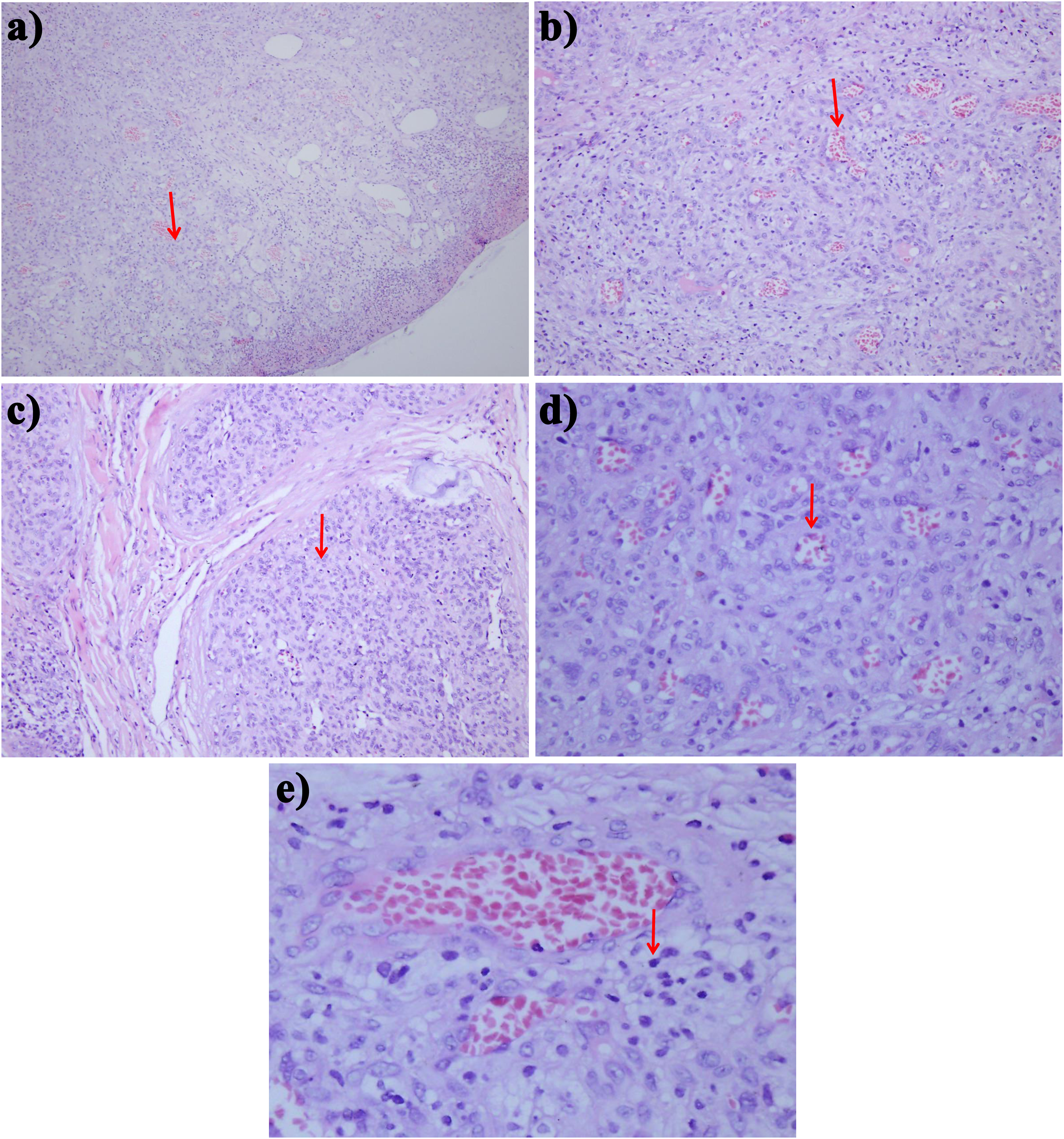
Histopathological pictures of hematoxylin and eosin (H&E) staining. (a) The lesion is located in the dermis showing large amounts of proliferating capillaries (red arrow, 100 × ); (b) The endothelial cells of capillaries are multi-layered, and a large number of red blood cells can be seen in the lumen (red arrow, 100 × ); (c) Capillaries in deep dermis are lobulated or arranged in nodules (red arrow, 200 × ); (d) Capillary endothelial cell proliferation and nutrient vessels with larger lumen are seen in the lobules (red arrow, 400 × ); (e) A small amount of lymphocyte infiltration can be seen around capillaries (red arrow, 400 × ).
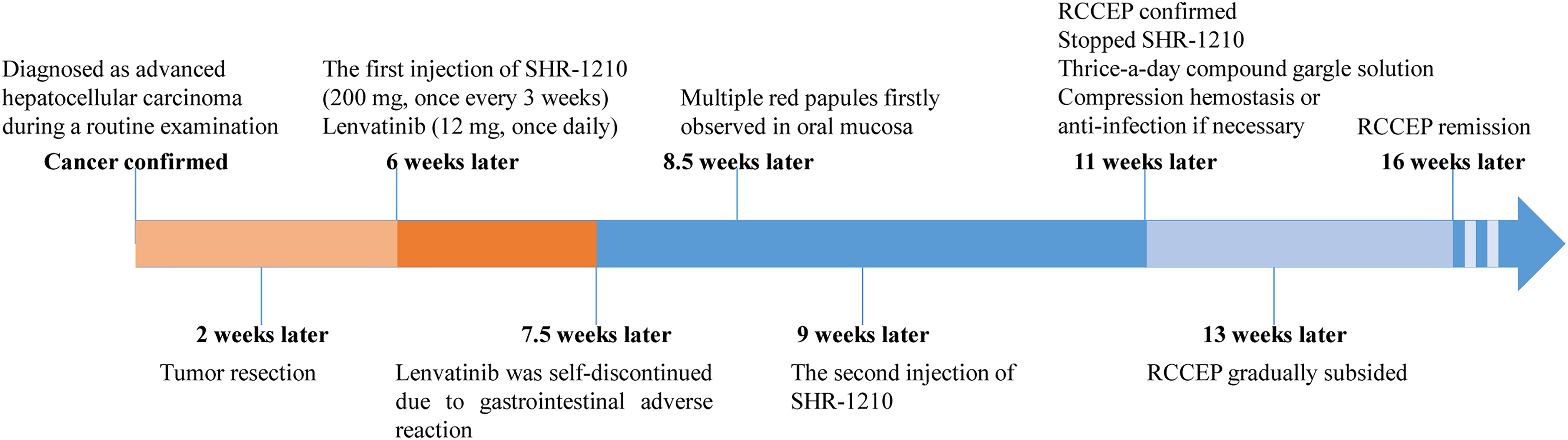
The timeline of different treatments for the patient with reactive cutaneous capillary endothelial proliferation (RCCEP).
Naranjo's assessment scale in the evaluation of adverse drug reactions.
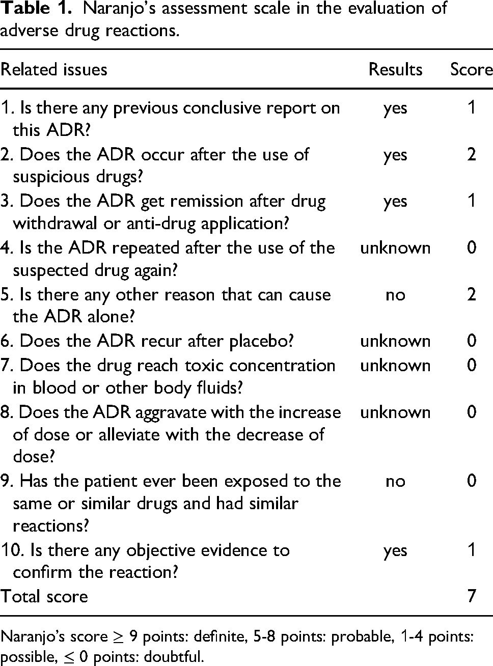
Naranjo's score ≥ 9 points: definite, 5-8 points: probable, 1-4 points: possible, ≤ 0 points: doubtful.
Discussion
The development of hepatocellular carcinoma can be modulated by the immune system. 10 Immunotherapy with immune checkpoint inhibitors has demonstrated strong antitumor activity in various tumors 11 and has or will soon become a first-line therapy for hepatocellular carcinoma. Liver tumors can evade antitumor immune responses by expressing the corresponding ligands in stromal cells and tumors. 12 Immune checkpoint inhibitors block the interaction of checkpoint proteins with their ligands, thereby preventing the inactivation of T cells. PD-1 is a coinhibitory receptor for programmed cell death ligand 1 and ligand 2 (PD-L1 and PD-L2) and is mainly expressed by activated T cells, B cells and myeloid cells. 13 Anti-PD-1 monoclonal antibodies bind specifically to PD-1, block the interaction between PD-1 and its ligands PD-L1/PD-L2, and induce T cells to recover their immune response to tumors and kill tumor cells. Among the immune-related adverse events induced by the current PD-1/PD-L1 monoclonal antibodies, lichenoid dermatitis, eczema and vitiligo are the most common adverse skin reactions, which are mostly Grade 1 or Grade 2. 14 The anti-PD-1 agent SHR-1210 is a fully humanized and high-affinity selective IgG4-κ monoclonal antibody. In a Phase 2 study, SHR-1210 showed good results, with an objective response rate of 14.7%, a disease control rate of 44.2% and a 6-month overall survival rate of 74.4% in patients with previously treated advanced hepatocellular carcinoma. 15 However, the majority of camrelizumab-treated patients (66.8%, 145/217) exhibited Grade 1/2 RCCEP on the skin surface of the head, face and trunk divided into “red-nevus-like,” “patch-like,” “mulberry-like,” “pearl-like” and “tumor-like” types. 8 Although gingival reactive capillary hemangioma was reported in a patient with lung cancer during SHR-1210 treatment, RCCEP in the oral mucosa was a very rare occurrence. Here, we reported a rare case of Grade 3 RCCEP that first manifested in the oral mucosa and was highly related to SHR-1210 administration.
It has been reported that treatment with low-dosage antiangiogenic drugs, such as apatinib and lenvatinib, could improve the symptoms of RCCEP.8,16 In our case, several red erythematous nodules were first reported in the oral mucosa on the 18th day after the first injection of SHR-1210 by the patient at the oncology department in our hospital, and lenvatinib had been stopped 8 days prior. No lesions at other sites were observed. Subsequently, an increasing number of red papules emerged and developed at sites such as the lip, face, scalp, neck, foot, calf, abdomen and groin. Lenvatinib was excluded as the cause. First, lenvatinib use was interrupted due to the common adverse gastrointestinal reaction. Second, lenvatinib functions as an oral multikinase inhibitor by blocking signal transduction after binding to vascular endothelial growth factor. As opposed to other PD-1 inhibitors, RCCEP is an immune-related adverse event unique to SHR-1210 treatment. Histological examination revealed capillary hyperplasia and capillary endothelial hyperplasia. RCCEP remission was observed after SHR-1210 was stopped for 7 weeks. Spontaneous regression of capillary hemangioma was reported after the termination of SHR-1210. 17 In addition, RCCEP was evaluated by the Naranjo scale adverse drug reaction score sheet, which supported our conclusions. Thus, it was speculated that RCCEP was induced by SHR-1210.
To date, the underlying mechanism of RCCEP induced by SHR-1210 is still unclear. A possible explanation is that SHR-1210 excessively activated immune function and disturbed the balance of proangiogenic factors and antiangiogenic factors, thus promoting vascular endothelial growth factor-2 expression and the proliferation of capillary endothelial cells.8,18 SHR-1210 may promote interleukin-4 secretion and stimulate recombinant cluster of differentiation (CD163) and M2 macrophage differentiation by activating CD4+ T cells, thus promoting vascular proliferation with the release of vascular endothelial growth factor A. 8 Histopathological analysis of the lesion tissue revealed strong staining of CD31 and high expression of Ki-67, vascular endothelial growth factor A and vascular endothelial growth factor receptor 2. Moreover, the enhanced activity of T cells against antigens may increase the levels of inflammatory cytokines and original autoantibodies. In Qing Yu's study, a rare reactive capillary hemangioma in the gingiva induced by SHR-1210 was caused by inadequate oral hygiene, which enhanced the interaction between gingival tissue and the drug.6,19 Alternatively, many immune-related adverse events, such as cytokine release syndrome, were proven to be autoinflammatory, caused by autoreactive T cells that acted on normal tissues after being released, rather than autoimmune.20,21 Similarly, acute mastitis that occurred after RCCEP might be a new immune-related adverse event caused by SHR-1210. 22 Considering that the combination of SHR-1210 and lenvatinib did not inhibit the occurrence of RCCEP in our patient, more studies are required to confirm the underlying mechanism of RCCEP.
According to the severity of symptoms, RCCEP is classified from Grade 1 to Grade 5. 8 In general, no special action should be taken for Grade 1 or Grade 2 RCCEP induced by SHR-1210. The majority of RCCEP lesions regress spontaneously within 1 to 2 months after SHR-1210 treatment. If the lesion is large or shows a high risk of bleeding, laser or minor resection should be considered. In Grades 3–5 RCCEP, generalized nodules develop throughout the body, which can cause skin infection, life-threatening conditions, and even death. In cases of rupture and skin infection, local or systemic antibiotic treatment should be considered. In our patient, Grade 3 RCCEP developed. In particular, lesions in the oral mucosa not only caused problems in the patient's daily life during eating and speaking but also imposed a huge psychological burden due to the fear of suffocation caused by their rupture and bleeding. Thus, SHR-1210 treatment was discontinued. Fortunately, the Grade 3 RCCEP spontaneously and gradually subsided after SHR-1210 withdrawal. The patient agreed with the inferred diagnosis and was satisfied with the treatment result.
In conclusion, the specific mechanism of RCCEP needs to be further explored, especially the link between antitumor immunity and antiangiogenesis. Although RCCEP occurrence positively correlates with good outcomes of SHR-1210 treatment, there is a need for therapies that avoid the occurrence of RCCEP or, more so, effective therapies to treat patients with Grade 3 to 5 RCCEP in clinical practice. Moreover, considering that RCCEP is rarely reported and easily misdiagnosed as pyogenic granuloma, hemangioma, angiosarcoma, aphthous stomatitis, mucosal blister, oral mucosal erythema, oral polyps or oral cancer, oral examination should be performed regularly during SHR-1210 treatment.
Footnotes
Authors’ Contributions
LY provided input into the concept and design of the study. LY and LH collected and assembled the data, wrote the article and critically revised the article. All authors have read and approved the final version at the time of submission.
Availability of Data and Materials
All data generated or analyzed during this study are included in this enclosed article.
Declaration of Conflicting Interests
The author(s) declared no potential conflicts of interest with respect to the research, authorship, and/or publication of this article.
Funding
The author(s) received no financial support for the research, authorship, and/or publication of this article
Ethics Approval and Consent to Participate
The study was conducted in accordance with the principles of the Declaration of Helsinki and Good Clinical Practice guidelines. The study was approved by the Ethics Committee of The Third Central Hospital of Tianjin (TCH20220325). Written informed consent was obtained from individual.
Informed Consent
Written informed consent was obtained from the patient for publication of this case report and any accompanying images. A copy of the written consent is available for review by the Editor of this journal.
Trial Registration
This article is a case report, which does not contain any clinical trials.
