
Abstract
Objectives. Emmprin (CD147/BSG) protein is estimated to play a key role in cell migration and chemoresistance in viral carcinogenesis. However, there are very limited studies investigating the CD147 in the oncogenesis of Kaposi's sarcoma-associated herpesvirus. This study aims to reveal the relationship between CD147 expression with histopathological parameters, disease pattern, and recurrence in Kaposi's sarcoma (KS). Methods. The study included 67 patients diagnosed with KS between January 1982 and September 2023. Clinical and histopathological features were analyzed retrospectively. HHV-8, CD31, and CD147 expressions were evaluated by immunohistochemistry. Results. Sixteen (24%) female and 51 (76%) male patients with median age of 64 (10-86) were included in the study. CD147 was positive in 57 (85%) cases and associated with nodular pattern (P = .001), presence of solid/fibrosarcomatous area (P = .005), and high mitotic activity (P = .035). The disease relapsed in 17 (27%) of the 63 patients with median 2 (0-12) years follow-up. While a 5-year relapse-free survival was 48.5% in the CD147 diffuse positive group, it was 83.4% in focal positive and 100% in negative cases (P = .029). Conclusion. Our study exhibited the relationship between CD147 overexpression and recurrence in KS, but the inhomogeneity of the treatment groups and the small number of patients should also be considered. These findings may provide insight into the pathogenesis of KS and the development of targeted therapies in the future.
Keywords
Introduction
Kaposi's sarcoma (KS) is a low-grade vascular tumor caused by “Kaposi's sarcoma-associated herpesvirus/human herpesvirus-8” (KSHV/HHV-8). It was first described by the Hungarian dermatologist Moritz Kaposi in 1872. 1 Four different clinical/epidemiological subtypes of the disease have been defined as classical (European), epidemic (acquired immunodeficiency syndrome-related), endemic (African), and iatrogenic. In 1994, Chang et al suggested that HHV-8 is the cause of all KS types. 2 Although there is no microscopical difference between clinical types, three stages can be observed macroscopically/clinically called patch type, plaque-type, and nodular lesion. KS is mostly encountered in men as a slow-growing lesion in the form of purple-colored macules or nodules in the distal parts of the extremities. The virus can be transmitted by saliva, solid organ transplantation, drug use, blood transfusion, or sexual.
Recent studies suggest that emmprin (CD147/BSG) protein plays an important role in cell migration and chemoresistance in viral carcinogenesis and overexpression of this is related to an advanced stage, malignant progression, and recurrence in solid tumors.3,4 The overexpression of CD147 has previously been reported to be associated with the progression of KS. 5 The aim of this study is to examine the relationship between CD147 expression and histopathological parameters, disease pattern, and recurrence risk in KS.
Materials and Methods
Patient Selection
A total of 79 patients whose biopsies were diagnosed with KS between January 1992 and September 2023 were included in the study. After immunohistochemical examination, 12 HHV8 negative/undetermined cases or cases with limited tissue were excluded yielding 67 cases. Demographic and clinical characteristics of the patients were achieved from the computer network data entry system and patient files archive. Ethics committee approval for the study was obtained from the local ethics committee (Decision number: 14/17-30).
Pathological Examination
Hematoxylin–eosin (HE) stained sections were evaluated with an optical microscope for the following 15 morphological properties: type of biopsy (punch/incision/excision); growth pattern (nodular/superficial); subcutaneous involvement; periadnexal extension; presence of central miniature veins, peripheral ectatic veins, solid-fibrosarcomatous area, papillary glomeruloid area, honeycomb-like area, and lymphangioma like area; presence of dissection in collagen fibers; presence of vessels parallel to the epidermis; presence of hyaline globules; and mitotic activity/10 high power fields (HPF). Sections were stained for HHV-8, CD31, and CD147 using the Leica Biosystems, Bond-Max Fully Automated Immunohistochemistry (IHC) device. The appropriate staining pattern (nuclear and cytoplasmic) for each marker was considered. For HHV-8 and CD31, which are used for diagnostic confirmation, the staining was evaluated as negative or positive (Figure 1). For CD147, the staining distribution was also categorized into three groups (no staining; focal staining; diffuse staining) (Figure 2).

Images of hematoxylin & eosin stained tumoral tissue (A, 200×) and HHV-8 positive immunohistochemical staining (B, 200×).

CD147 immunohistochemical staining patterns. (A) Cytoplasmic CD147 expression, control tissue-liver (IHC, 200×). (B) CD147, negative staining (IHC, 200×). (C) CD147, focal cytoplasmic staining (IHC, 200×). (D) CD147, diffuse cytoplasmic staining (IHC, 200×).
Statistical Analysis
Statistical analysis was performed by SPSS® 27.0 for Windows software. Descriptive statistics were presented as frequency (percentage), mean ± standard deviation, or median (min-max). The χ2 and Fisher's exact tests were used to compare the proportions in different categorical groups. Survival estimates were calculated with the Kaplan–Meier method. The log-rank test was used to identify the independent effects on survival. Prediction of relapse by CD147 expression was specified using Cox regression analysis. An overall type-1 error level was used to infer statistical significance.
Results
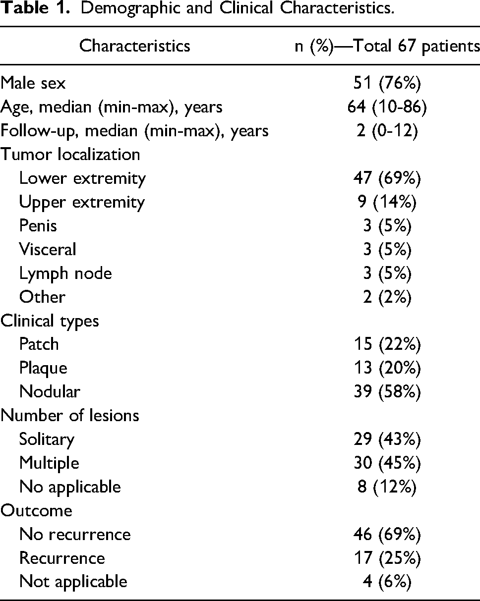
A total of 67 cases (median age 64 [10-86]), 16 (24%) female and 51 (76%) male, were included in the study. Eighteen patients had a follow-up of more than 5 years, and 45 had a follow-up of fewer than 5 years (median follow-up 2 years, range 0-12); follow-up data could not be obtained for four patients. The most common location of the tumor is in the lower (69%) and upper (14%) extremities. Twenty-eight (42%) patients clinically had patch-/plaque-type and 39 (58%) had nodular-stage disease. While 29 (43%) patients had solitary lesions, 30 patients presented with multiple (45%) lesions. Demographic and clinical characteristics of the patients were summarized in Table 1.
Demographic and Clinical Characteristics.
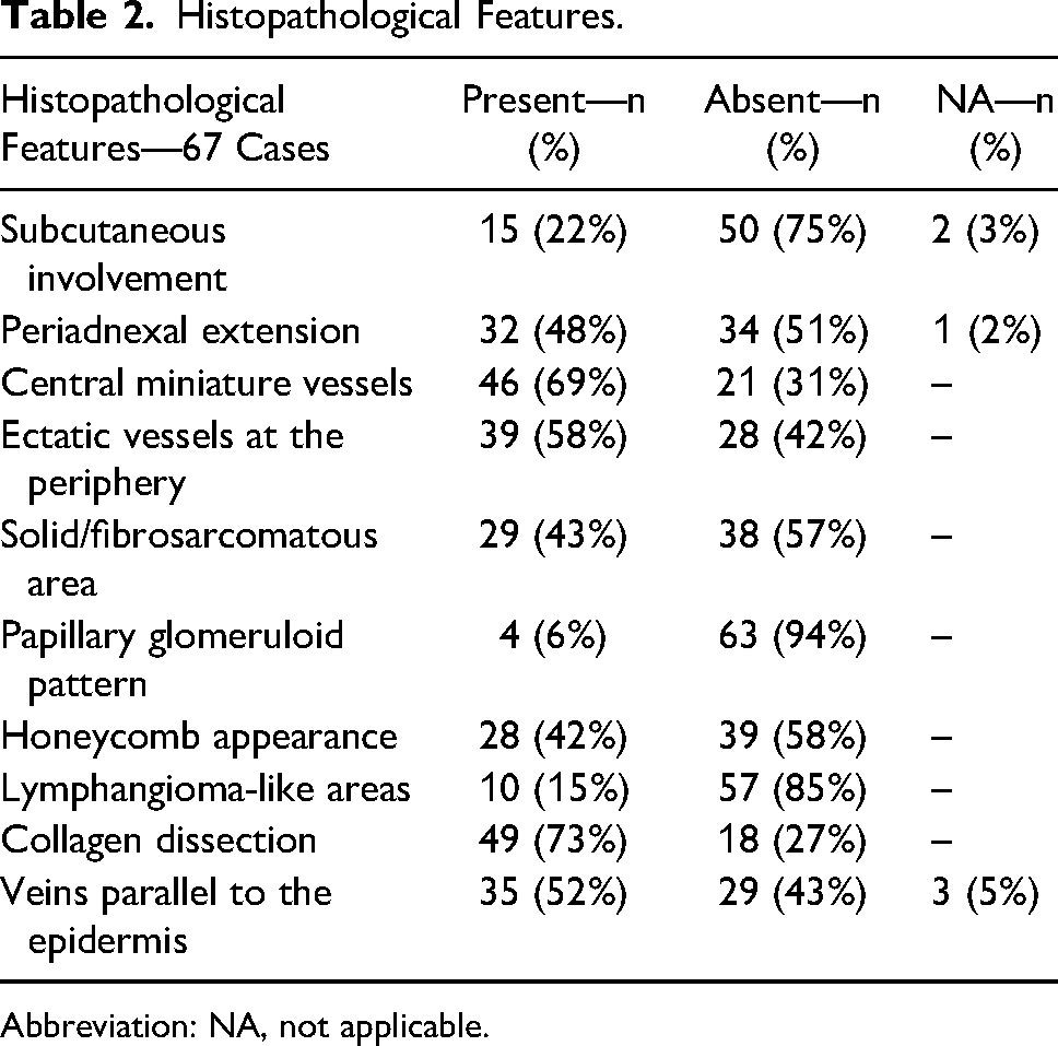
The samples consisted of 35 (52%) punch biopsies, 6 (9%) incisional biopsies, and 26 (39%) excisional biopsies. Mitotic activity was 0/10 HPF in 21 (31%), 1/10 HPF in 36 (54%), and ≥2/10 HPF in 10 (15%) samples. Other morphological features are presented in Table 2.
Histopathological Features.
Abbreviation: NA, not applicable.
All cases were positive for HHV8, while CD31 was positive in 65 (97%) and CD147 in 57 (85%) lesions. Statistically significant relations were found between CD147 expression with nodular pattern (P = .001), presence of solid/fibrosarcomatous area (P = .005), and high mitotic activity (P = .035), but no correlations were revealed with other histopathological indicators (Table 3).
Relationship Between CD147 Expression and Histomorphological Findings.
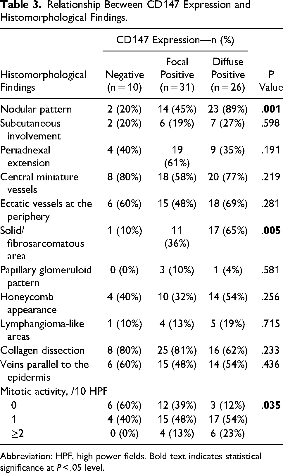
Abbreviation: HPF, high power fields. Bold text indicates statistical significance at P < .05 level.
The disease recurred in 17 (27%) of the 63 patients and the median relapse-free survival (RFS) was 9 (95%CI: 6.7-11.3) years. In seven patients, the disease relapsed 5 years after diagnosis. Five- and 10-year estimated RFS was 70.6% (95%CI: 54.6-86.6) and 31.8% (95%CI: 9.1-54.5), respectively. During the entire follow-up period, the frequency of relapse was 36% (9/25) in CD147 diffuse positive patients, 17.9% (5/28) in CD147 focal positive patients, and 30% (3/10) in CD147 negative patients (Figure 3). While a 5-year estimated RFS was 48.5% (95%CI: 22.3-74.7) in the CD147 diffuse positive group, it was 83.4% (95%CI: 60.4-106.5) in focal positive and 100% in negative cases (P = .029, Figure 4). Diffuse positive staining with CD147 was found to predict the recurrence risk at 5-year follow-up (HR: 5.9, 95%CI: 1.25-27.9, P = .025).
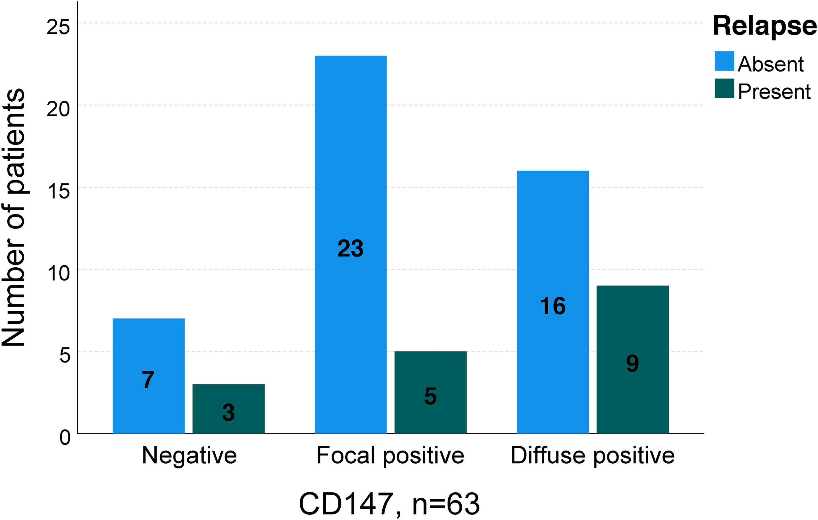
Disease recurrence rates according to CD147 positivity.
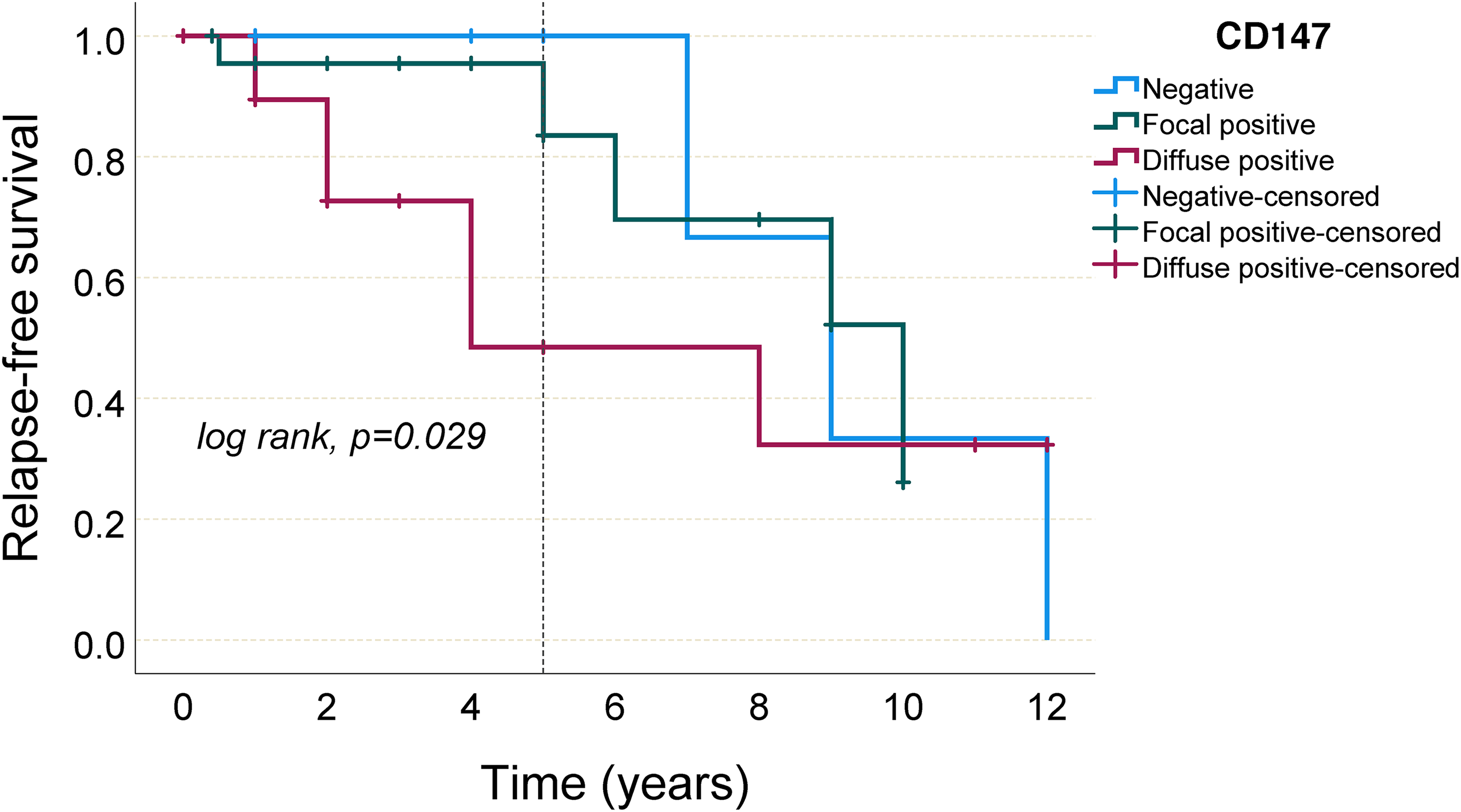
Kaplan–Meier curves of 5-year relapse-free survival by CD147 expression.
Discussion
KS is a multicentric angioproliferative spindle cell tumor of endothelial origin with marked pathological and clinical heterogeneity.6,7 Differential diagnoses of Kaposi sarcoma include angiosarcoma, hobnail hemangioma, spindle cell hemangioma, kaposiform hemangioendothelioma, and severe vascular stasis. The male-to-female ratio varies from 3:1 to 15:1, which was 3.2:1 in our study.8,9 Classical KS is characterized by multiple skin lesions and often localized distal of the lower extremities. Rarely, it can be seen in the upper extremity and visceral organs. In other clinical/epidemiological types, head and neck, lymph node, and mucosal surface involvement are more common.10,11 Although the most common involvement in our cohort was the extremities, extraordinary sites such as the lung and colonic submucosa were also encountered.
Emmprin is a multifunctional heavy transmembrane glycoprotein that is synthesized in high amounts by tumoral and microenvironment stromal cells. It stimulates the production of matrix metalloproteinases. 3 It is also expressed in some normal tissues and functions in the blood–brain barrier, T cell activation, menstruation, and tissue repair. Emmprin regulates pathogenic events such as signal transduction, chemoresistance, tumoral invasion, and metastasis by interacting with CD44, cyclophilin A, monocarboxylate transporters, and ATP-binding cassette transporters on the cell surface.12,13 Studies showed that CD147 protein plays a key role in cell migration in viral tumor pathogenesis, and overexpression of this protein is associated with advanced stage and recurrence in solid tumors. 4 For this reason, CD147 expression has been investigated in various malignancies in the literature. In a study, it was suggested that CD147 expression could be used in the differential diagnosis and prognosis prediction of uterine smooth muscle tumors. 14 Another study from Japan concluded that CD147 may have a role in the invasion process of breast cancer. 15 Futamura et al showed that emmprin expression is a significant prognostic factor for progression-free survival in patients with high-grade osteosarcoma. 16 In another study, 50 radical nephrectomy specimens performed for renal cell carcinoma were examined, and it was revealed that CD147 expression plays an important role in disease progression and resistance to sunitinib treatment. 17 All these findings indicate that emmprin may have a place in targeted therapy. Although its contribution to cancer progression has been extensively studied, studies demonstrating the role of emmprin in viral oncogenesis are very limited. 18 In one study, it was shown that de novo infection with KSHV leads to fibroblast and endothelial cell invasion by emmprin upregulation, and this is mediated by latency-associated nuclear antigen. 19 Another study revealed the contribution of CD147 to KSHV-related cell invasion by increasing ADAMTS1 and 9 regulation. It was also discovered that targeting CD147 with RNA interference and downstream of ADAMTS in the nude mouse model significantly suppressed pro-inflammatory, pro-angiogenic cytokine production, endothelial cell invasion, and KSHV+ telomerase-immortalized human umbilical vein endothelial cell tumorigenesis. 20
Our study revealed the relationship between CD147 overexpression and disease recurrence in KS cases, but the inhomogeneity of the treatment groups, mixed clinical stages, small number of patients, and the short follow-up period should also be considered. Detailed histopathological analysis showed that CD147 overexpression was associated with nodular growth pattern, solid fibrosarcomatous areas, and high mitotic activity. These findings may provide insight into the pathogenesis of KS and the development of targeted therapies in the future.
Footnotes
Acknowledgments
None.
Declaration of Conflicting Interests
The author(s) declared no potential conflicts of interest with respect to the research, authorship, and/or publication of this article.
Funding
The author(s) disclosed receipt of the following financial support for the research, authorship, and/or publication of this article: This study was granted by Hacettepe University Scientific Research Unit (Grant number: 348-12/2013).
Ethics Approval
Ethics committee approval for this study was obtained from the Hacettepe University Non-interventional Clinical Researches Ethics Board (Decision number: 14/17-30).
Trial Registration
N/A.