
Abstract
Background
Gastrectomy with lymphadenectomy is the standard of care for noncurative endoscopic resection (ER) (endoscopic curability C2; eCuraC2) for early gastric cancer (EGC); however, this strategy may be excessive for most patients because few patients have lymph node metastasis (LNM). In addition, whether EGCs with undifferentiated components (undiff-components) in the eCuraA group are suitable for ER remains controversial. The aim of this study was to stratify the eCuraC2 group according to LNM risk, and to verify the safety of conservative treatment for patients with undiff-components of the eCuraA group.
Methods
We retrospectively analyzed 272 patients with submucosal invasion or with undiff-components who underwent initial ER for EGC. These specimens were classified into eCura A, B, C1, and C2 groups according to the Japanese Gastric Cancer Association (JGCA) guidelines, and the rate of LNM in each group was analyzed. The key risk factors were identified by analyzing the correlation between different combinations of risk factors and LNM, and according to the LNM risk, further graded the eCuraC2 group.
Results
Among the 162 eCuraC2 patients, 9 (5.6%) had LNM. But no patients in the other groups, including all 57 patients of the eCuraA group (T1a, < 2 cm in diameter and no ulceration) with undiff-components, had LNM. A tumor diameter >3 cm (1.7% vs 12.2%, P = .005), positive for vertical margins (1.6% vs 20.0%, P < .001), submucosal invasion (≥500 μm) (0.7% vs 6.6%, P = .012), undiff-components type dominance (0% vs 11.9%, P < .001) and lymphovascular infiltration (LVI) (1.3% vs 16.7%, P < .001) were significantly correlated with LNM. When the patients in the eCuraC2 group were divided into 4 groups based on the presence of LVI and undiff-components, the LNM rate in each group was 0/81 patients (0%) in the LVI (−) undiff-components (−) group, 3/45 patients (6.7%) in the LVI (−) undiff-components (+) group, 0/15 patients (0%) in the LVI (+) undiff-components (−) group, and 6/21 patients (28.6%) in the LVI (+) undiff-components (+) group. Finally, based on these 2 factors, eCura C2 patients were classified into 3 LNM risk grades: low (LVI (−) undiff-components (−), LNM 0%), intermediate (LVI (+) or undiff-components (+), LNM 5%), and high (LVI (+) undiff-components (+), LNM 28.6%).
Conclusion
Based on LVI and histological differentiation, eCuraC2 patients were classified into 3 LNM risk grades, and approximately half of the eCuraC2 patients were reclassified into the low-risk group. No LNM was found in patients in the eCuraA group with undiff-components.
Keywords
Introduction
Endoscopic resection (ER) has increasingly become a fundamental treatment for early gastric cancer (EGC) because of the minimally invasive nature of this procedure. Therefore, effectively screening patients with high-risk lymph node metastasis (LNM) via pathological examination is very important. Based on histologic risk factors, the European Society of Gastrointestinal Endoscopy (ESGE), Japanese Gastric Cancer Association (JGCA), and American Society for Gastrointestinal Endoscopy (ASGE) have classified patients who undergo ER into curative and noncurative groups. The JGCA uses endoscopy curability (eCura) A, B, C1, and C2 to name these groups, where endoscopic curability C2 (eCuraC2) is equivalent to the noncurative ER group. Gastrectomy with lymphadenectomy is the standard of care for noncurative ER (eCuraC2), but LNM was observed in only 4.41% to 10% of patients who undergo radical surgery.1–7 Therefore, this recommendation for most patients in the eCuraC2 group may be excessive. Radical gastrectomy may lead to only limited improvements in prognosis in patients with a low rate of LNM after noncurative ER,5,8 and the postoperative mortality rate was ∼0.5% in patients who undergo radical gastrectomy for EGC.9,10 Therefore, it is necessary to further stratify the current eCuraC2 group based on the LNM risk. In addition, ASGE recommends radical surgery for the undiff-components type of EGC regardless of the tumor size, whereas JGCA and ESGE recommend conservative treatment for the T1a undiff-components type (diameter <2 cm) of absent ulceration. The patients suitable for ER remain controversial.
In this study, we proposed a risk grading method for LNM in the eCuraC2 group. Second, we verified the safety of conservative treatment for patients with undiff-components of eCuraA group.
Methods
Study Population
We retrospectively investigated patients who underwent ER for EGC at 6 large general hospitals in Ningbo, China, from 2018 to 2023, and all the pathological features of the samples have been independently rereviewed. In this study, we focused on the LNM rate of patients with undiff-components or with submucosal invasion. The inclusion criteria were as follows: T1 EGC patients who (1) underwent ER as first-line treatment, (2) had undiff-components or submucosal invasion, and (3) underwent gastrectomy with lymphadenectomy or follow-up for ≥ 3 years after ER. The exclusion criteria were as follows: patients who (1) had synchronous or metachronous advanced cancer, (2) were lost to follow-up or had an unknown LNM status, (3) had neuroendocrine tumors, (4) received any adjuvant therapy, and (5) died because of other diseases.
Assessment of Clinicopathological Factors and Correlation Analysis With LNM
We collected data on age, sex, tumor size, tumor location, and ulceration status from the hospital digital records and operation records of each patient. All the pathological slides were reevaluated by one experienced gastrointestinal pathologist, and a professorial consultation was conducted on specimens of disagreement with the original pathological diagnosis. The evaluated histologic factors included tumor size, histologic differentiation, lymphovascular invasion (LVI), ulceration condition, and depth of submucosal invasion (DSI). The histological types were classified into the pure differentiated type, the differentiated type with undiff-components, and the undiff-dominant type. And according to the sixth edition of the JGCA Guidelines, the cutoff value for distinguishing between the differentiated type with undiff-components and the undiff-dominant type was set at ≥50% undiff-components. The former category included mild-to-moderate adenocarcinoma, and the undiff-components included poorly differentiated adenocarcinoma, signet ring cell carcinoma, mucinous adenocarcinoma, and undiff-component. The DSI was measured from the lower border of the muscular mucosae according to the guidelines and categorized as SM1 (<500 μm) or ≥ SM2 (≥500 μm). When LVI was difficult to judge, patients were evaluated by immunostaining with antibodies against CD31 and D2-40. The lymph node status was obtained directly from the examination of radical surgical specimens or from follow-up visits. The patients who underwent ER alone with no evidence of recurrence during the follow-up period (≥3 years) were regarded as negative for LNM. After the initial treatment, the patients were recommended to be followed up with abdominal computed tomography and endoscopy every 6 to 12 months for at least 5 years. Clinical decisions and management followed the “Guidelines of the Chinese Society of Clinical Oncology, Gastric Cancer” in all 6 hospitals. The follow-up of these patients was performed according to routine clinical care protocols. 11
Evaluation of Endoscopic Curability (eCura)
According to the sixth edition of the JGCA guidelines, 272 patients were classified as eCuraA, eCuraB, or eCuraC, and the LNM/REC rate in each group was analyzed. 12 The evaluation criteria for the eCura system are as follows: resection is classified as eCuraA when all of the following conditions are fulfilled, provided that the cancer has no ulcerative findings: en bloc resection, any tumor size in patients of histologically differentiated type-dominant, tumor size ≤2 cm in patients of histologically undifferentiated type-dominant, pT1a, negative horizontal margin (HM0), negative vertical margin (VM0), and no lymphovascular infiltration (Ly0, V0). However, if the undifferentiated component of the lesion exceeds 2 cm in length, endoscopic curability is classified as eCuraC2. When the cancer is ulcerative, the resection is still classified as eCuraA when all of the following conditions are fulfilled: en bloc resection, tumor size ≤ 3 cm, histologically differentiated type-dominant, pT1a, HM0, VM0, Ly0, and V0; eCuraB: en bloc resection, histologically differentiated type-dominant, pT1b1 (<500 μm from the muscularis mucosae), HM0, VM0, and LV0, tumor size ≤3 cm. However, if the undiff-component invades the submucosal layer, endoscopic curability is classified as eCuraC2. Endoscopic curability is classified as eCuraC when it does not fulfill the conditions described above and is classified as either eCuraA or eCuraB. Endoscopic curability is classified as eCuraC1 when it is a histologically differentiated type that is either not resected en bloc or has a positive horizontal margin despite fulfilling other criteria to be classified into either eCuraA or eCuraB. All other eCuraC resections are subclassified as eCuraC2 (Figures 1 and 2).
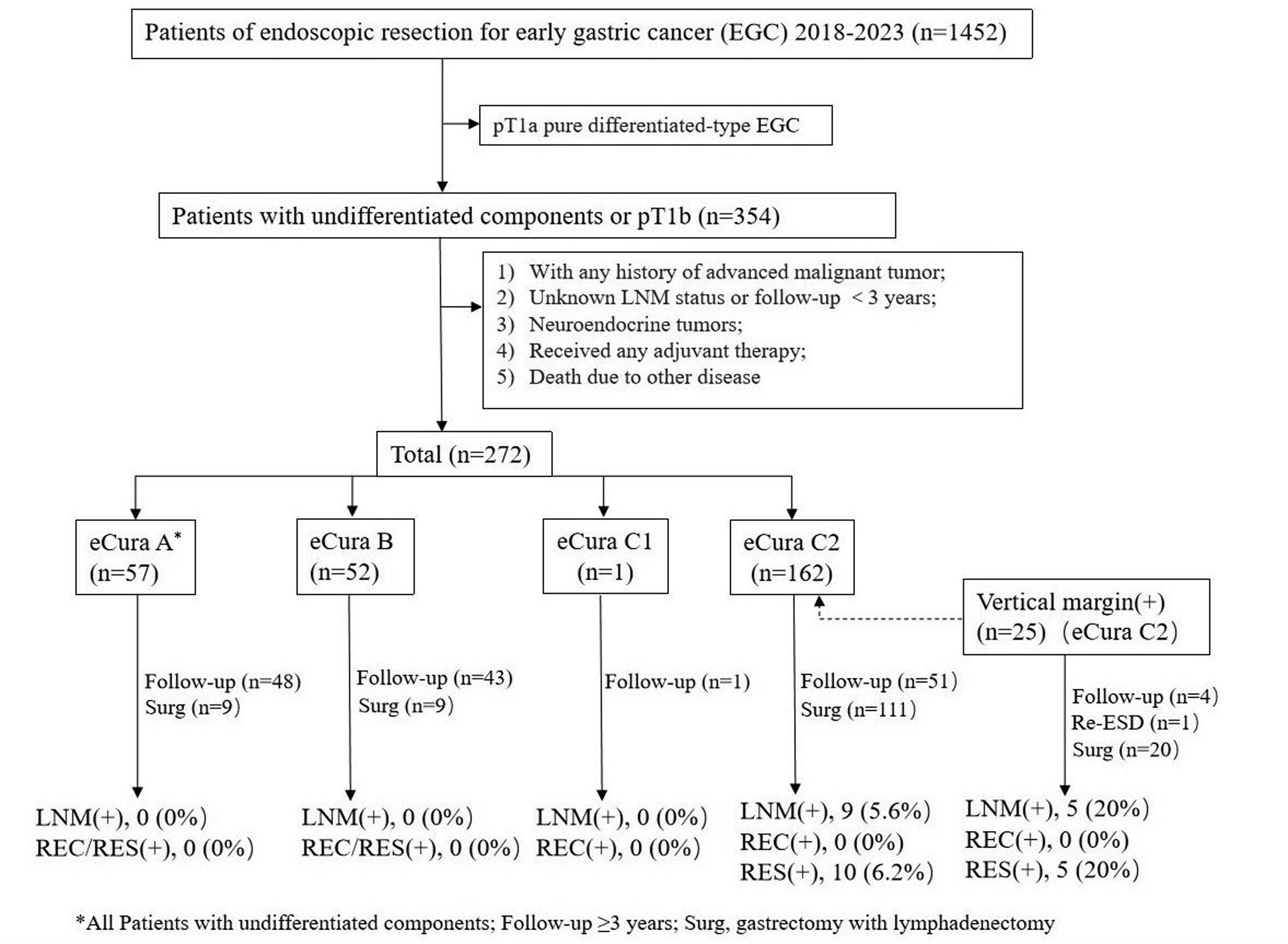
Treatment and lymph node metastasis, recurrence, and residual rates in 272 patients. eCura system, endoscopic curability system; LNM, lymph node metastasis; RES/REC, residual/local recurrence.
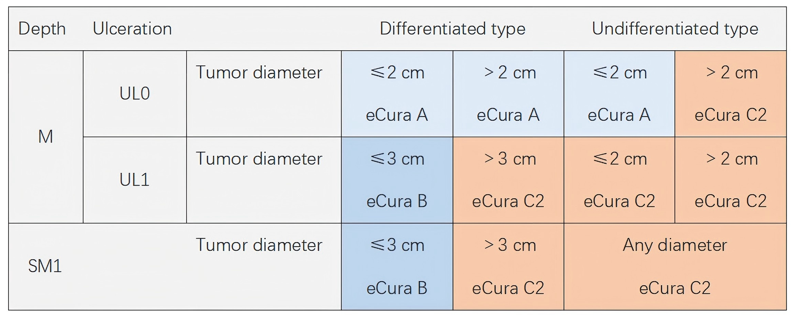
The evaluation criteria of the Japanese gastric cancer association guidelines 2021 (sixth edition) for the Endoscopic Curability (eCura) system. 12 HM0, Negative Horizontal Margin; VM0, negative Vertical Margin; Ly0, V0, No Lymphovascular Infiltration.
LNM Risk Grading for the Noncurative ER Group (eCuraC2)
Based on the different combinations of LNM-associated critical risk factors, we further stratified eCuraC2 tumors.
Statistical Analysis
Statistical analyses were performed via SPSS software (Chicago, IL, USA). Differences in the rates of LNM and recurrence between the groups were estimated via the χ2 test or Fisher's exact test. Statistical significance was defined as P < .05.
Results
Clinicopathological Characteristics
We included 1452 patients who underwent ESD as a first-line treatment; among them, 354 patients had either positive submucosal invasion or undiff-components. The 82 patients were excluded according to the exclusion criteria, and 272 patients were ultimately included in this study. The included patients were classified as eCuraA (57 patients, 21.0%), eCuraB (52 patients, 19.1%), eCuraC1 (1 patient, 0.3%), or eCuraC2 (162 patients, 59.6%) according to the JSGA guidelines. The patients in the eCuraC2 group underwent additional gastrectomy with lymphadenectomy (111 patients, 68.5%) and Re-ESD (1 patient, 0.6%) within 1 to 2 months after ER, and the remaining 50 patients (30.9%) chose to undergo follow-up. The 9 patients (5.6%) in the eCuraC2 group were positive for LNM (the number of peri-intestinal lymph nodes, median 22, IQR 17-44), and none of the patients recurred. The 25 patients with positive vertical margins underwent additional gastrectomy with lymphadenectomy (20 patients, 80%), Re-ESD (1 patient, 4%), and follow-up (4 patients, 16%). LNM (5 patients, 20%) and residual cancer (5 patients, 20%) were found in these patients. LNM and recurrence were not detected in any of the patients in the other groups (eCuraA, B, and C1) (Figure 1).
Correlation of Histopathological Parameters With Lymph Node Metastasis
Table 1 summarizes the clinicopathological characteristics and correlations of each parameter with LNM. The patients included 197 men and 75 women, ranging from 33 to 87 years old (median 65 years). The lesion locations were as follows: upper third, 31 (11.4%); middle third, 72 (26.5%); and lower third, 169 (62.1%). A tumor diameter >3 cm, ulceration, a positive vertical margin, a depth of submucosal invasion ≥ SM2, LVI (+), and undiff-components were observed in 41 (15.1%), 16 (5.9%), 25 (9.2%), 121 (44.5%), 36 (13.2%), and 135 (49.6%) patients, respectively. A tumor size >3 cm (1.7% vs 12.2%, P = .005), positive vertical margin (1.6% vs 20.0%, P < .001), ≥SM2 (0.7% vs 6.6%, P = .012), undiff-component type dominance (0% vs 11.9%, P < .001), and LVI (1.3% vs 16.7%, P < .001) were significantly correlated with LNM. LNM was not associated with age, sex, ulceration status, or tumor location. When we classified the patients into LVI (−) and LVI (+) groups, we found a significantly greater LNM rate in the LVI (+) group (16.7% vs 1.3%, P < .001), irrespective of the other parameters. However, among eCuraC2 patients, the LNM rate of the undiff-components (+) group was significantly greater than that of the control group in both the LVI (−) and LVI (+) groups (Table 2).
Correlations of Each Parameter With Lymph Node Metastasis.
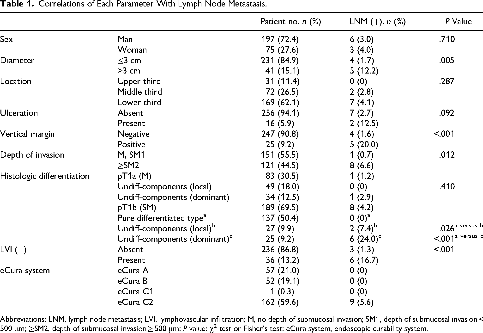
Abbreviations: LNM, lymph node metastasis; LVI, lymphovascular infiltration; M, no depth of submucosal invasion; SM1, depth of submucosal invasion < 500 μm; ≥SM2, depth of submucosal invasion ≥ 500 μm; P value: χ2 test or Fisher's test; eCura system, endoscopic curability system.
Correlations Between Lymphovascular Infiltration Status and the Rate of Lymph Node Metastasis.
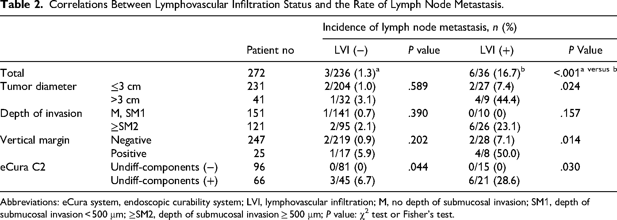
Abbreviations: eCura system, endoscopic curability system; LVI, lymphovascular infiltration; M, no depth of submucosal invasion; SM1, depth of submucosal invasion < 500 μm; ≥SM2, depth of submucosal invasion ≥ 500 μm; P value: χ2 test or Fisher's test.
Details of the Patients With Undiff-Components in the eCuraA Group
According to the JGCA guidelines, 57 of the 135 patients with undiff-components were classified into the eCuraA group. All 57 patients in the eCuraA group had varying proportions of undiff-components because only pT1a EGC patients with undiff-components were included in this study. Among the 57 patients with eCuraA, 14 patients (24.6%) had undiff component dominance, and the remaining 43 patients (75.4%) had at least one focal undiff component. None of the 57 patients had LNM or recurrence.
Composition of the Noncurative ER Group (eCuraC2) and LNM/REC Rate
Figure 3A shows the proportions of risk factors in the eCuraC2 group in this study. According to the eCura evaluation system, 162 (59.6%) patients were classified into the eCuraC2 group, but only 9 (5.6%) of these patients were positive for LNM. Most patients (n = 75) were classified into the eCuraC2 group due to deep submucosal invasion alone, but none of the patients had LNM or recurrence. The remaining 87 patients (53.7%) had at least one other risk factor, and LNM was found in 9 patients (10.3%).
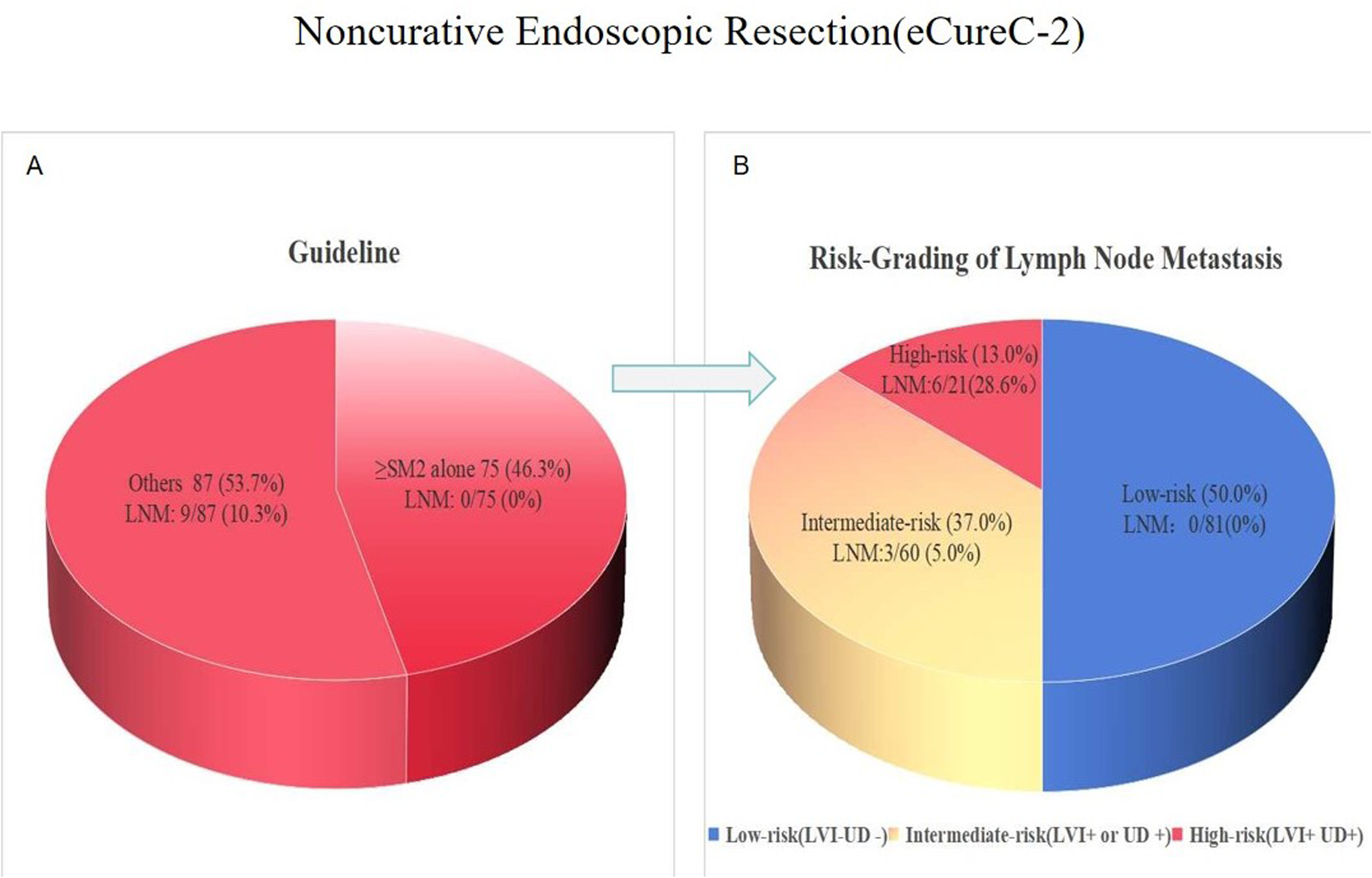
Lymph Node Metastasis (LNM) rate in eCuraC2 group. Seventy-five patients (46.3%) were classified into the eCuraC2 group due to the depth of submucosal≥500 μm alone, but none of the patients had LNM or recurrence. (B) The risk grading of the present study for the eCuraC2 group. Based on lymphovascular infiltration and histological differentiation, 50% of patients of the eCuraC2 group were classified as low-risk group, and the LNM/recurrence Was 0%. JGCA Guideline, Japanese Gastric Cancer Treatment Guidelines; DSI, Depth of Submucosal Invasion; LNM, Lymph Node Metastasis; LVI, Lymphovascular Infiltration; UD, Undifferentiated Component.
Risk Grading of LNM for the Noncurative ER (eCuraC2) Group
Figure 3B shows the risk grading of the present study for the eCuraC2 group. When we classified the patients with eCuraC2 into 4 groups based on the different combinations of LVI (Figure 4B) and undiff-components (Figure 4A), the rates of LNM in each group were as follows: 0 of 81 patients (0%) in the LVI (−) undiff-components (−) group, 3 of 45 patients (6.7%) in the LVI (−) undiff-components (+) group, 0 of 15 patients (0%) in the LVI (+) undiff-components (−) group, and 6 of 21 patients (28.6%) in the LVI (+) undiff-components (+) group. Finally, we divided the patients into 3 groups: low-risk (LVI (−) undiff-components (−), LNM rate 0%), intermediate-risk (LVI (+) or undiff-components (+), LNM rate 5%), and high-risk (LVI (+) undiff-components (+), LNM rate 28.6%). The low-risk group accounted for 50% of the eCuraC2 patients according to our risk-grading methods, and the LNM rate was 0%.

The key risk factors for lymph node metastasis in early gastric cancer. Tumor cells contain undifferentiated components (A, 200 ×). Positive lymphovascular invasion (LVI) (B, 200 ×).
Discussion
The pathological examination of ER samples is the gold standard for determining whether additional surgery is necessary. Gastrectomy with lymphadenectomy is the standard of care for noncurative ER (eCuraC2), but LNM occurs in only 4.41% to 10% of patients undergoing radical surgery.1–7 Therefore, this recommendation for most patients in the eCuraC2 group may be excessive. In the present study, 59.6% of patients were classified into the eCuraC2 group according to the JSGA guidelines, but only 9 (5.6%) patients were positive for LNM.
The eCura scoring system was proposed based on a Japanese multicenter study. The purpose of this system is to further stratify patients who fail to meet the curative criteria after ER, especially elderly individuals who cannot benefit from additional radical surgery.12,13 The eCura scoring system has 3 risk categories based on 5 clinicopathological factors that predict LNM in patients undergoing radical surgery after noncurative ESD. 13 The JGCA recommends that the scoring system be applied to elderly patients or those with severe comorbidities with noncurative ER, provided that patients fully understand the risk. The eCura scoring system for noncurative ER consists of 5 clinicopathological factors, which are scored as follows: 1 point each for a tumor size >30 mm, positive vertical margin, venous invasion, SM2, and 3 points for lymphatic invasion. The total risk score is categorized into 3 LNM risk groups: low (0-1 point: 2.5% risk), intermediate (2-4 points: 6.7%), and high (5-7 points: 22.7%). The eCura scoring system is a useful aid for selecting the appropriate treatment strategy after noncurative ER for EGC.7,14 However, there are several issues with the eCura scoring system, as follows: (1) undiff-component type is an independent risk factor for LNM in EGC, although it is not considered a risk factor in the eCura scoring system; therefore, applying this system to the undiff-component type carries a certain degree of risk; (2) vascular infiltration is an independent risk factor for LNM in EGC, but it accounts for only 1 point on the eCura scoring system; therefore, it carries risk in patients with vascular infiltration, especially those accompanied by undiff-components; and (3) there is still a 2.5% risk of LNM in the low-risk group. If the system can reduce the risk of LNM in the low-risk group, its application could be extended to a broader population beyond just elderly individuals.
The undiff-component was an independent risk factor for LNM. Jung et al 15 reported that the undiff-components in the SM1 layer were an independent risk factor for LNM. The JSGA guidelines recommend that undiff-components with submucosal invasion be classified into the eCuraC2 group. In this study, when we classified patients with submucosal invasion into the pure differentiated type, differentiated type with undiff-components, and undiff-components dominant, the LNM rate in the latter 2 groups was significantly greater than that in the former group (0% vs 7.4%-24.0%, P < .001). However, the eCura scoring system does not weight the undiff-components type as a risk factor; therefore, the application of this scoring system in patients with undiff-components in the eCuraC2 group has risks that cannot be ignored. In 2017, the research team of the eCura scoring system performed an internal validation for the eCura scoring system using 1969 patients. The authors suggested that the scoring system be used for patients of differentiated type because there was a limited proportion of the undiff-component type in their development cohort for the eCura scoring system. 8
Both lymphatic infiltration and vascular infiltration are independent risk factors for LNM.3,16,17 However, lymphatic invasion is weighted 3 points, whereas vascular invasion is weighted only 1 point in the eCura scoring system. 13 According to this scoring system, patients with vascular invasion in the eCuraC2 group are classified as having a low risk for LNM when other risk factors are absent. EGC metastasizes through vascular and lymphatic vessels, regardless of which route of metastasis is associated with a poor prognosis. We considered it questionable that the eCura scoring system uses different scores for vascular and lymphatic infiltration, since the 2 vasculature systems are not independent. 18 In addition, distinguishing vascular infiltration from lymphatic infiltration via hematoxylin and eosin (HE) staining alone is difficult.12,19 The JSGA guidelines recommend the use of Elastica-van Gieson staining and D2-40 immunohistochemical staining to identify vascular and lymphatic infiltration, respectively.12,19 The elastica-van Gieson staining can stain the elastic layer of arterial and vein walls; therefore, it is helpful for detecting arterial and venous invasion, but cannot stain capillaries. It is very important to choose an effective method to detect LVI because it is often easily missed on HE slides. CD31 and D2-40 immunohistochemical staining were the most conventional methods for identifying LVI; therefore, in this study, we used these 2 antibodies to identify LVI. The development of the eCura scoring system was based on the pathological diagnosis of multiple institutions, whereas the assessment method for lymphatic and vascular infiltration was not clear, such as whether immunohistochemical or specific staining was used, or only HE slides were used.
A depth of submucosal invasion ≥500 μm is considered an important risk factor for the evaluation of endoscopic curability in the guidelines, but the measurement of submucosal invasion depth markedly differs because of differences in the method of measurement and the condition of the muscular mucosa. The depth of submucosal invasion was measured from the lower edge of the mucosal muscle to the deepest point of invasion. However, the mucosal muscle often breaks or becomes irregular due to cancer invasion, inflammation, and ulcers, which results in significant differences in the measurement of submucosal infiltration depth.20,21 In the present study, we also observed that the LNM rate increased with increasing depth of submucosal invasion. However, when patients with LVI and undiff-components were excluded, the LNM rate in patients with a depth of submucosal invasion ≥500 μm was 0%. In our cohort, most patients (n = 75) were classified into the eCuraC2 group because of submucosal infiltration ≥500 μm alone, accounting for 46.3% of the eCuraC2 group, but none of these patients had LNM or recurrence. According to our grading method, these patients were classified into the low-risk group for LNM. The eCura scoring system, developed in a Japanese multicenter study, revealed that a depth of submucosal invasion ≥500 μm was not an independent risk factor for LNM, and according to this scoring system, patients with a depth of submucosal invasion ≥500 μm but no other risk factors could be classified into the low-risk group.
The eCura scoring system is helpful for the management of patients with noncurative ER; however, patients in the low-risk group still have a 2.5% risk of LNM. If the system can reduce the risk of LNM in the low-risk group, its application can be extended to a broader population beyond just elderly individuals. Although the depth of submucosal invasion, tumor size, and positive vertical margin were associated with LNM in the present study, when we classified these patients according to the different combinations of LVI and undiff-components, we observed that the undiff-components and LVI were the key risk factors for LNM. Regardless of other risk factors, the LNM rate of patients in the eCurac2 group was 0% when both the undiff-components and LVI were absent. Based on the different combinations of the 2, eCuraC2 could be classified into low-risk (LVI (−) undiff-components (−), LNM rate 0%), intermediate-risk (LVI (+) or undiff-components (+), LNM rate 5.0%), and high-risk (LVI (+) undiff-components (+), LNM rate 28.6%). Our risk-grading methods provided more options for approximately half of the eCuraC2 patients.
The ASGE guidelines recommend radical surgery for the undiff-component type irrespective of lesion size, whereas the JSGC guidelines recommend conservative treatment for patients with undifferentiated T1a cancer <2 cm without ulceration.11,12,22,23 Multicenter prospective studies conducted by the Japan Clinical Oncology Group (JCOG) 1009/1010 to evaluate T1a patients with undiff-components EGC revealed that the 5-year overall survival (OS) of ESD was comparable to that of surgical gastrectomy when the tumor diameter was ≤2 cm and ulcerations were absent. 24 Consequently, these lesions are integrated into the eCuraA group according to the sixth JSGA guidelines. 12 In the present study, no LNM or recurrence was observed in patients in the undiff-components eCuraA group. However, we considered that caution was still necessary in such patients, because some researchers have reported that the incidence of LNM (2.6%-2.9%) in these patients was higher than the classic absolute indication.25,26 In addition, the incidence of LNM in T1a gastric cancers in large Asian series was reported to be 2% to 5%, but Western series had reported LNM rates ranging from 4% to 13%.27–35 Choi et al 36 reported that the LNM rate of T1a gastric cancer was significantly associated with race; that was, Asian/Pacific Islanders (APIs) presented the lowest rate of lymph node metastases (5.2%), followed by Hispanics (7.0%), whites (9.7%), and blacks (10.9%). Therefore, we believe that differences in risk based on race/ethnicity should be considered when assessing the risk of LNM in T1a patients with undiff-components.
This study has several limitations. First, our study has a retrospective design, and the small number of specimens may limit the generalizability of the results. Second, although the patients in this study were collected from multiple centers, to improve the accuracy of diagnosis, all slides were reevaluated by one expert gastrointestinal pathologist; therefore, further validation in more realistic multicenter patients is needed. Third, the risk-grading methods of this study are based on only 2 key risk factors; therefore, the application of this grading method requires high accuracy of pathological diagnosis of risk factors.
In conclusion, LVI and the undiff-components are the most critical risk factors for LNM in EGC patients. Based on LVI and histological differentiation, eCuraC2 patients can be further classified into 3 LNM risk grades: low (LNM, 0%), intermediate (LNM, 0%-6.7%), and high (LNM, 28.6%). No LNM or recurrence was observed in patients in the undiff-components eCuraA group.
Footnotes
Ethical Approval
Ethical approval for this study was waived.
Informed Consent
Informed consent was waived.
Funding
The authors disclosed receipt of the following financial support for the research, authorship, and/or publication of this article: This work was supported by the Public Welfare Technology Application Research Project of Ningbo, Zhejiang Medical and Health Technology Project, Ningbo Top Medical and Health Research Program (grant numbers 2025S183, 2023KY1131, and 2023010211).
Declaration of Conflicting Interests
The authors declared no potential conflicts of interest with respect to the research, authorship, and/or publication of this article.
Trial Registration
Not applicable, because this article does not contain any clinical trials.