
Abstract
Large B-cell lymphomas may exhibit morphology typical of a diffuse large B-cell lymphoma (DLBCL) or a Burkitt lymphoma. Rarely, they may exhibit either a blastoid morphology or features that are intermediate between a DLBCL and Burkitt lymphoma (the so-called intermediate/blastoid morphology); the term high-grade B-cell lymphoma (HGBL) is used to describe the latter. The presence of MYC and BCL2 rearrangements can be seen either in HGBL or in lymphomas with DLBCL morphology (so-called double-hit lymphoma) and rarely in plasmablastic lymphomas. Herein, we report a rare occurrence of a double-hit lymphoma with plasmablastic features in a patient with a history of follicular lymphoma (FL) aberrantly expressing keratin in the transformed component posing a diagnostic conundrum. Immunohistochemistry and molecular work-up in conjunction with clinical history were essential in arriving at the correct diagnosis and thus illustrating a diagnostic pitfall.
Keywords
Introduction
Plasmablastic lymphoma is a rare type of aggressive large B-cell lymphoma that demonstrates plasmablastic/immunoblastic morphology and expresses several plasma cell markers with absent or reduced B-cell marker expression.1–4 This lymphoma typically occurs in immunocompromised patients and presents as extranodally (nasal and oral cavity, gastrointestinal tract, bone and soft tissue, skin, etc.). This lymphoma is composed of medium to large immunoblastic and plasmablastic appearing cells. It can also demonstrate a “starry-sky” appearance often seen in the context of Burkitt lymphoma. It typically lacks B-cell (CD20, PAX5, OCT2 (POU2F2)) markers, although CD79A may be retained in 40% of tumors, reflecting its expression from early B-precursors through plasma cell differentiation. Plasma cell (CD38, CD138, IRF4/MUM1, VS38c, BLIMP1 (PRDM1/ZNF683), XBP1) markers are typically expressed, supporting the plasmablastic phenotype.
The presence of concurrent MYC and BCL2 translocations in a large cell lymphoma with either a diffuse large B-cell morphology or high-grade morphology (blastoid/intermediate) is diagnostic of a diffuse large B-cell lymphoma (DLBCL)/high-grade B-cell lymphoma (HGBL) with MYC and BCL2 rearrangements (the so-called “double-hit” lymphomas).1,2 These are rare lymphomas with poor prognosis and often requiring dose-intensified chemotherapy regimens. Almost all of these lymphomas express pan-B-cell and germinal center markers (CD10 or BCL6). While MYC rearrangements are common in plasmablastic phenotype, double-hit phenomenon with a plasmablastic phenotype is extremely rare and is almost exclusively limited to rare examples of transformed follicular lymphomas.5–7
Plasmablastic lymphoma can aberrantly express keratin in 9% to 15% of specimens depending on the particular keratin. 8 In addition, CD45 (PTPRC, pan hematopoietic marker) is typically negative or reduced; however, CD45 expression can be detected in a subset of tumors, including the present patient.1,2 The usual absence of CD45 expression, although present in this specimen, together with keratin positivity could represent a diagnostic trap for surgical pathologists who may not entertain plasmablastic lymphoma as a possibility. We describe the first reported example of a keratin expressing HGBL with MYC and BCL2 translocations, with a plasmablastic phenotype that likelytransformed from the patient's known follicular lymphoma.
Case Report
A 44-year-old male patient, previously diagnosed with classic/low-grade (grade 1–2) follicular lymphoma, presented with pancytopenia three months following completion of his lymphoma treatment. There were no described lytic lesions on imaging studies or monoclonal serum proteins. He underwent a bone marrow biopsy, touch preparation and aspiration as part of his workup for pancytopenia. The bone marrow touch preparation demonstrated a discohesive group of cells with deep blue moderate cytoplasm, occasional cytoplasmic vacuoles, round to oval eccentric nuclei (with occasional binucleation), coarse to fine chromatin and prominent nucleoli (Figure 1A). Normal bone marrow elements were markedly reduced and cytologically appeared unremarkable. The bone marrow core biopsy demonstrated near total replacement by a neoplastic population of large undifferentiated cells with plasmablastic/immunoblastic morphology; amphophilic cytoplasm, eccentric vesicular nuclei and prominent eosinophilic nucleoli. These neoplastic cells in some areas appeared to be clustering and cohesive, while in other areas appeared more discohesive (Figure 1B and C). Areas of necrosis were also noted.

(A) Bone marrow touch preparation demonstrates medium to large, atypical cells with dense blue cytoplasm, and round to oval nuclei with prominent nucleoli, ×100 magnification. (B) Core biopsy demonstrates sheets of cohesive atypical cells with a pseudo-glandular appearance, ×2 magnification. (C) At higher magnification, these cells are large in size with vesicular nuclei and prominent nucleoli, ×50 magnification.
Immunohistochemical studies on bone marrow biopsy (Figure 2) demonstrated that the neoplastic cells were positive for AE1/AE3 (A), CAM5.2 (B), CD45 (C), CD30 (D) and negative for CD19 (E) and CD138 (F). Additional immunohistochemical

Immunohistochemical studies on bone marrow biopsy demonstrate that the neoplastic cells are positive for AE1/AE3 (A), CAM5.2 (B), CD45 (C), CD30 (D) and negative for CD19 (E) and CD138 (F), x10 magnification.
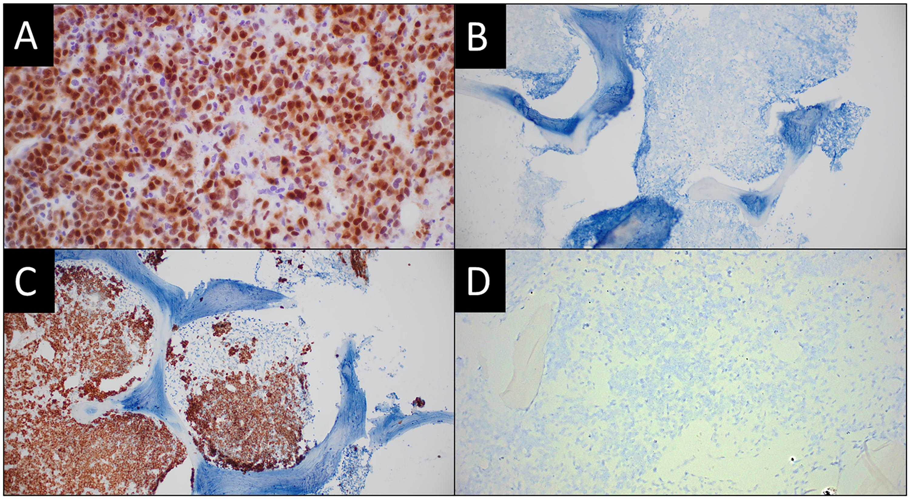
Immunohistochemical studies demonstrate that the neoplastic cells are positive for IRF4/MUM1, x40 magnification (A). In-situ hybridization studies demonstrate positivity for lambda (C) and negativity for kappa (B) and EBV, x10 magnification (D).
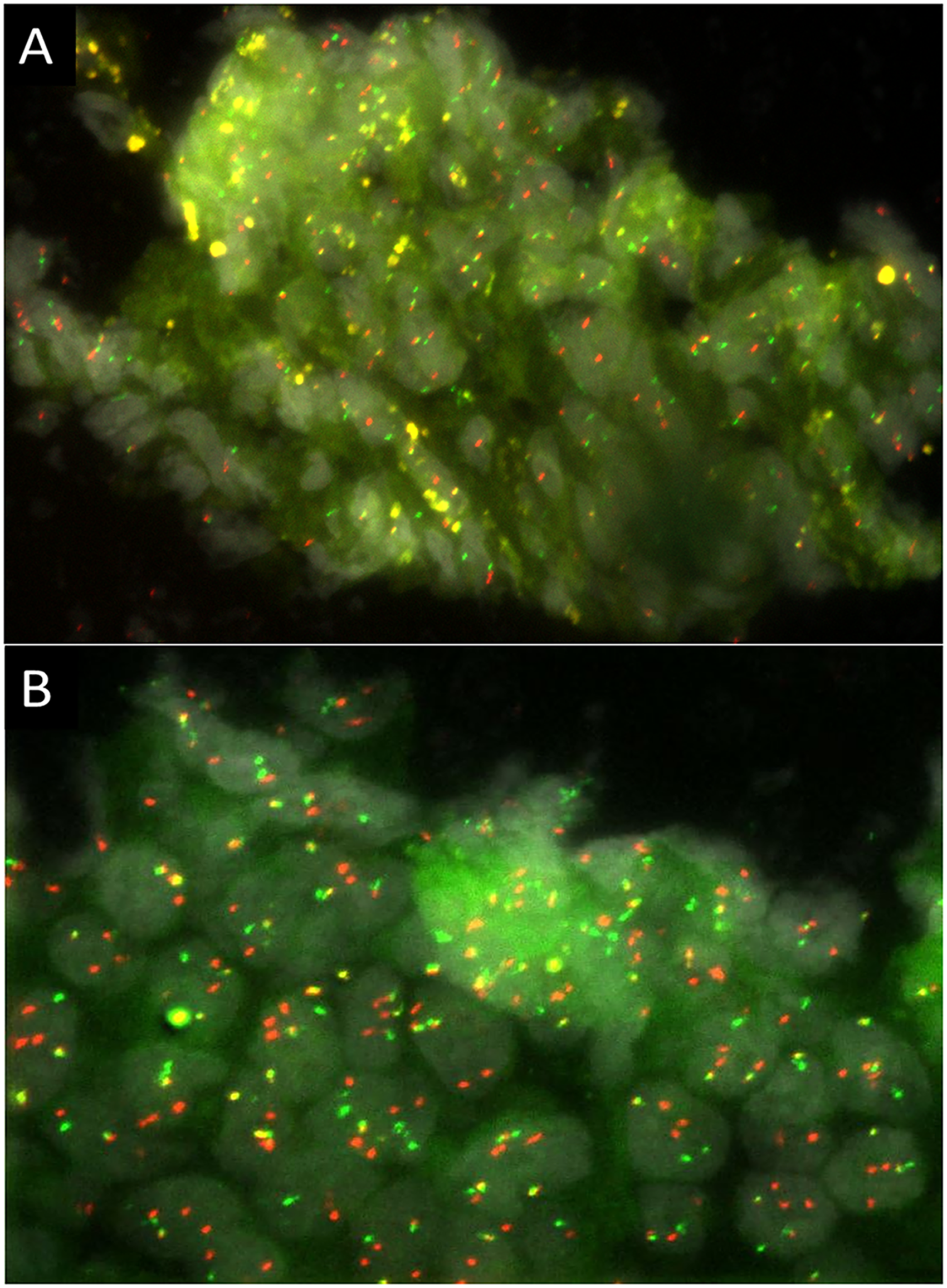
Fluorescence in-situ hybridization studies demonstrate (A) MYC translocation with break-apart differentially labeled probes targeting the upstream (5′) and downstream (3′) flanking regions of the MYC gene (A) and (B) IGH::BCL2 FISH shows fused signals consistent with IGH::BCL2 fusion (B).
Discussion
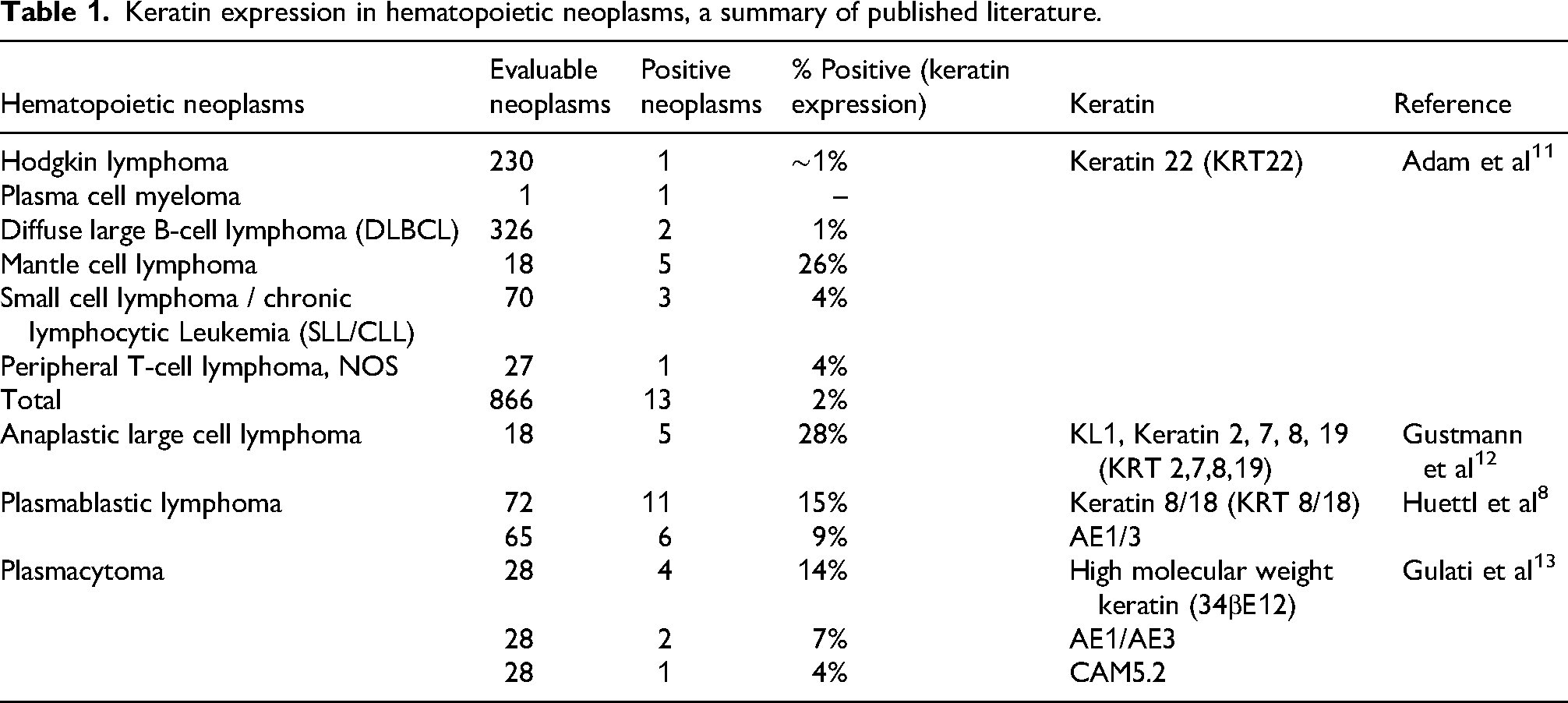
CD45 is perceived to be a reliable pan-hematopoietic marker albeit with some notable exceptions; these examples are either consistently negative, or dim to negative in a subset of specimens, for example, Classic Hodgkin lymphoma, plasmablastic lymphoma, acute lymphoblastic leukemia/lymphoma, myeloid sarcoma, etc.1,2 In tumors with poorly differentiated or undifferentiated morphology, absence of CD45 might erroneously lead to elimination of hematopoetic neoplasms from diagnostic consideration. Conversely, the presence of CD45 in a tumor that otherwise expresses keratins raises an interesting set of differentials. Aberrant expression of CD45 has been reported rarely in carcinomas, particularly when they are undifferentiated or demonstrate neuroendocrine differentiation.9,10 On the other hand, aberrant expression of keratin, albeit rare, is well documented in hematopoietic neoplasms (Table 1).8,11–13 Keratin expression in hematological disorders presents a diagnostic challenge, as they may morphologically and immunophenotypically resemble metastatic poorly or undifferentiated carcinoma. Particularly, in a large study by Adam et al, keratin expression was found in less than 5% of specific B and T-cell lymphomas that were included (DLBCL, chronic lymphocytic leukemia/lymphoma, peripheral T-cell lymphoma, not otherwise specified (PTCL, NOS)); the notable exception was mantle cell lymphoma which had a prevalence of keratin expression in 26% of specimens (Table 1). 11 In a separate study looking at plasmablastic lymphoma in particular, Huettl et al demonstrated aberrant expression of AE1/AE3 in 9% and keratin 8/18 (KRT8/18) in 15% of tumors. 8 The other lymphoma with a relatively high degree of keratin expression is anaplastic large cell lymphoma (27%). 12 In addition, individual patient reports have demonstrated keratin expression in T-cell lymphoma, primary cutaneous anaplastic large cell lymphoma, ALK positive large B-cell lymphoma, and primary effusion lymphoma.14–18
Keratin expression in hematopoietic neoplasms, a summary of published literature.
We describe a HGBL with plasmablastic features that also demonstrated concurrent MYC and BCL2 translocations; the so-called “double-hit lymphoma.” Interestingly, our patient had a history of follicular lymphoma which triggered the addition of CD45 to the initial panel of immunostains. This initial panel also included various keratins because of the cohesive and pseudo-glandular look in some areas. The combination of CD45 positivity with co-expression of AE1/AE3 and CAM5.2 was intriguing and required a further extended panel of immunostains. All specific hematopoietic markers for B-cell, T-cell, myeloid, histiocytic, mast cell lineage etc were negative. The presence of CD30 raised other differential diagnoses such as embryonal cell carcinoma and anaplastic large cell lymphoma (a type of T-cell lymphoma). However, CD30 can also be expressed in a subset of aggressive B-cell lymphomas including plasmablastic lymphoma. IRF4/MUM1 (interferon regulatory factor 4/ multiple myeloma - originally described in a multiple myeloma cell line) was positive. IRF4/MUM1 expression typically indicates a more differentiated B cell phenotype and is positive in plasma cell neoplasms, plasmablastic lymphomas and a variety of other B cell lymphomas and some T-cell lymphomas (including anaplastic large cell lymphoma).The complete lack of any B-cell marker, albeit with IRF4/MUM1 expression (especially given the history of follicular lymphoma) prompted us to add kappa and lambda in-situ hybridization test. This demonstrated lambda light chain restriction and clinched the diagnosis. The morphology (large cells with eccentric amphophilic cytoplasm and round to oval nuclei with prominent nucleoli) and the immunophenotype (lack of B-cell markers, IRF4/MUM1 positivity and light chain restriction) supported a diagnosis of a HGBL with plasmablastic features. Plasmablastic lymphoma expresses CD38, CD138, IRF4/MUM1, VS38c, BLIMP1 (PRDM1/ZNF683), and XBP1. They are negative for CD20 and usually demonstrate reduced or absent expression of PAX5 and CD45.1–4 CD79A can be expressed in approximately 40% of specimens. While lack of CD138 is unusual in plasmablastic lymphoma, it is not exclusionary given that 10% to 42% of plasmablastic lymphoma specimens can be negative for CD138.3,4 IRF4/MUM1 and CD38 are positive in most plasmablastic lymphoma specimens and hence, are more sensitive markers. Other helpful immunohistochemical markers such as BLIMP1 (PRDM1/ZNF683), XBP1 and VS38c to demonstrate plasmablastic/plasmacytic differentiation are unfortunately not readily available in most immunohistochemistry laboratories.
The patient's history of follicular lymphoma, characterized by the presence of IGH::BCL2 rearrangement, suggests high-grade transformation of his indolent lymphoma. Compared to previously reported 2% to 3% annual risk of transformation (pre-rituximab era), the current annual risk appears to be less (approximately 1%) based on population databases and trial results. 19 The gold standard for diagnosis of follicular lymphoma transformation is histologic confirmation via tissue biopsy, and the morphologic subtypes of a transformed follicular lymphoma are more commonly DLBCL or HGBCL/DLBCL with MYC and BCL2 translocations.19,20 Less commonly, follicular lymphoma can transform to plasmablastic lymphoma, Hodgkin lymphoma, lymphoblastic lymphoma and can rarely transdifferentiate to a histiocytic or dendritic cell neoplasm. 19 Based on literature review, there are only six reported follicular lymphoma neoplasms that transformed to plasmablastic lymphoma.6,7,21,22 Only two of these transformed follicular lymphomas (FLs) with plasmablastic phenotype had concurrent MYC and BCL2 rearrangements (double-hit).6,7 Interestingly, all six tumors were EBV negative similar to our patient's sample; an unusual finding given that 67% of plasmablastic lymphomas are EBV positive. 23 Another way to look at this is to analyze the prevalence of a double-hit lymphoma with plasmablastic phenotype. While MYC translocations are common in plasmablastic lymphoma, 24 concurrent MYC and BCL2 rearrangements are extremely uncommon with only three case reports so far in the literature.5–7 Two out of these three case reports are transformed FLs6,7 while the third one is reportedly a de novo double-hit HGBCL. 5 Molecular clonality comparison between the patient's antecedent follicular lymphoma and the current high-grade neoplasm could not be performed because the original follicular lymphoma specimen was unavailable, and the bone marrow sample was reviewed in consultation. Therefore, transformation in this specimen was inferred based on the patient's clinical history, morphologic features, and the presence of the IGH::BCL2 rearrangement consistent with underlying follicular lymphoma.
In conclusion, we describe a very rare neoplasm with an extremely uncommon constellation of findings ie plasmablastic lymphoma with MYC and BCL2 rearangements transforming from an underlying follicular lymphoma and expressing aberrant keratins. The undifferentiated morphology and presence of keratins might erroneously lead to a metastatic carcinoma diagnosis; while inaccurate, this could be construed as a somewhat reasonable thought process for most surgical pathologists who are not practicing hematopathology on a regular basis. However, in our specimen the presence of CD45 should prompt an additional panel of markers. Albeit, as mentioned above, CD45 can be negative in several lymphomas including plasmablastic lymphoma and anaplastic large cell lymphoma, which tend to have a relatively higher prevalence of keratin expression. In these specimens a careful examination of clinical history, morphology, immunohistochemical findings (e.g., CD30 or IRF4/MUM1 expression, etc.) should prompt utilization of additional immunohistochemical markers. To the best of our knowledge, this is the first reported double-hit lymphoma (likely transformed from underlying FL) with plasmablastic phenotype and aberrant keratin expression.
Footnotes
Ethical Considerations
ARUP Laboratories and University of Utah School of Medicine do not require IRB approval for a case report.
Funding
The authors received no financial support for the research, authorship, and/or publication of this article.
Declaration of Conflicting Interests
The authors declared no potential conflicts of interest with respect to the research, authorship, and/or publication of this article.