
Abstract

Ependymomas are common CNS tumors in children. DNA methylation and transcription profiling have proven that ependymomas arising from different anatomical locations are genetically distinct and that molecular-based risk stratification is superior to histological grading. 1 Posterior fossa ependymomas are classified into two main groups: posterior fossa group A (PFA) and posterior fossa group B (PFB), based on DNA methylation profiling. 2 PFAs are characterized by global loss of H3 p.K28me3 (K27me3) similar to diffuse midline gliomas, H3 K27-altered. However, the loss of trimethylation in PFAs occurs predominantly due to EZHIP overexpression rather than histone H3 gene mutations. Mutations involving EZHIP and histone H3 (H3 p.K28M (K27M)) genes account only for 9% and 4% of PFAs, respectively. 2 In this report, we describe a PFA ependymoma with H3 K27M mutation and present a review of H3 K27M-mutant PFAs from the literature.
A 10-year-old girl presented with headache and non-projectile vomiting of 2-month duration without history of seizures, drop attacks or loss of consciousness. Magnetic resonance imaging (MRI) of the brain showed a well-defined heterogeneously enhancing mass lesion arising in the fourth ventricle measuring 4 × 2.3 × 1.8 cm, suggestive of ependymoma (Figure 1A-D). There was no diffusion restriction (Figure 1E), and a computed tomography (CT) scan showed focal calcification (Figure 1F). Gross total decompression of the tumour was performed. Microscopy showed a glial neoplasm of typical ependymal morphology with round to oval cells with cytoplasmic processes arranged in perivascular pseudorosettes (Figure 2A). The cells had mildly pleomorphic nuclei and granular chromatin. Multiple cellular nodules exhibiting increased pleomorphism and mitotic figures were present. There were hyalinized blood vessels, focal microvascular proliferation, geographic necrosis, and dystrophic calcification. Immunohistochemistry showed GFAP positivity with focal perivascular accentuation (Figure 2B), EMA paranuclear dot positivity (Figure 2C), and OLIG2 negativity. H3 p.K27me3 showed loss of nuclear expression (Figure 2D), K27M-mutant H3 was diffusely positive (Figure 2E) and EZHIP was negative (Figure 2F). ATRX showed intact nuclear expression with focal p53 positivity (about 10%). Ki-67 (MKI67) labelling index was about 15%–20% in the cellular nodules. Patient received adjuvant radiotherapy and at 3 years’ follow-up was clinically and radiologically stable. There was no evidence of recurrence.
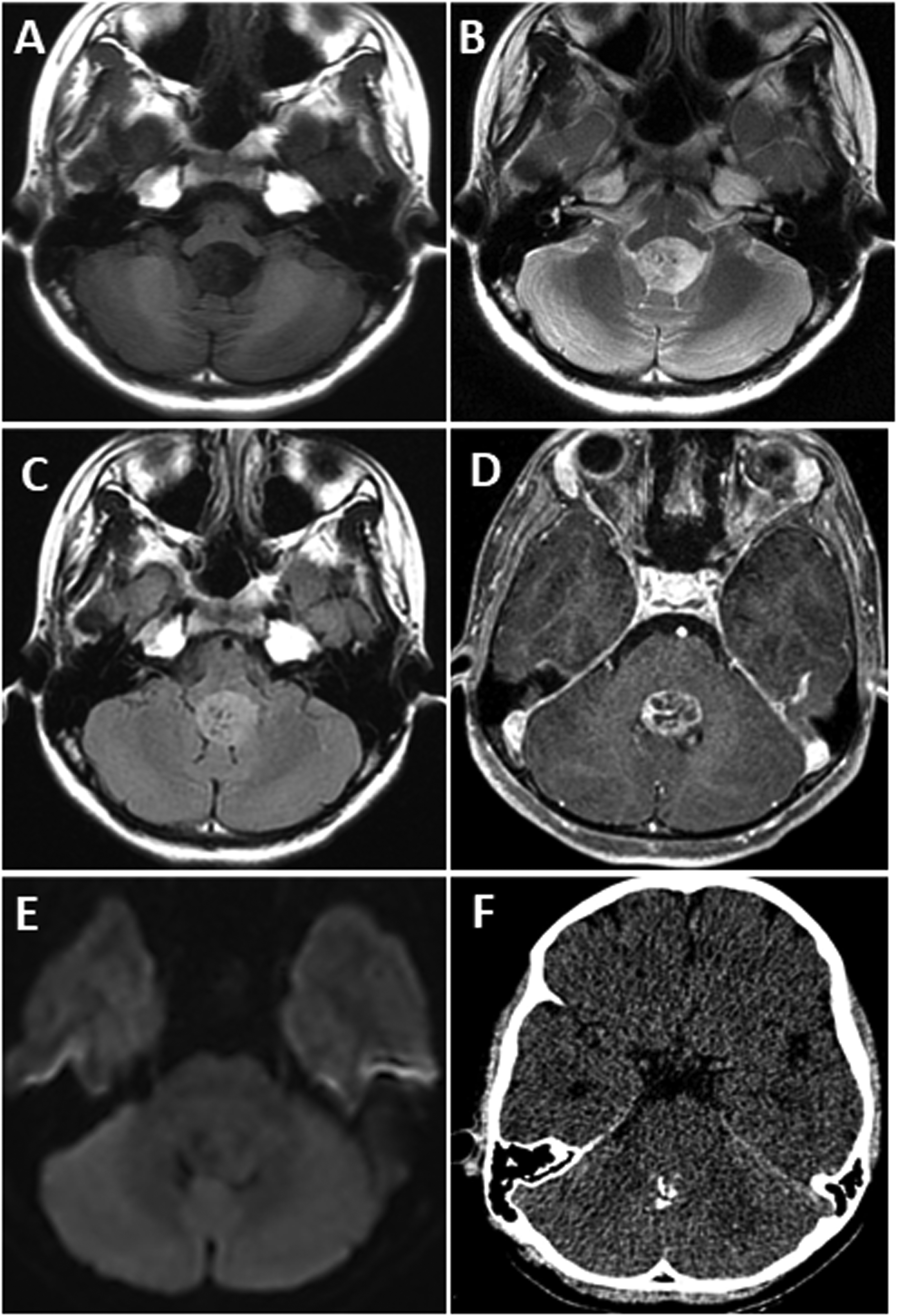
Axial T1 (A), T2 (B), FLAIR (C), post-contrast T1 (D), diffusion-weighted (E), and plain CT (F) images show a fourth ventricular mass with heterogeneous signal intensity and enhancement (A–D). There is no diffusion restriction (E). Calcification is present in the lesion (F).

Ependymoma with perivascular pseudorosettes (A) and GFAP immunoreactivity with perivascular accentuation (B). EMA shows typical paranuclear dot positivity (C). There is loss of nuclear expression for H3 p.K27me3 (D) with diffuse nuclear immunoreactivity for H3 p.K27M-mutant protein (E) and negativity for EZHIP (F). (A: H&E; B–F: Immunoperoxidase. Magnification = scale bar (A: 50 μm; B–F: 20 μm)).
H3 p.K27M mutation in PFAs was first described by Gessi et al in 2016. 3 To the best of our knowledge, only 25 patients have been reported in the English literature. The incidence of H3 p.K27M mutation ranges from 0.6% to 6% in different studies.4–6 PFA ependymomas with H3 p.K27M mutation in the literature3–8 along with the present tumor are enumerated in Table 1. The summary is as follows: mean age at presentation was 6 years (range: 1.5 to 12 years) with an equal male:female ratio. Genes encoding for histone 3.1 protein (HIST1H3C (H3C3), HIST1H3B (H3C2), and HIST1H3D (H3C4)) were most frequently mutated (17 of 23, 74%), while 26% of mutations involved the H3F3A (H3-3A) gene encoding for histone 3.3. Tumor recurrence was noted in 54% (7 of 13) of patients, of whom four had spinal recurrence. Overall, the median progression-free survival was 79 months (median overall survival: not reached). There was no statistically significant difference in the progression-free survival between tumors harboring mutations in histone 3.1 and histone 3.3 genes (79 months vs 18 months, p = .601).
Literature Review—Clinicopathological and Molecular Features of PFA Ependymomas With H3 p.K27M Mutation.
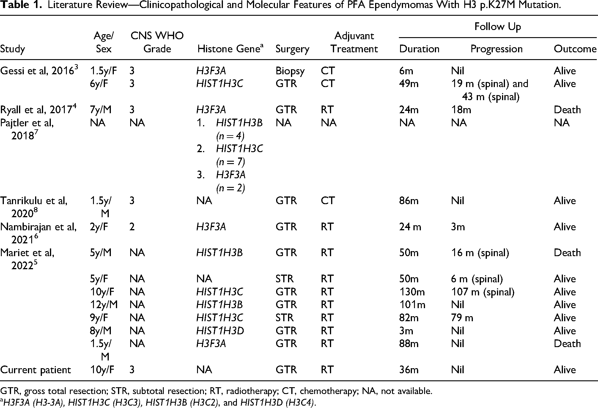
GTR, gross total resection; STR, subtotal resection; RT, radiotherapy; CT, chemotherapy; NA, not available.
H3F3A (H3-3A), HIST1H3C (H3C3), HIST1H3B (H3C2), and HIST1H3D (H3C4).
In a recent study by Mariet et al, H3 p.K27M-mutant PFAs were compared with their EZHIP-overexpressing counterparts. The former were found to have older age at presentation (6 years vs 2.4 years), were more midline in location and less likely to have lateral extension into the foramen of Luschka on MRI. However, the clinical outcome and histopathology did not differ significantly between the two. 5
To conclude, the occurrence of H3 p.K27M mutation is a rare event in PFA ependymomas. Histone 3.1 genes are more commonly involved than histone 3.3 genes. Although they share the global loss of trimethylation signature with H3 K27-altered diffuse midline gliomas, they appear to have a better prognosis. Hence, correlating histomorphology and immunohistochemistry is essential for accurate histopathological diagnosis.
Footnotes
Ethical Approval
Our institution (SCTIMST, Trivandrum) does not require ethical approval for reporting individual case reports.
Informed Consent
Informed consent for information published in this article was not obtained because this is a report in which the data is anonymized without any use of patient identifiers.
Author Contributions
MSS: manuscript preparation, editing, and data collection. DN: manuscript review and editing. GD: manuscript review and editing. KC: manuscript preparation, editing, and data collection. RP: conception, manuscript editing, and final approval.
Funding
The authors received no financial support for the research, authorship, and/or publication of this article.
Declaration of Conflicting Interests
The authors declared no potential conflicts of interest with respect to the research, authorship, and/or publication of this article.
Data Availability Statement
Not applicable.