
Abstract
Thirty-five patients undergoing a Bröstrom procedure for ankle instability were studied retrospectively as to the presence or absence of spurs and loose bodies, outcome, and mortise relationships. 100 adult volunteers had their ankles radiographically and clinically examined for spurs, loose bodies, and laxity. 100 patients’ ankles with computed axial tomography were examined to define malleolar relationships.
The AOFAS Hindfoot scores on the Bröstrom patients with or without spurs were not different. Patients undergoing a Bröstrom procedure had a 3.37 times incidence of spurs and/or loose bodies compared to normal adult population. The incidence of asymmetric but asymptomatic ankle laxity in normal adults was 11%. The fibula has a 38° range of position relative to the axis of the talus and the medial malleolus. A posterior fibular position may predispose to injury.
Keywords
INTRODUCTION
Impingement spurs and loose bodies are commonly seen in the ankle joints of athletes. 4,5,8,13,17,18,20 However, these osseous findings may not always cause painful symptoms or interfere with function. Impingement spurs are believed to be the result of repetitive microtrauma during athletic activity, wherein forced dorsiflexion leads to subperiosteal hemorrhage and subsequent ossification. 24,27 Some patients with anterior impingement spurs have also been noted to have moderate arthritic ankle joint narrowing. 29 After debridement these arthritic patients had a poorer prognosis than those with impingement spurs alone. 29
Harrington believed that chronic ankle instability led to degenerative arthritis of that joint. 11 The association of progressive degenerative arthritis with joint instability has been confirmed in other joints such as the shoulder, hip and knee. 6,14,21,25 It is therefore reasonable to believe that the arthritic changes associated with chronic instability at the ankle are different from the tibial impingement spurs associated with repetitive dorsiflexion microtrauma. This difference seems obvious, but as yet has not been thoroughly documented.
In patients with chronic ankle sprains there are several anatomic variations that have been associated with recurrent instability. Varus of the tibial plafond and a varus hindfoot have been described in association with recurrent instability. 19,28 However, we have also observed that patients with chronic ankle instability seemed to have variations in the position of the fibula relative to the medial malleolus which may predispose to instability. We therefore decided to take a fresh look at mortise relationships in both normal ankles and in unstable ankles to determine the incidence of abnormal mortise findings. We also looked at the incidence of spurs and loose bodies in a patient population with unstable ankles, comparing this incidence to the incidence of spurs, loose bodies, and abnormal laxity in the ankles of 100 normal adult volunteers. We also studied lateral and anteroposterior roentgenograms in the unstable ankle population as well as the computed axial tomographic (CAT) relationships of the mortise in 100 adults presenting for an ankle CAT scan. We wanted to see if there was variation in axial malleolar relationships. With these studies we believed we could better define the following: 1. The incidence of spurs, loose bodies, and abnormal laxity in the normal adult population. 2. The incidence of spurs and loose bodies in those patients with chronically unstable ankles. 3. Determine if the presence of spurs affected surgical outcome of an ankle ligament reconstruction. 4. Describe the normal range of malleolar axial relationships. 5. Describe the type and incidence of abnormal mortise variations seen in a patient population with chronic instability.
MATERIALS AND METHODS
Medical records were reviewed to collect patient data on individuals who had had surgery between January 1992 and September 1997 in which a modified Bröstrom procedure was performed to correct chronic, symptomatic ankle instability. 3,7 These individuals were identified, roentgenograms retrieved, and charts and operative reports reviewed. The patients were contacted and brought in for examination. Two were out of state and were contacted by telephone. Five could not be contacted.
The patients’ roentgenograms in this unstable ankle population were reviewed to determine the following information: the incidence of loose bodies or spurs, the incidence of varus of the heel or plafond tilt, and the incidence of what appeared to be a posteriorly positioned fibula relative to the medial malleolus. These patients were also examined and questioned to determine the outcome of their stabilization procedure using the AOFAS Hindfoot Score. 15
Statistical consultation was obtained to establish an adequate sample size in the adult population to determine the incidence of spurs, loose bodies, and abnormal laxity in the adult population at large. 12 This would enable us to meaningfully compare the incidence of spurs and loose bodies in our unstable ankle patient group to that of the normal adult population, using standard statistical techniques (a logistic regression model estimated risk, confidence intervals, and chi-square analysis). 12 Two separate radiologic studies were therefore employed to learn more about normal adult ankles. The first study was carried out to determine the incidence of spurs, loose bodies, and abnormal laxity. One hundred adult volunteers had a clinical and fluoroscopic (FluoroScan Imaging, Systems, Northbrook, Illinois) examination of each ankle in the anteroposterior and lateral planes. Costs and decreased radiation exposure led us to choose this method of examination over conventional roentgenograms. This study required approval by the Radiation Safety Committee and the Institutional Review Board (IRB). All study subjects signed an Informed Consent before radiographic and clinical examination of their ankles was conducted. This gave us 200 ankle examinations in 100 adults to provide a statistical incidence of spurs or loose bodies in adults. The incidence of ankle stability in these patients was determined by side-to-side comparison utilizing varus and anterior drawer stress testing performed by two of the authors (PES and JEM). On a scale of 0 to +3, the ankle had to rate +2 by both examiners to be regarded as pathologically lax.
Two additional radiologic studies were carried out to determine malleolar relationships in both the normal adult population as well as our separate patient population wherein the ankles were unstable. The anteroposterior and lateral roentgenograms of those patients that underwent stabilizing ankle surgery were reviewed. The anteroposterior roentgenogram measurements gave us an estimation of the incidence of either a varus tibial plafond or a varus hindfoot in this patient population. (Figure 1A & 1B) The lateral roentgenogram (true lateral with the foot resting on the x-ray cassette) was assessed to determine whether or not there appeared to be a posteriorly positioned fibula. ((Figure 2A) As our radiologic technique was identical for all patients seen in our office in this series, we compared these patients, dividing the series into those where the lateral roentgenogram did or did not appear to show posterior positioning. (Figure 2B)
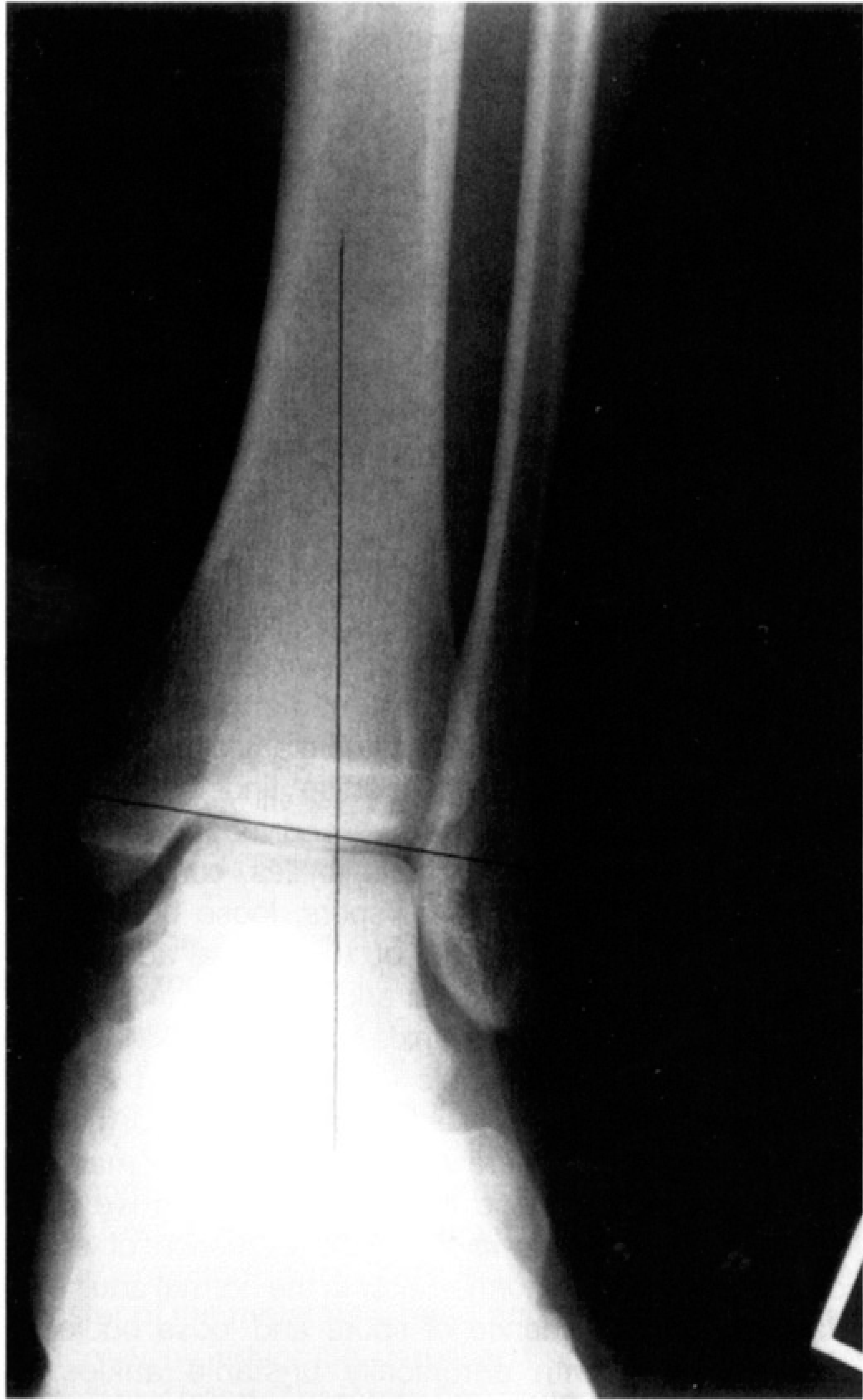
A standing anteroposterior roentgenogram of the ankle of one of the instability patients illustrating varus of the lower limb. The angle between the plane of the ankle joint is acute relative to true vertical.

A posterior view of the varus hindfoot in a patient with chronic ankle instability.
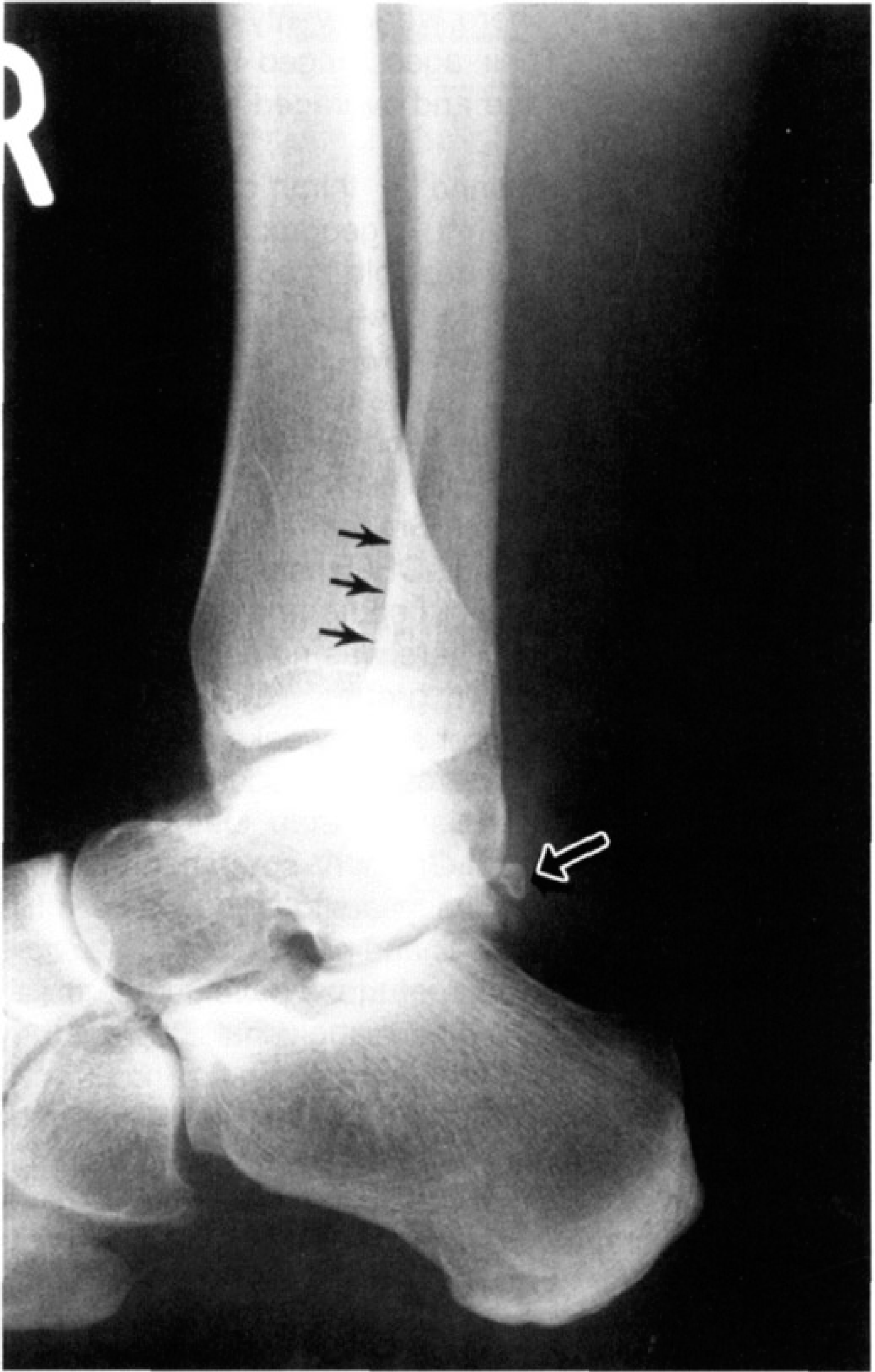
A lateral roentgenogram of the ankle of one of the instability patients, illustrating apparent posterior positioning of the lateral malleolus. The arrows point to the anterior border of the fibula. The posterior arrow points to an os trigonum.
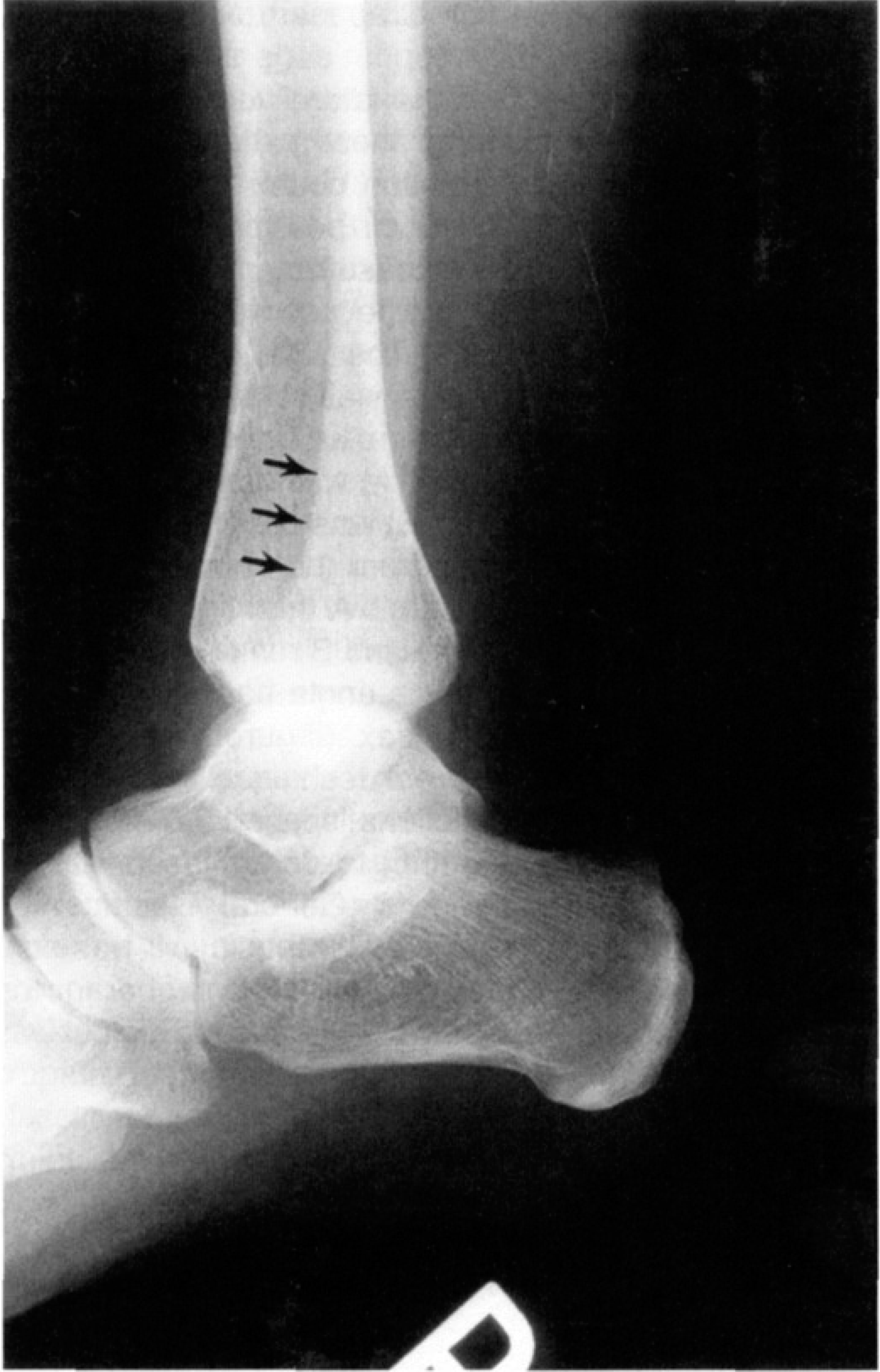
A lateral roentgenogram of an ankle of a normal adult, illustrating the more anterior position of the lateral malleolus. The arrows point to the anterior border of the fibula, more anterior than that of 2A.
In spite of the fact that the lateral roentgenographic technique was the same for these patients, we recognized that even a 10° rotation could mislead us into believing there was a posterior positioned fibula. We decided a more accurate measurement in variation of malleolar relationships would be to review 100 patients’ CAT studies of the ankle. The axial malleolar index (transverse plane of the talus) was calculated in the following manner: the plane of the articular surface of the talus was determined, a line (A) was drawn that bisected this axis. A second line (B) was drawn, running perpendicular to the talar axis from the anterior border of the medial malleolus, laterally. A third line was drawn (X) from the intersection of A and B running to the anterior border of the fibula. The angle between B and X represented the malleolar index. (Figure 3) Starting at the talar articular surface, our technique involved four serial axial, 1-mm thick sections, spaced 0.8-mm apart, reformatted in bone algorithm to determine the transverse plane of the talar axis (General Electric High Speed Advantage, Milwaukee, Wisconsin). It was constant for each patient tested. A positive number for the malleolar index meant that the lateral malleolus was posterior to the horizontal plane of the medial malleolus. A negative number meant that the lateral malleolus was actually anterior to the horizontal plane of the medial malleolus.
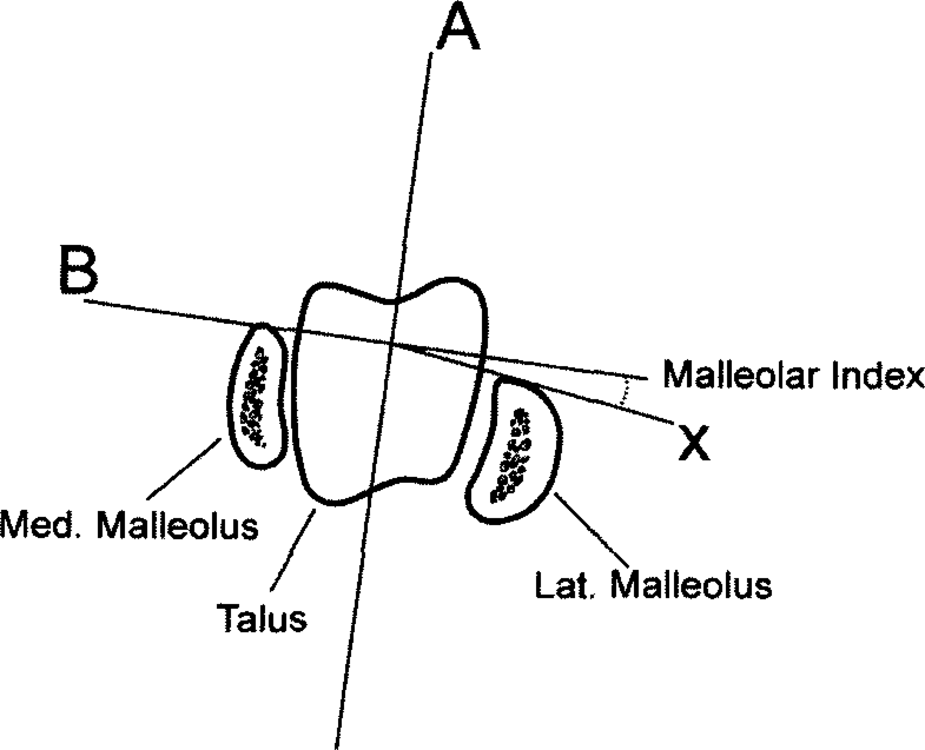
A diagrammatic illustration of the technique for measuring the axial malleolar index.
RESULTS
Clinical Studies
Between January 10, 1992 and September 8, 1997, thirty-five patients had thirty-seven (two bilateral) modified Bröstrom procedures for chronic, symptomatic ankle instability. 3,7 There were twenty-one males and sixteen females. Their ages ranged from sixteen to sixty-three years of age and averaged thirty-four years of age.
Follow-up was obtained in thirty of the thirty-five patients. In five patients a geographical move had occurred and it was not possible to contact them. Two of the thirty patients had a one-year ten months and a one-year eleven months follow-up, respectively. The remaining twenty-eight patients had two years or greater (up to seven years) and averaged 3.5 years follow-up. The nineteen patients that had spurs and/or loose bodies, ranging from Grade ll-IV, were seen in follow-up and averaged an AOFAS score of ninety-one. The eleven patients without spurs and/or loose bodies had a score of ninety. There was no difference in outcome between these two groups. One patient went back to competitive soccer at six weeks post-operative against our advice. Her Bröstrom procedure failed, but the second Bröstrom was successful. One patient with severe loose bodies and Grade IV spurs had a wound infection. He is now five years post-operative. His ankle is stable, but stiffness and pain resulted in a score of fifty-nine. Overall, each of these patients had a satisfactory result and indicated that given the choice they would have the procedure again.
Radiologic Studies
Roentgenographic review in all thirty-five patients revealed the following information: Loose bodies were present pre-operatively in fifteen patients; Spurs were present in eighteen patients; Loose bodies and/or spurs were present in twenty of the thirty-five patients (57%). In the group of thirty-five chronically unstable ankle patients, twelve were sent to us with outside roentgenograms, but twenty-three had satisfactory roentgenograms that were taken by our office's standard technique. We had reproducible, standardized, anteroposterior, lateral, and mortise planes that were utilized to assess the tibial plafond and mortise relationships. We used all the thirty-five patients’ roentgenograms to determine the presence or absence of spurs and loose bodies, but we used only our twenty-three patients’ x-rays for purposes of determining mortise relationships. Varus of the hindfoot or of the tibial plafond as described by Sugimoto was present in five of twenty-three ankles (21.7%). Twelve of the twenty-three ankles had, what we believed, was a posteriorly positioned fibula, relative to the medial malleolus (52.1%). One of these patients had a cavovarus foot with both a varus plafond and the posteriorly positioned malleolus.
The incidence of spurs and ankle laxity occurring naturally in an adult population of 100 volunteer adults was determined by using an anteroposterior and lateral fluroscan image of each of their ankles. This population sample was taken from volunteer hospital employees, visitors, construction workers, office employees, as well as adult patients who presented for evaluation of medical complaints other than an ankle problem. In addition, each volunteer had a varus stress test and anterior drawer test performed on both ankles by each of us. Their age, gender, whether they had symptoms of ankle pain or history of previous injury to their ankle was also recorded. There were thirty-one males and sixty-nine females. Their ages ranged from twenty-one to seventy-seven, averaging 41.4. The incidence of spurs or loose bodies was 17% or seventeen of 100 volunteers. Of these 100 individuals 11% had at least a +2/+3 asymmetric laxity in at least one ankle.
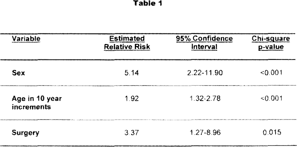
A logistic regression model was used for statistical analysis to compare the surgical and non-surgical groups with respect to the incidence of bone spurs or loose bodies, adjusting for differences in age and sex. These parameters were evaluated using the Wald statistic, comparing to a standard chi-square distribution. The results of this analysis are seen in Table 1. The key statistic in this table is expressed in the “surgery” variable. Simply put, if two subjects are the same sex and age, a patient that undergoes a Bröstrom procedure has a 3.37 times increased likelihood of having bone spurs or loose bodies, compared to an adult that does not have an unstable ankle (p>0.015).
The CAT scan results of 100 consecutive patients that presented at Providence Hospital for radiographic study were reviewed. These were not our patients, but rather referred by a variety of physicians, for a variety of diagnoses. On chart review these diagnoses included: ankle sprain, avulsion fracture, loose bodies, tendonitis, ruptured tendon, non-specific foot or ankle pain, plantar fasciitis, etc. These CAT scans were reviewed to determine the normal adult variation in the malleolar index. The age range of the patients was eleven to sixty-eight years. The average malleolar index was 9.3° with a standard deviation of 6.5°. The spread in this 38° variation is seen in the graph in (Figure 4. The extremes in axial variation ranged from −12° to +26°. (Figure 5A & 5B) There were twenty-three patients that had a malleolar index of +15° or greater. We reviewed these patients’ charts and found that 15 patients or 65% had a history of ankle sprain with chronic pain leading to the subsequent CAT scan.
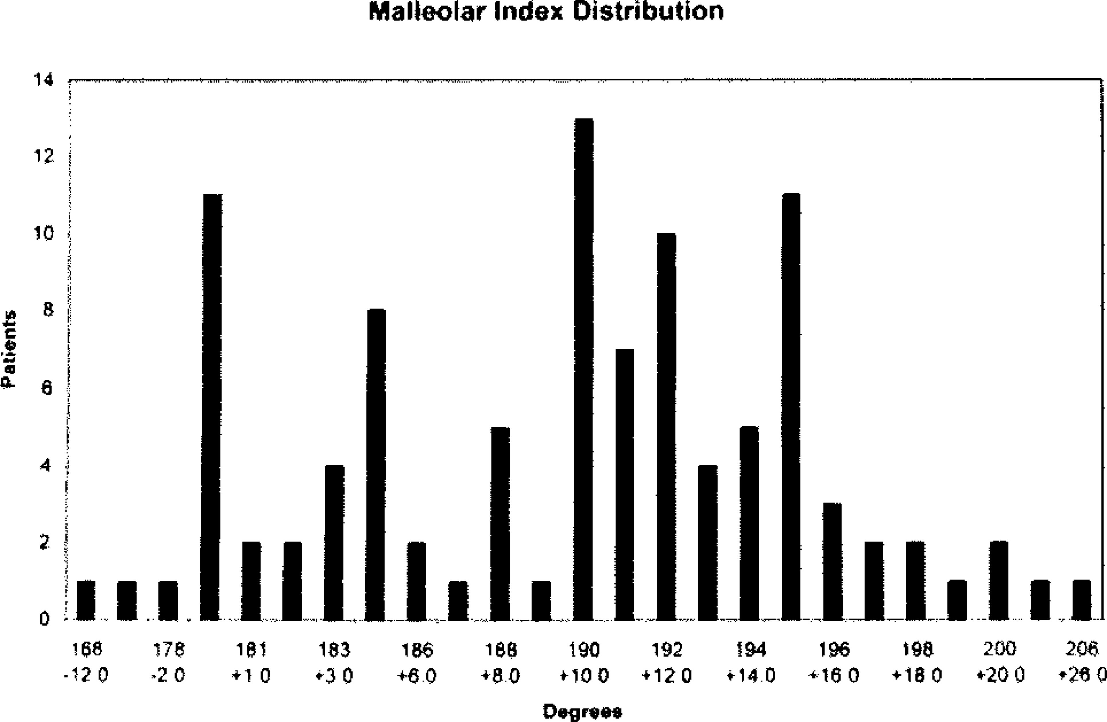
A graph illustrating the range of variation of the axial malleolar indices.
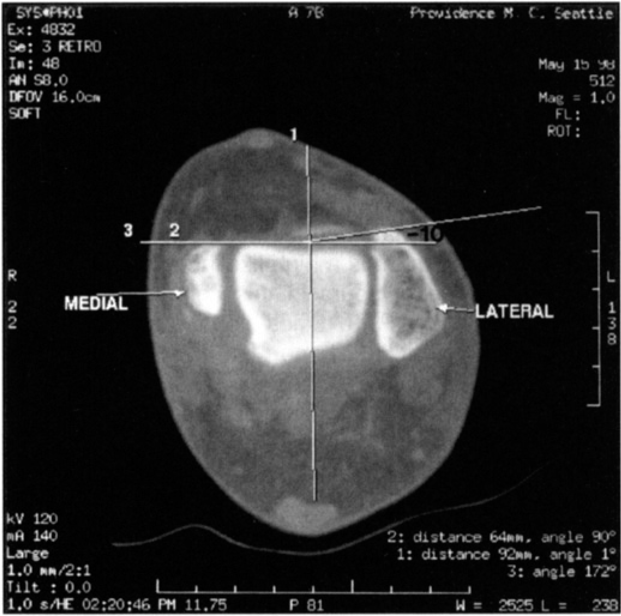
Computed axial tomographic illustration of the right ankle of a patient with a lateral malleolus border that is anterior to that of the medial malleolus (malleolar index −10°), relative to the transverse plane of the talus.
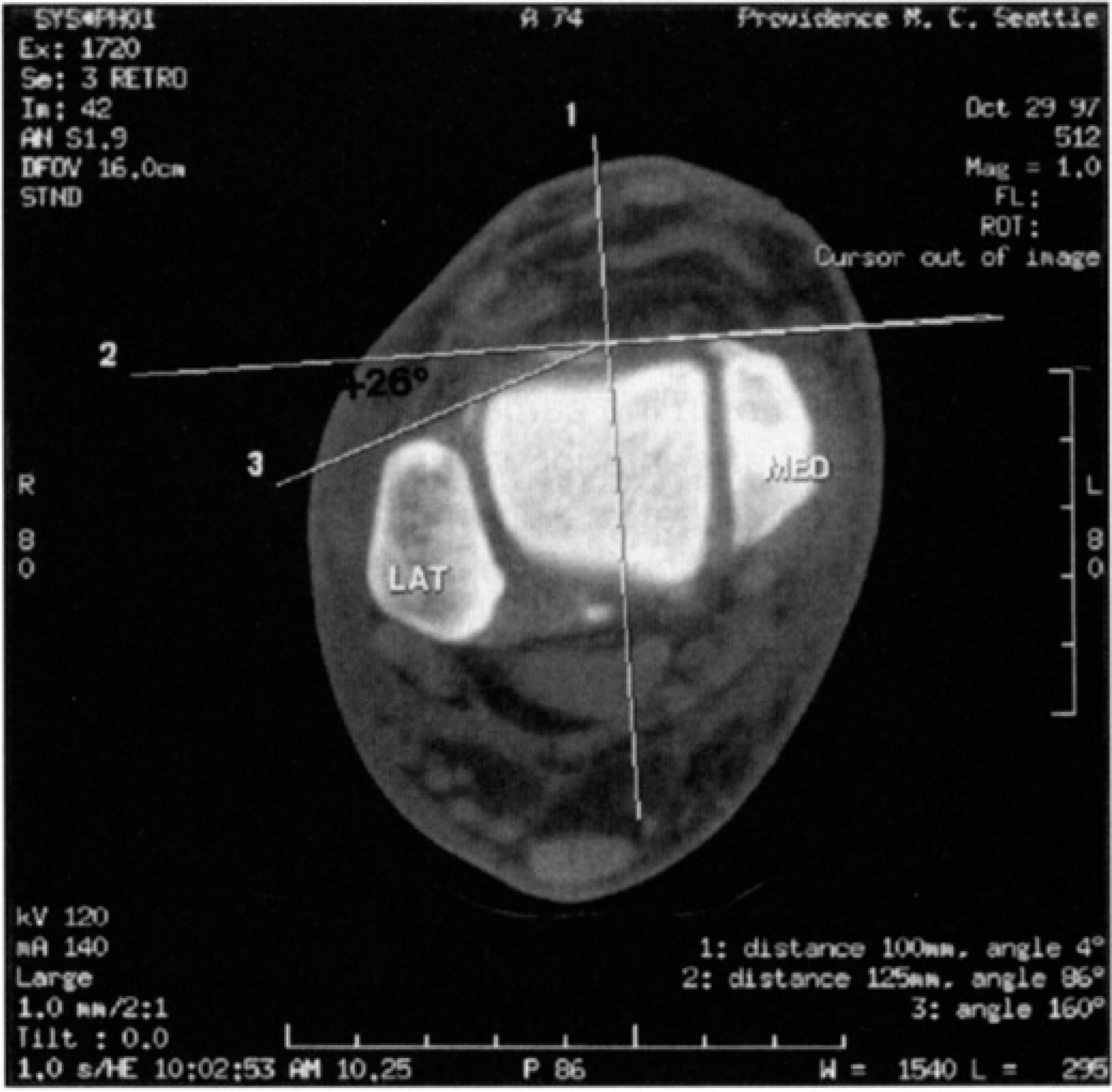
Computed axial tomographic illustration of the left ankle of a patient with the lateral malleolar border that is posterior to the medial malleolus (malleolar index +26°), relative to the transverse plane of the talus.
DISCUSSION
There is an overlapping relationship between impingement spurs seen at the ankle in competitive athletes versus spurs and loose bodies seen in patients with chronic ankle instability. Ankle impingement spurs have been well documented by a variety of authors from 1943 on. 17,18,20 However, Stoller and associates were first to attribute their formation to repetitive dorsiflexion at the ankle joint, microtrauma, subperiosteal hemorrhage, and subsequent ossification. 27
We believe these anterior ankle impingement spurs associated with athletics are different from degenerative spurs associated with chronic ankle ligament instability. We believe both impingement and chronic instability can produce spurs in the same patient. The tibial spurs of impingement are anterior, whereas the degenerative spurs are more global, as presented in our previously published classification. 24 Rubin and Whiten described the association between chronic ankle instability, synovitis and arthritic symptoms. 22 However they did not comment on the treatment of these patients, and they did not speculate that if untreated these ankle joints might go on to progressive arthritic deterioration. Harrington described patients with chronic ankle instability wherein degenerative arthritic findings were present. He treated some of these patients with a lateral stabilization procedure, but he did not comment as to whether or not he attempted spur resection. He did note that with ligament stabilization fourteen of twenty-two patients had some improvement out of the series of thirty-six. Sammarco also reported good results in a small patient subgroup of stabilized ankles, wherein some arthritic change was noted. 23
In 1992 we reported on the results of open or arthroscopic debridement of forty-three ankles in thirty-seven patients. 24 A classification system for spurs was proposed, ranging from Grade I to Grade IV. The grade was dependent upon the size and number of ankle spurs present. Our designation of Grade IV represented ankle spur formation beyond an impingement etiology with the description “pantalo-crural arthritic destruction.” van Dijk and associates corroborated our differentiation when they described sixty-two patients that underwent ankle arthroscopic debridement. 29 They divided these patients into two groups: those with impingement spurs and those with degenerative arthritis. However they performed spur resection only, not commenting on the presence or absence of ankle instability. At the two-year follow-up they reported that those patients with degenerative changes had not done as well as those with impingement spurs only.
Recurrent sprains and ankle instability are commonly associated with sports. 3,4,8,27,30 Proprioceptive response and peroneal tendon strength are clearly associated with sprain recurrences and instability. 9,16 Untreated athletes can have re-injury rates as high as 25%. 13 It is logical to assume both anterior tibial impingement spurs and degenerative spurs or loose bodies can be seen in association with chronic instability. Our retrospective analysis of thirty-five chronically unstable ankle patients revealed that 57% had spurs and/or loose bodies. Many of these patients played sports and it was not always possible to differentiate the etiology of the spur. For example, loose bodies can occur from fragmented spurs on the tibial or talar side, or by avulsion from the talar dome or malleoli. In essence, we report that whether spurs or loose bodies are present or not, symptomatic chronic ankle instability should be corrected. Further, if an adequate debridement is carried out at the time of surgery, there is a high likelihood of success.
The incidence of spurs in this chronically unstable patient population is 3.37 times higher than in a comparable adult population. These thirty-five unstable ankle patients had a 57% incidence of spurs and/or loose bodies compared to 17% seen in 200 ankles of 100 randomly selected individuals. The difference between these incidences is significant at p>0.015. The 11% incidence of ankle laxity in this random population is higher than the 5% reported by Bonnin in 1944. 2 His study population is not described. We intentionally spread out the vocations and age to reflect a broader spectrum. Our higher incidence may also reflect a more vigorous lifestyle in our time and region.
Sugimoto and associates have commented that a varus hindfoot or a varus tibial plafond is a possible predisposing factor towards chronic ankle instability. 28 The relationship between the varus heel of a cavus foot and chronic instability has also been well documented. 19 We describe a variation in the position of the fibula which we believe may predispose patients to sprains or recurrence. In our unstable ankle patient population we observed what appeared to be a more posteriorly positioned fibula, relative to the medial malleolus. The incidence was 52.1% in this patient population. This anatomic variation of a more “open” ankle mortise may predispose an athlete that has experienced an ankle sprain to a greater likelihood of recurrence. We noted this variation in twelve of our twenty-three patients wherein we believed our lateral roentgenogram was a true lateral film. Sigvard T. Hansen, MD has also noted this posteriorly positioned fibula in patients with severe arthritis and a history of ankle sprains that undergo total ankle replacement. 10
In spite of our radiographic technique being standardized for lateral roentgenograms, we believed we needed to verify through the CAT scan the actual variation in the malleolar axial relationship. We wanted to rule out the possibility that the apparent posterior position of the lateral malleolus in the unstable ankles was artifact, due to external rotation of the foot during the lateral roentgenogram. We therefore initiated the axial malleolar measurements relative to the transverse plane of the talus on 100 consecutive patients to investigate the variation of malleolar index. In fact, we confirmed that the malleolar index does vary from −12° to +26°, a range of 38°. This finding does not provide proof that those patients with unstable ankles in our series had a more vulnerable mortise with a posteriorly positioned lateral malleolus, but the correlation is strong. Further 65% of the twenty-three patients in the CAT scan study with a malleolar index greater than +15° had a history of an ankle sprain.
CONCLUSIONS
On the basis of this work we conclude the following:
Patients undergoing ankle stabilization procedures may have tibial impingement spurs as well as degenerative arthritic spurs with or without loose bodies. In fact, the likelihood of spurs and loose bodies in patients with unstable ankles is 3.37 times that of the normal adult population. The presence of spurs or degeneration in a patient's ankle is not a contraindication to a stabilization procedure. The incidence of spurs or loose bodies in the normal adult population is 17%, and the incidence of abnormal laxity in normal adults was 11%.
Patients with unstable ankles that have spurs should have a debridement at the time of ankle stabilization. If properly carried out, these patients can expect the high likelihood of a satisfactory outcome from ankle stabilization.
We confirm the observations of Myerson and Sugimoto regarding the presence of limb, heel, or plafond varus in association with chronic ankle instability. We also describe for the first time the malleolar index and the posteriorly positioned fibula. We note that malleolar relationships can vary by as much as 38°. We believe the posteriorly positioned fibula is an anatomic variant that may predispose patients to chronic ankle instability. A simple lateral roentgenogram, taken with the non-weight bearing ankle resting medially on the plate, will reveal posterior positioning of the fibula. CAT scanning is optional.
Patients presenting with an ankle sprain that have a posterior fibular position, a varus plafond, or a cavus foot at the time of their initial sprain may require a greater degree of post-sprain brace protection and rehabilitation.
Footnotes
ACKNOWLEDGEMENTS
The authors thank Anita Rocha, MS for assistance in the statistical analysis and Deborah A. Amacker for coordinating and conducting the clinical and one of the radiologic studies.