
Abstract
A primary function of the peroneus longus and peroneus brevis is to provide the eversion moment necessary to balance the opposing inversion moments. Surgeons often deal with the loss of or need to sacrifice one of these tendons. This study compares the evertor mechanisms of the peroneus brevis and peroneus longus muscle. This is accomplished in a cadaver model in which the performance of each of the muscle tendons during early heel rise of gait is assessed utilizing the same tendon loads in each so that force is not a variable. Six fresh-frozen cadaver foot-ankle specimens were studied during a simulated early heel rise phase of the gait cycle. The study compared the effect of the peroneus brevis and peroneus longus by separately applying the same load to the each of the tendons. At the talonavicular joint, the peroneus brevis loaded condition externally rotated the navicular 2.1° more than when the peroneus longus was loaded. At the subtalar joint, the peroneus brevis loaded condition resulted in 0.9° more calcaneus valgus relative to the talus than was present during the peroneus longus loaded condition. The experimental data support the hypothesis that the peroneus brevis tendon mechanism is more effective than is the peroneus longus mechanism in rotating the navicular externally and the calcaneus into valgus. This has clinical implications for assisting surgeons in trying to preserve evertor function.
INTRODUCTION
A primary function of the peroneus brevis and peroneus longus is to provide the eversion moment necessary to achieve a balance with the opposing invertors and the external moments resulting from the ground reaction force. Evertor function is altered when the peroneus longus or brevis tendon is ruptured or used for a soft tissue reconstruction. Consequently, it is essential to understand how the actions of these tendons affect both the talonavicular and subtalar joints, two joints that have key roles in the progression of acquired flatfoot deformity. This knowledge will provide greater insight into the functional ramifications of conditions or procedures that alter the roles of the peroneus longus or peroneus brevis.
A stronger evertor may refer to the muscle that can generate the greater force at its tendon, or it may refer to the muscle tendon that generates the greater moment about the joint of interest. The ability to evert is a function of both the force-generating capacity of a muscle and the effective moment arm about the joint. The force-generating capacity is generally accepted to be proportional to the physiologic cross-sectional area of the muscle. For the peroneus longus, this is nominally twice that for the peroneus brevis. 9,10 If one considers tendon strength to be an indicator of muscle force utilized, the demonstration that the failure strength of the peroneus brevis and peroneus longus tendons is no different is consistent with the same force levels being utilized by these muscles. 1 Thus, the inconsistency in the physiologic cross-sectional area results versus the tendon strength data makes it difficult to estimate the functional force levels used by these muscles. The moment arms are more difficult to assess because each of the tendons acts across multiple joints.
The peroneus brevis inserts on the fifth metatarsal, while the peroneus longus inserts on the first metatarsal. Compared to the peroneus brevis, the peroneus longus has a more distributed effect as a result of its lateral tether and its traversing of the plantar aspect of the foot prior to attaching at the base of the first metatarsal. There is some dispute in the literature as to which of the peroneal tendons is the stronger evertor, with some sources citing the peroneus brevis 6 and others favoring the peroneus longus. 9 Knowledge of the contributions of each of the muscles specifically to talonavicular joint and subtalar joint rotation will aid in selection of which tendon to harvest for transfer or reconstruction purposes.
This study examines the functional contributions of the peroneus longus and peroneus brevis to external rotation at the talonavicular joint and valgus at the subtalar joint. Secondarily, plantarflexion and abduction at these joints are documented. An understanding of the relative roles of the two tendons in plantarflexion and abduction at the subtalar and talonavicular joints will provide further insight into actions of the two muscles. Furthermore, it is important to understand the roles of these muscles at a time when they are under their greatest demand. Based on electromyographic (EMG) activity, this occurs in the early heel rise phase of the gait cycle. 7 Therefore, our objective is to delineate the functional differences of the peroneus longus tendon and peroneus brevis tendon mechanisms at this critical time. The study examines the hypothesis that a force in peroneus brevis tendon, with its insertion at the base of the fifth metatarsal, will affect the talonavicular and subtalar joints differently than an equal force in the peroneus longus tendon during early heel rise.
METHODS
Six fresh-frozen cadaver foot-ankle specimens thawed overnight at room temperature were utilized. With the foot plantigrade and a 220 N vertical load applied to tibia, weightbearing mediolateral, anteroposterior, and dorsal radiographs were obtained to screen for preexisting deformity. Exclusion criteria included prior surgery, an excessively high or low medial longitudinal arch, and poor bone quality noted on radiographs. Dissection consisted of removal of all soft tissue superior to the malleoli while preserving tendon lengths. The portion of the fibula superior to the ankle mortise was removed, and the proximal tibia was potted in epoxy.
Three-dimensional joint motions were recorded using electromagnetic motion sensors (Polhemus Fastrak, Colchester, VT) rigidly attached to the talus, calcaneus, navicular, and cuboid bones via a pair of carbon vinyl ester rods. Each specimen was placed in a custom testing apparatus (Fig. 1) with the tibia oriented 17° anterior of vertical, a position consistent with early heel rise (i.e., 40% of the gait cycle). The apparatus has been described previously. 2 Three stepper motors (Industrial Devices Corp., Novato, CA) in series with force transducers (Sensotec, Columbus, OH) applied orthogonal ground reaction force components to the plantar surface of the foot via a Plexiglas footplate. Four stepper motors in series with force transducers generated the necessary pull on the Achilles, posterior tibial, peroneus longus, and peroneus brevis tendons. The tendons were gripped with cryoclamps. 8 A custom-programmed software control interface (LabView v4.1, National Instruments, Austin, TX) was used to coordinate the closed-loop feedback control system.
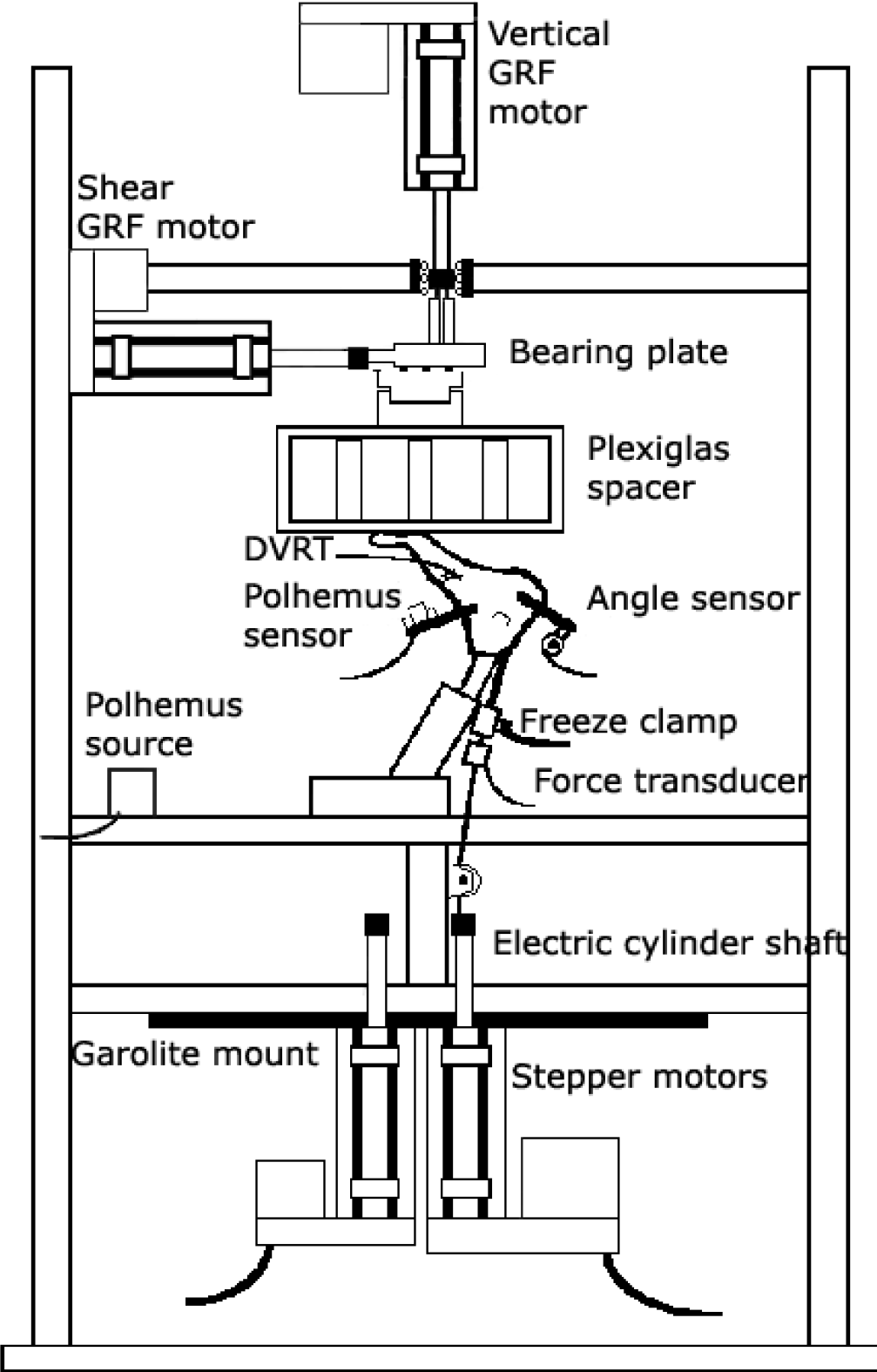
Foot testing apparatus.
A reference position was defined using a vertical ground reaction force of 357 N (i.e., 50% nominal body weight) and no tendon loading. Joint orientations under this set of loads were defined as 0°. Loading trials consisted of simulating the early heel rise position, when the peroneus longus and peroneus brevis both demonstrate their greatest electromyo-graphic activity. This occurs at 40% of the gait cycle. The loading trials consisted of loading the Achilles tendon under calcaneal position feedback mode until 10° calcaneal plantarflexion was achieved. Simultaneously, the stepper motors applied ground reaction force components corresponding to 50% of full scale (357 N vertical, 27 N anterior, 13 N medial) and applied tendon loads based on physiologic cross-sectional area and EMG profiles 7 to the posterior tibial tendon (223 N) and either the peroneus brevis or the peroneus longus tendon (259 N). Three trials were completed in which the load of 259 N was applied to peroneus brevis and no load was applied to peroneus longus during loading to heel rise. Subsequently, three trials were completed in which this 259 N load was applied to peroneus longus and no load to peroneus brevis. The order was randomized.
Terminology describing the rotations in the foot can be confusing. The terms eversion and inversion are generally used with respect to the foot rotating about the subtalar axis, an axis that is not perpendicular to any of the anatomical planes. Rotations of the talonavicular joint will be defined in terms of navicular rotation with respect to the talus: external rotation is about an axis perpendicular to the coronal plane; plantarflexion is about an axis perpendicular to the sagittal plane; and abduction is about an axis perpendicular to the transverse plane. At the subtalar joint, the definitions will be the same, with the exception that rotation about an axis perpendicular to the coronal plane will be termed valgus.
To compare the effects of load in each of the muscles, each specimen was tested in each of two conditions: peroneus longus only loaded and peroneus brevis only loaded. Each specimen served as its own control. The primary outcome variables were the difference in external rotation at the talonavicular joint and the difference in calcaneal valgus at subtalar joint for the two muscle loaded conditions. For the two muscle loaded conditions, comparisons were made of external rotation at the talonavicular joint and valgus at the subtalar joint using a Student paired t test. A two-tailed test was used with an alpha level for significance set at p < .05. A Bonferroni correction was used to adjust for the two primary outcome variables. This reduces the alpha level for each test to .025 to bring the alpha level overall back to .05.
RESULTS
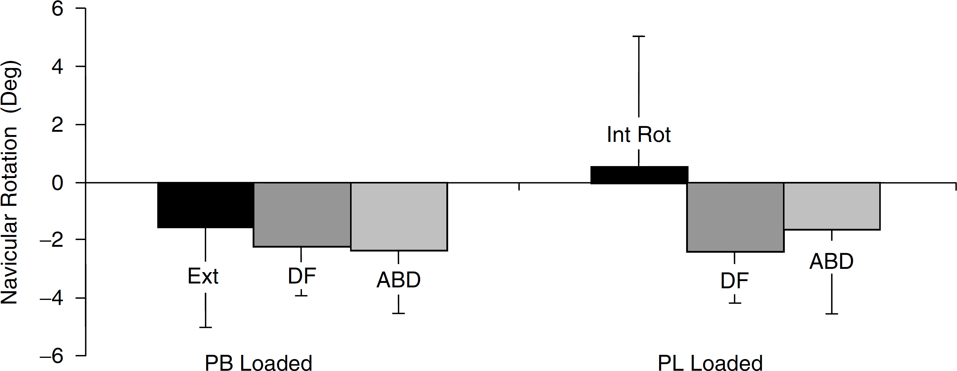
External rotation at the talonavicular joint (i.e., the navicular externally rotated relative to the talus) was 2.1° ± 1.5° greater (Fig. 2) when the peroneus brevis was loaded (p = .018). In addition, when the peroneus brevis was loaded, navicular plantarflexion averaged 0.2° ± 0.2° greater than for the peroneus longus loaded condition and was not significant (p = .08). When the peroneus brevis was loaded, the navicular was abducted on average 0.8° ± 1.0° more than for the peroneus longus loaded condition, but this difference was not significant (p = .11). Thus, the one significant effect at the talonavicular joint in early heel rise is the greater external rotation associated with peroneus brevis load.

The bar heights indicate the greater amount of each rotation from flatfoot to heel rise for the peroneus brevis loaded condition relative to the peroneus longus loaded condition, with one exception: the navicular does not plantarflex for either condition, but the difference is shown as more plantarflexed position to reflect that it dorsiflexes less. Only the external rotation and valgus rotation differences are significant.
At the subtalar joint, peroneus brevis load resulted in calcaneal valgus that was 0.9° ± 0.6° greater (Fig. 2) at heel rise compared to the peroneus longus loaded condition (p = .017). In addition, when the peroneus brevis was loaded, the calcaneus was more dorsiflexed an average 0.4° ± 0.4° (p = .041) and more abducted an average 0.7° ± 0.4° (p = .041) than for the peroneus longus loaded condition. Thus, the predominant effect at the subtalar joint in early heel rise is greater valgus associated with peroneus brevis load.
DISCUSSION
The peroneal tendons can be harvested for use in such procedures as tendon transfers or ligament reconstruction. Selection of which of the two tendons to use may be based on surgical considerations and, to some degree, on published data of physiologic cross-sectional area of the muscle or tendon diameter, variables that are highly dependent on measurement technique and that are known to vary widely from individual to individual. In deciding which tendon to sacrifice, surgeons strive to follow the paradigm of muscle balance; that is, an attempt is made to balance antagonistic muscle groups so that there is comparable inversion and eversion power. Knowledge of the actions of the peroneal tendons at the talonavicular joint and subtalar joint provides objective criteria for making this decision.
The difference measured in external rotation at the talonavicular joint was approximately 2°. This may appear small and irrelevant, but such a shift in talonavicular joint orientation may be clinically significant, considering that a normal range of motion is 24° 5 (i.e., about a 10% change) and that the force needed to generate more external rotation against the antagonist (posterior tibialis) may be quite significant. Considering that both muscles are primarily hindfoot evertors, a difference of 2° is reasonable. Furthermore, the finding of greater external rotation of the navicular with the peroneus brevis loaded was consistent across all six specimens.
At both the talonavicular joint and subtalar joint, the peroneus brevis loaded condition demonstrated greater rotations predominantly in the coronal plane and secondarily in the transverse plane. The navicular was more externally rotated and abducted and, similarly, the calcaneus was in more valgus and abduction than for the peroneus longus loaded condition. Despite the small angular changes measured, the results represent a greater tendency for the peroneus brevis mechanism to rotate the foot into a direction associated with acquired flatfoot deformity. This result is consistent with the role of the peroneus brevis in which posterior tibial tendon deficiency did not manifest itself when peroneus brevis function was absent. 6
A further observation of interest was noted when examining the rotational changes that occurred when going from the plantigrade position to the heel rise position. With the 259 N peroneus brevis load, the talonavicular joint externally rotated 1.6° ± 3.5° when going into the heel rise position (Fig. 3); however, with the 259 N load on the peroneus longus, the talonavicular joint changed direction and internally rotated 0.5° ± 4.5°. The rotations about the other two axes (i.e., dorsiflexion and abduction) were in the same direction during heel rise, regardless of which tendon was loaded. Thus, besides being more externally rotated at heel rise when the peroneus brevis was loaded, the direction of rotation was reversed with the peroneus longus loaded. For the subtalar joint, there was no reversal in the direction about any of the axes for the two tendon loading conditions. This may be a manifestation of the method used to define the reference position.
Talonavicular joint rotation change about each axis from plantigrade position to heel rise position. Shown are the results for the peroneus brevis loaded condition and the peroneus longus loaded condition.
Studies have been performed to estimate the muscle moment arm lengths of the extrinsic foot muscles. For the inversion-eversion and flexion-extension, axes of the foot moment arms have been estimated from tendon excursion measurements using a test apparatus with 6 df. 3 Relative to moment arm length of the tibialis posterior, the average evertor moment arm lengths for the peroneus longus and peroneus brevis were 82% and 85%, respectively. Although not specified as lengths in that study, the eversion moment arm lengths for the two muscles were on the order of 25 mm. In a different study, the moment arm lengths of the extrinsic foot muscles about the ankle and subtalar joints were estimated using the tendon excursion method. 4 At the subtalar joint, the peroneus longus and peroneus brevis moment arm lengths were calculated to be 21.8 mm and 20.5 mm, respectively. The different results for these two studies can be attributed, in part, to differences in experimental setups. The former study attached the foot to a footplate that was rotated and the latter study generated the rotation without a footplate to control the foot.
The estimates of peroneus brevis and peroneus longus moment arm lengths indicate that the moment arm lengths are approximately equal. 3,4 The muscle architecture studies 9,10 support the peroneus longus as being twice the strength as the peroneus and the equal tendon strengths for these two tendons 1 suggest the two muscles may well function at the same force levels. The larger muscle belly of the peroneus longus may be associated with requirements for greater excursion or velocity of shortening rather than strength. Thus, it is difficult to estimate the peroneus brevis and peroneus longus forces used at heel rise. In retrospect, it would have been beneficial to monitor the rotational changes while incrementally increasing the peroneus brevis and peroneus brevis muscle forces. This would have illustrated when lesser forces produce significant rotational changes and at what point increases in force can no longer produce rotational changes.
The approach taken in this study was to compare the actions of the peroneus brevis and peroneus longus tendon mechanisms with respect to their effects at the talonavicular and subtalar joints at a time during the gait cycle when both muscles have their peak EMG activity. The same force was applied by each of the muscles, in different trials, in order to compare the roles of the two tendon mechanisms. The peroneus brevis mechanism was more effective than the peroneus longus mechanism in producing external rotation and abduction at the talonavicular joint and valgus and abduction at the subtalar joint. Thus, although generally referred to as evertors of the foot, this approach demonstrated mechanisms for these two muscle tendons that differ in their effects on the talonavicular and subtalar joints.
CONCLUSION
The study compared the mechanisms of the peroneus brevis and peroneus longus by separately applying the same load to the each of the tendons. Differences in rotation at the talonavicular and subtalar joints were observed for the two scenarios that are independent of muscle force and, therefore, reflect the actions of the tendons with respect to these two joints. The experimental data support the hypothesis that peroneus brevis tendon mechanism is more effective than is the peroneus longus mechanism in rotating the navicular externally and the calcaneus into valgus. This has clinical implications for assisting surgeons in the selection of which tendon to harvest.
Footnotes
ACKNOWLEDGMENTS
Financial support from NIH 1R01AR44508 and OREF 98-020 and the Clark, Frese and Kirby Foundations is acknowledged.