
Abstract
Background: One of the challenges of total ankle arthroplasty continues to be achieving a solid distal fusion of the tibiofibular joint. Delayed union rates of 29% to 38% and the nonunion rates of 9% to 18% for syndesmotic fusion have been documented. The risk of tibial component migration has been reported to increase 8.5 times if a solid syndesmotic fusion is absent. Growth factors have been shown to accelerate bone healing and may enhance the fusion of the syndesmosis and, thereby, decrease the frequency of nonunion and subsequent tibial component migration. Methods: An autologous platelet concentrate was used to increase the amount of growth factors at the site of the distal tibiofibular joint fusion in 20 total ankle arthroplasties. Results: Our 6-month fusion rate was 100%. When compared to historical controls (6-month fusion rate of 62%) the difference was statistically significant (p < 0.0001). Conclusion: The improved rate of distal tibiofibular fusion may be attributable to the increased presence of growth factors provided by an autologous platelet concentrate.
INTRODUCTION
Total ankle replacement surgery has now been practiced for over three decades. Early in the evolution of this procedure, problems with component design, prosthesis fixation, surgical technique, and patient selection led to early failures. 9,21 Some recommended complete abandonment of total ankle replacement in favor of ankle arthrodesis. 5,10,11,22 During the mid to late 1980s, second generation systems corrected many of the flaws associated with their predecessors. These ankle replacement systems are still undergoing modifications as new technologies and research data are evaluated. 15,23 With improved surgical techniques and new component materials, total ankle arthroplasty (TAA) has returned as a viable option in the treatment of patients with debilitating ankle arthritis in whom conservative management has failed.
Since 1984, when the first Agility Ankle (DePuy Corp. Warsaw, Indiana) was used, this system has undergone five phases of development. 23,24 These modifications have improved function and ease of insertion. Distal tibiofibular joint arthrodesis is essential to the support of the tibial component and ultimately to the success of this ankle arthroplasty. In a review of the first 100 Agility ankle arthroplasties, the nonunion rate of the syndesmosis was found to be 9% and the delayed-union rate (defined as more than 6 months) was 29%. 23 Migration of the tibial component was related to nonunion and delayed union of the syndesmosis. Improving the rates of distal tibiofibular joint arthrodesis is expected to decrease the migration rates of the tibial component and improve the function and long-term survival of the prosthesis.
We believe that the use of growth factors supplied by an autologous platelet concentrate and increased biomechanical stabilization will improve the rate of distal tibiofibular joint arthrodesis and prosthesis ingrowth during TAA. Growth factors found in platelets are released during the initial inflammatory phase after injury. 25 Over the past 2 decades studies in both animals and humans have shown that growth factors contained in an autologous platelet concentrate have successfully induced bone formation. 6,16,17,18,27 This report presents our preliminary results from 20 consecutive patients who had total ankle arthroplasty and explains our protocol.
MATERIALS AND METHODS
Beginning in July of 2001, an autologous platelet concentrate was used in 20 consecutive TAA patients (20 ankles). Although institutional review board approval was not required at this hospital, all patients gave informed consent before participating in this study. The age range was 40 to 80 (mean 62) years. There were 11 males and nine females. The most common preoperative diagnosis (12 patients) was post-traumatic arthritis. Two patients had been diagnosed with rheumatoid arthritis, and six patients had osteoarthritis. In all patients at least 6 months of nonoperative treatment involving some type of bracing had failed to relieve symptoms. Twelve right ankles and eight left ankles were involved. Followup was from 6 to 21 months, with an average of 15 months.
The operative technique was as described by Alvine with some modifications. 2 An anterior ankle approach was used, along with the medial placement of an external fixation device. A lateral incision also was used to expose and prepare the distal tibiofibular joint for arthrodesis. After debridement of the soft tissue in the syndesmosis, a 2-mm drill was used to prepare the lateral surface of the tibia and the medial surface of the fibula for fusion by making multiple drill holes in the subchondral plate and cortical bone. Symphony PCS autologous platelet concentrate (Depuy Corp, Warsaw, Indiana) was sprayed on the bone surfaces of the syndesmosis and over the cut surfaces of the distal tibia and talus. The autologous platelet concentrate also was mixed in the local bone graft obtained from the joint preparation and applied to the porous coating of the prosthesis. After the TAA components were inserted and the external fixator was removed, the bone graft was packed in the distal tibiofibular joint. Two 4.5-mm corticle screws were used to carefully compress the fibula against the tibia for fusion (the original technique places two 3.5-mm cortical screws). A layered closure was done, and a sterile dressing and posterior bulky splint holding the ankle in neutral dorsiflexion were applied.
Dressings were removed at 2 weeks, and a short leg nonweightbearing cast was applied. Patients were kept nonweightbearing with the ankle held in neutral dorsiflexion for a total of 8 weeks (this differs from the original technique of allowing motion out of a splint each day and beginning weightbearing at 6 weeks). Patients who had less than 10 degrees of dorsiflexion at their 5-week postoperative appointment were enrolled in a physical therapy program and placed in a removable splint. The remainder of the patients who all had at least 10 degrees of dorsiflexion at their 5-week postoperative appointment were casted. Full weightbearing was begun at 8 weeks if the syndesmosis appeared to have fused and the components were stable in the ankle. If the syndesmosis was not completely fused at 8 weeks, the patient was placed in a cast walker boot for the first month of weightbearing. After weightbearing had commenced, followup examinations were done and radiographs were made at 1 month, 3 months, 6 months, and then yearly.
The Agility ankle prosthesis was used in all patients. This prosthesis is a semi-constrained ankle arthroplasty device with a fixed-bearing titanium-backed tibial component and a cobalt chrome talar component. The ultra-high-molecular weight polyethylene (UHMWPE) insert is concave to capture the talar component. Both tibial and talar components are porous coated. The prosthesis offers six sizes each of tibial and talar components, as well as a thicker UHMWPE insert. 2
Symphony PCS is a system in which a platelet concentrate is prepared from a 55 cc volume of autologous blood. This volume is centrifuged, and the platelet-rich layer is combined with thrombin to produce a concentrate gel with four to six times the normal concentration of platelets. This gel is then sprayed directly on the prepared bone surfaces and on the porous coating of the tibia and talar components.
Each patient was seen at 2, 5, and 8 weeks after surgery during the nonweightbearing phase of our protocol. Weightbearing was allowed at 8 weeks after surgery, and followup continued at 1 month, 3 months, 6 months and then yearly. Anteroposterior, lateral, and mortise radiographs were obtained at each visit. Complete syndesmotic fusion was defined as bony trabeculae spanning the distal tibiofibular joint seen in the anteroposterior view (Figures 1 and 2). Areas of lucency along the bone implant interface and subsidence of the prosthesis were recorded.
RESULTS
Of the 20 ankle prostheses implanted using our protocol, 17 (85%) were judged to have a solid distal tibiofibular fusion at 8 weeks after surgery. Two patients (10%) had successful fusion of the syndesmosis at 3 months postoperatively, and one patient (5%) had complete fusion of the syndesmosis at 6 months. Mean followup for these patients was 15 (6 to 21) months. There was no subsidence of any prosthesis and no change in position of the implants compared to that shown in the immediate postoperative radiographs.
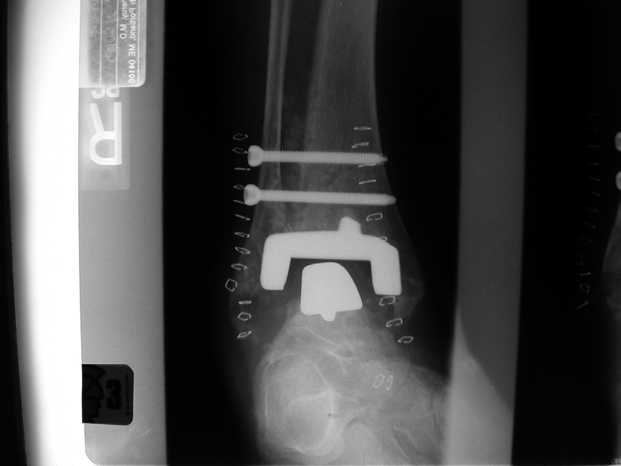
Anteroposterior radiograph at 2 weeks postoperatively
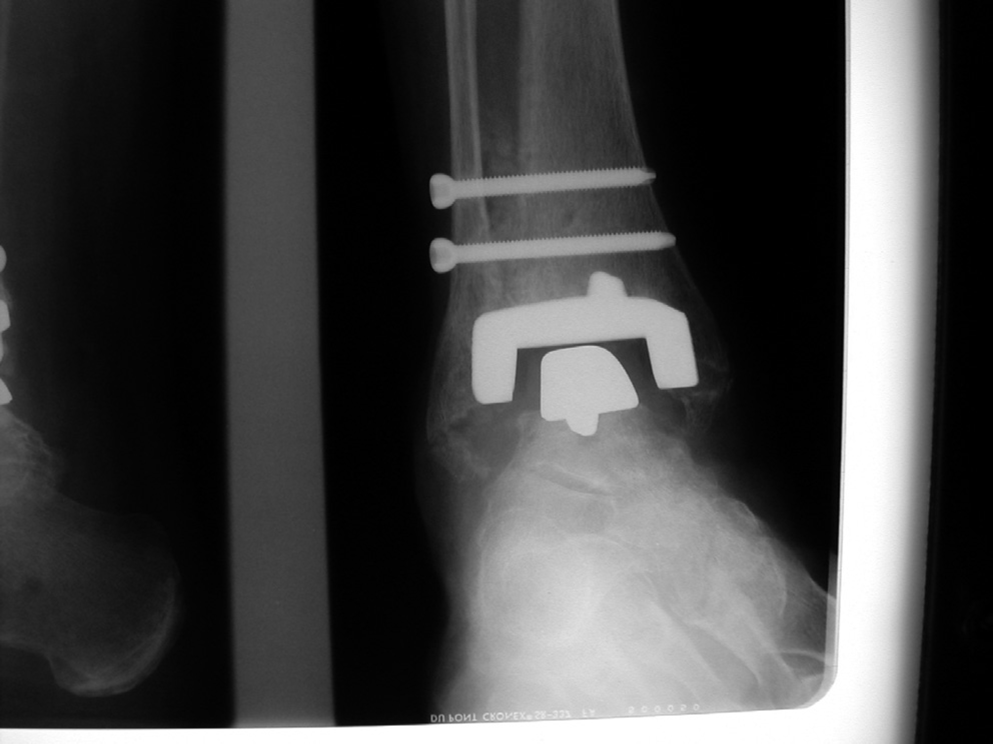
Anteroposterior radiograph at 8 weeks postoperatively showing bridging trabeculae.
Reported rates of distal tibiofibular joint fusion and nonunion
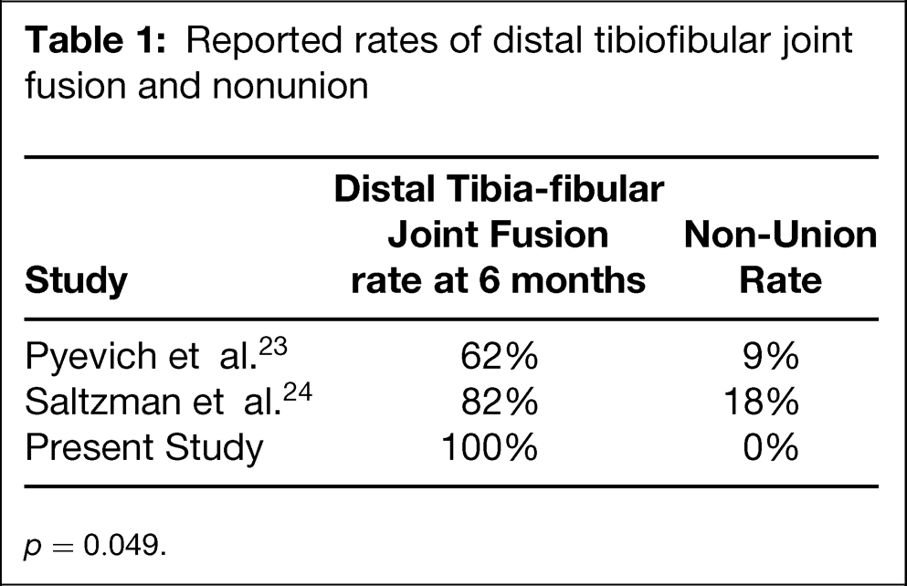
p = 0.049.
DISCUSSION
Total ankle replacement surgery was first done in the early 1970s with mixed results. These first procedures were aimed at avoiding the complications of ankle arthrodesis such as nonunion, malunion, infection, soft tissue slough, and nerve injury. 8,19 Because the loss of ankle motion is related to the development of arthrosis throughout the foot and to an abnormal gait, a successful TAA was thought to improve both the longevity and function of the foot. 3,14 Unfortunately, initial short-term success with TAA eventually led to unacceptable long-term failure rates that often ended in ankle arthrodesis, tibial-talar-calcaneal arthrodesis, or below-knee amputation. 3,12 Many physicians abandoned TAA as a treatment option. 5,10,11,13,22,26 From these early trials, however, valuable knowledge was obtained that led to significant improvements in second-generation ankle arthroplasty systems.
The Agility TAA used for this study incorporates several recent advances. One improvement, a metal-backed tibial component, decreased stress transfer to the cancellous bone of the distal tibia. 7 Porous coating on the tibial and talar components has been shown to be superior to cement fixation. 1,3,4 Finally, a less constrained design has been shown to protect the bone-prosthesis interface. 20,23 One of the challenges with this system is the requirement for syndesmotic fusion. To support the malleolar flanges that resurface the medial and lateral recesses, this prosthesis relies on a successful distal tibiofibular fusion. The tibial component has porous coating on three surfaces to increase the surface area for bone contact and to allow for load sharing with the fibula. Successful fusion of the syndesmosis is important for the long-term function of the prosthesis. 2 Pyevich et al. and Saltzman et al. found significant correlations between migration of the tibial component and delayed union or nonunion of the syndesmosis. 23,24 The reported risk ratio for migration of the tibial component in a patient with a distal tibiofibular joint nonunion compared to a solid fusion is 8.5. 23 Also, the prevalence of ballooning lysis in ankles with delayed union or nonunion was significantly higher than with solid fusion. 23
Our results demonstrate a statistically significant improvement in the distal tibiofibular joint fusion rate with the use of autologous platelet concentrate than that previously reported. 23,24 Our preliminary data show a 100% fusion rate by 6 months. The current literature reports delayed union of the syndesmosis in 29% to 38% of patients, and a nonunion rate of 9% to 18% (Table 1). 23,24 This finding is noteworthy because evidence exists that a distal tibiofibular joint nonunion is related to migration of the tibial component in the Agility total ankle arthroplasty. 23 The excellent short-term results in this series may be attributable to our protocol, which uses an autologous platelet concentrate, increases the size of the syndesmotic screws, and lengthens the time of immobilization.
Another explanation for the improved rate of healing of the syndesmotic fusion might be different operative techniques to obtain the fusion and a prolonged nonweightbearing protocol. To study this possibility, the authors currently are engaged in a prospective study comparing the syndesmotic fusion rates in total ankle arthroplasties using the same operative technique with and without the use of autologous platelet concentration. Early results are demonstrating improved fusion rates when an autologous platelet concentration is used.