
Abstract
Background:
The objective of this study was to report the results of a new minimally invasive Achilles reconstruction technique and to assess the perioperative morbidity, medium- to long-term outcomes, and functional results.
Methods:
Our series was comprised 14 patients (11 men and 3 women), with a mean age of 45.6 years at surgery. Each patient had a chronic Achilles tendon rupture. The mean interval from rupture to surgery was 5.5 months (range, 2-10). The mean total follow-up was 30.1 months (range, 12-78). All patients were operated with a central turndown flap augmented with free semitendinosus tendon graft and percutaneous sutures in a minimally invasive approach assisted by endoscopy. The patients underwent retrospective assessment by clinical examination, the American Orthopaedic Foot & Ankle Society (AOFAS) ankle and hindfoot score, and the Achilles Tendon Total Rupture Score (ATRS). Paired t tests were used to assess the preoperative and postoperative AOFAS scores, ATRS scores, and ankle range of motion.
Results:
The length of the defect ranged from 3 to 8 cm (mean, 5.1), while the length of the turndown flap ranged from 8 to 13 cm (mean, 10.1). The mean AOFAS score improved from 64.5 points preoperatively to 96.9 points at last follow-up. The mean ATRS score improved from 49.4 preoperatively to 91.4 points at last follow-up. None of the patients developed a wound complication. No patient had a rerupture or sural nerve damage.
Conclusion:
All patients in our study had a favorable outcome with no complications. We believe that with this triple-repair technique, one can achieve a strong and robust repair such as in open surgery while at the same time reducing the incidence of complications.
Level of Evidence:
Level III, retrospective comparative study.
The Achilles tendon is the most frequently ruptured tendon in the human body, 28 usually seen in middle-aged men taking part in recreational sport activities. 48 The rupture leads to loss of plantarflexion strength. Functionally, it translates to gait disturbances; inability to stand on the toes, run, and play sports; and difficulty in climbing stairs. 22 Although most Achilles tendon ruptures can be easily diagnosed, 20% of these can be missed by the first examining surgeon. 27 After an initial sense of discomfort, the patients experience a decrease in the level of pain, leading to delay in seeking medical attention. 48 Thus, there is an increasing number of patients requiring management of a chronic neglected rupture. Although an exact time frame is not defined, a general consensus exists that patients diagnosed with a rupture 4 to 6 weeks after injury would be classified as having a chronic rupture. 48
Management of a chronic Achilles tendon rupture is a challenge for an orthopaedic surgeon. Although it is clear that surgery gives the best results,5,19,37 a clear consensus is lacking pertaining to the technique. Many operative procedures have been described by various authors over the years with successful outcomes. The procedures aim to bridge the gap created between the tendon ends, restoring the physiologic tension in the gastroc-soleus complex. 48 Traditional open surgeries for reconstruction of the Achilles tendon involve a high risk of infection and morbidity. 54 The precarious blood supply of the skin at the site of the Achilles tendon rupture leads to increased chance of wound breakdown and infection. 21 The open procedures involve extensive tissue dissection, causing damage to the paratenon and the subcutaneous tissues further compromising the tenuous blood supply of the region. 34 Wound complications include superficial infections, deep infections, and wound breakdown possibly requiring plastic surgical coverage for significant soft tissue defects.1,36,63
Minimally invasive surgeries have the advantage of faster rehabilitation, reduced hospital stay, and enhanced recovery. 34 The minimal tissue dissection is particularly suited for reconstruction of the Achilles tendon. Few authors have reported minimally invasive techniques for chronic ruptures.25,31,36 Many studies have described the turndown flap and its various modifications, but none have described a minimally invasive approach for the same.40,41,50,52,57
In this study, we present a new minimally invasive technique that included endoscopic debridement, and then reconstruction of the Achilles tendon was done by combining 3 prevailing methods of reconstruction: central gastrocnemius turndown flap, ipsilateral free semitendinosus augmentation, and percutaneous sutures. The principal objective of this study was to report the results of this minimally invasive technique and to assess the perioperative morbidity, medium- to long-term outcomes, and functional results.
Methods
Between 2010 and 2015, the authors’ triple-repair technique was used on 16 patients. One case was lost to follow-up and 1 was excluded from the study due to less than 1 year of follow-up. Thus, 14 patients were included in our study and underwent retrospective assessment by clinical examination, the American Orthopaedic Foot and Ankle Society (AOFAS) ankle and hindfoot score, 18 and the Achilles Tendon Total Rupture Score (ATRS). 47
Our series included 11 men and 3 women, with a mean age of 45.6 years (range, 27-63) at surgery (Table 1). Each patient had a chronic Achilles tendon rupture. Patients with local infection, acute ruptures, and reruptures following prior operative management were excluded from our study. The mean interval from rupture to surgery was 5.5 months (range, 2-10). The mean total follow-up was 30.1 months (range, 12-78). The average preoperative AOFAS and ATRS scores were 64.5 and 49.4, respectively. The average follow-up of our series was 30.1 months (range, 12-78).
Patient Characteristics.
There were 8 right and 6 left tendons involved with no bilateral cases. Eight patients presented primarily with pain. All patients were unable to stand on their toe tips and had difficulty climbing stairs. On clinical examination, Thompson’s test was positive and a palpable defect was felt in the Achilles tendon in all patients. All patients had preoperative ankle radiographs and magnetic resonance imaging (MRI).
In 11 cases, the initial diagnosis of an acute rupture was missed, and 3 cases were treated conservatively with a cast. None of the cases had previous surgery. Seven had previous symptoms of Achilles tendinopathy. Three cases had history of local steroid injections at the Achilles insertion. Four cases were traumatic in origin. One was involved in recreational sporting activity, 2 had a history of trauma with a sharp object, and 1 was a diabetic.
Operative Technique
The patient was positioned prone under spinal anesthesia. A pneumatic tourniquet was applied at the mid-thigh level. The ankle was positioned over the edge of the table to aid in proper tensioning.
Semitendinosus graft harvest
A 2.5- to 3-cm skin incision made over the pes anserinus at the level of the tibial tuberosity. The incision lay midway between the tibial tubercle and the posterior border of the tibia. The semitendinosus tendon was identified and dissected from the other hamstring tendons and fascial attachments. The tendon was released from the insertion and its proximal part harvested using a tendon stripper. Whip stitches were placed on either end of the tendon graft with No. 2 nonabsorbable sutures.
Endoscopy portal placement
Skin markings were made as shown in Figure 1. Two midline portals and 1 superolateral portal were used. The proximal midline portal was made at the level of the free end of the ruptured Achilles tendon (Figures 2B and 3A). It was primarily used as the viewing portal. The superolateral portal was made 3 cm proximal to the first portal and was used to debride the free end of the proximal Achilles tendon stump and also for passage for the semitendinosus graft. The distal midline portal was made at the insertion of the Achilles tendon, which was used to debride the retrocalcaneal area and the distal tendon stump.
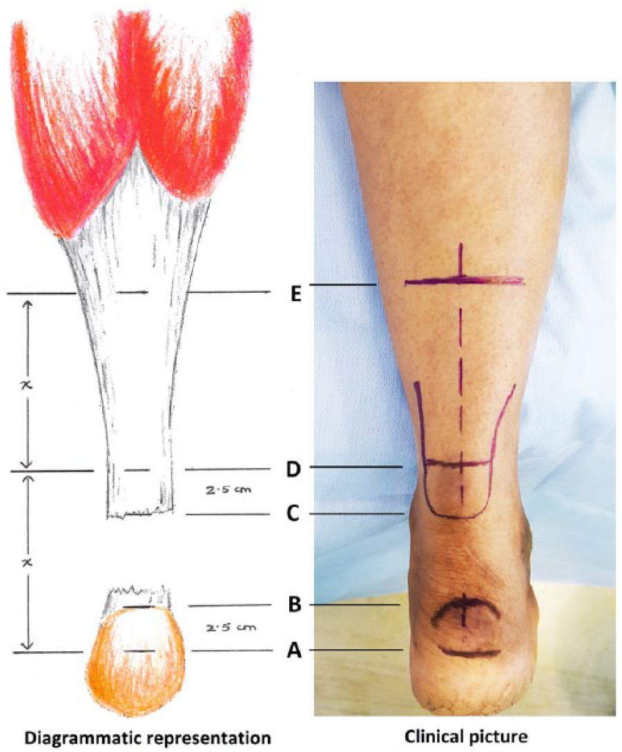
Markings to determine the length of the turndown flap: mark A, 2.5 cm from B; mark B, Achilles tendon insertion; mark C, distal end of proximal stump; mark D, 2.5 cm from C, with X = distance between A and D; mark E, length “X” from D.
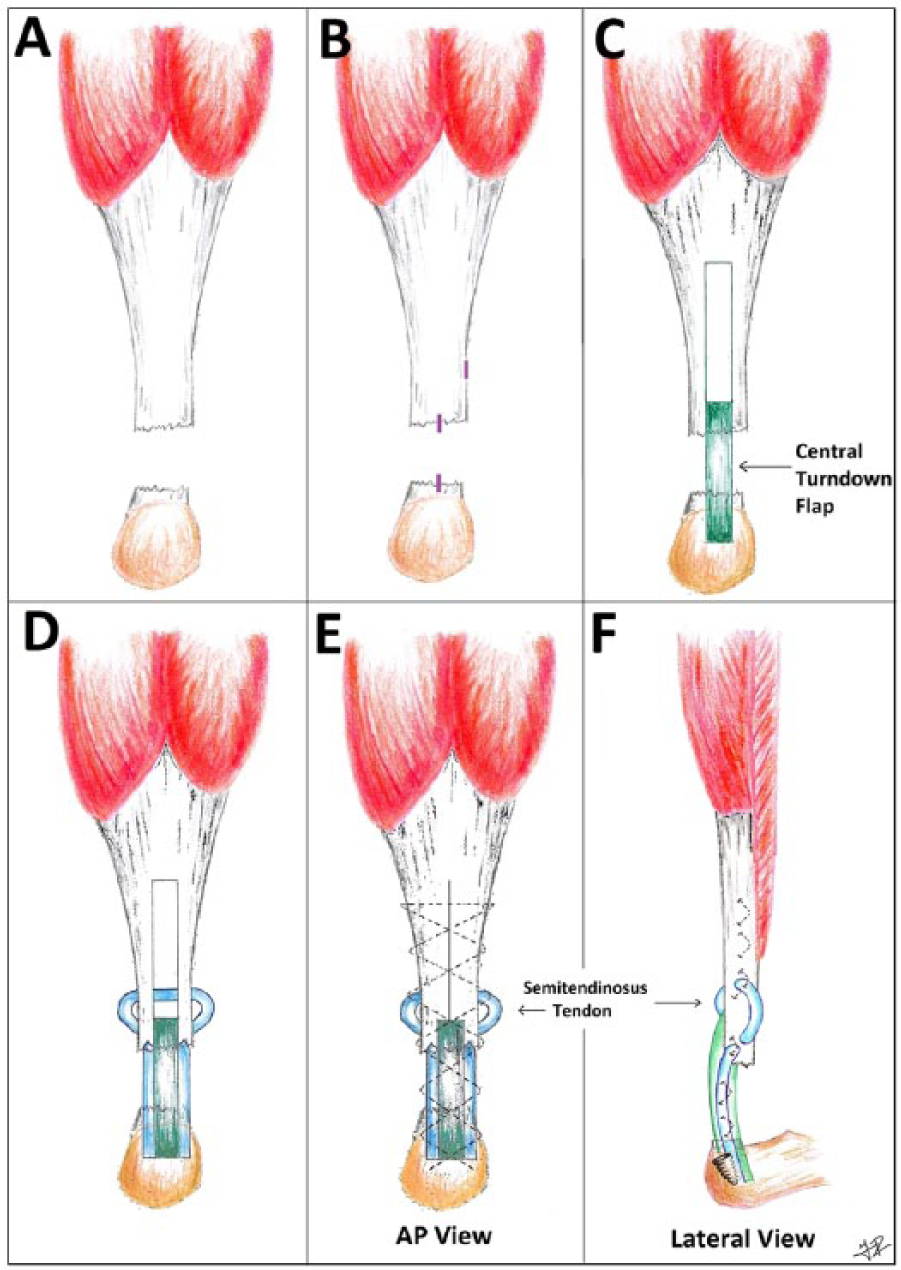
Diagrammatic representation of the method. (A) Achilles tendon rupture with gap between the tendon ends. (B) Arthroscopy portals: 2 midline and 1 superolateral. (C) Central gastrocnemius turndown flap. (D) Passage of free semitendinosus graft through the proximal stump. (E) Percutaneous sutures extending from the proximal stump to the distal end of the turndown flap and semitendinosus graft. (F) Lateral view of the reconstructed tendon. AP, anteroposterior.
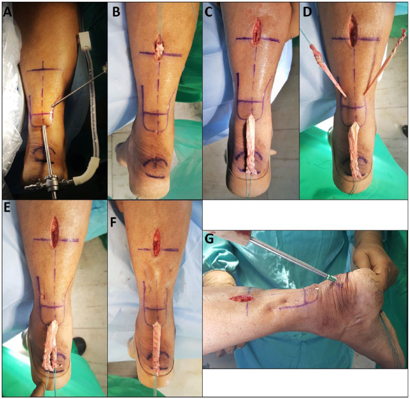
Operative views of the method (right leg). (A) Arthroscopic debridement of the proximal tendon stump. (B) Creation of the central turndown flap. (C) Turndown flap brought out through the proximal midline arthroscopy portal. (D) Passage of free semitendinosus graft through proximal stump. (E) The semitendinosus graft delivered through the proximal arthroscopy portal. (F) Percutaneous sutures with creation of a single flap-graft tendon mass. (G) Insertion of interference screw.
Central turndown flap
The foot was placed in 30 degrees of plantarflexion, and length of the defect was measured. The length of the turndown flap (X) was measured as shown in Figure 1. A 3-cm midline incision was made over the mark “E” indicating the proximal extent of the turndown flap (Figure 3B). The proximal part of the flap was demarcated and raised from the middle third of the Achilles aponeurosis. A whip stitch with a No. 2 nonabsorbable suture was tied to the proximal flap. Sizing of the raised flap was done and a tendon stripper used to create the distal portion of the flap up to 2.5 cm (Figure 1, mark D) from the ruptured tendon end. The tendon stripper was felt percutaneously to determine its position. Markings on the graduated tendon stripper were used to confirm the length of the turndown flap. The tendon stripper was not moved further to prevent detachment of the flap. This flap was turned down and delivered out through the proximal midline arthroscopy portal (Figure 3C).
Semitendinosus tendon augmentation
A 1- to 2-cm incision was made opposite the posterolateral portal on the medial side. A mosquito forceps was passed through the substance of the Achilles tendon in a mediolateral direction, and the semitendinosus tendon graft was passed through this tract (Figure 3D). The graft ends were again inserted through the proximal Achilles stump distal to the attachment of the turndown flap in a crisscross manner. The graft ends were now brought out through the proximal midline portal (Figure 3E).
Percutaneous sutures
The residual gap in the Achilles aponeurosis (created due to the turndown flap) was closed through the midline turndown flap incision with a No. 2 nonabsorbable suture. The sutures were then continued distally in a percutaneous and crisscross manner, incorporating the turndown flap as well as the semitendinosus grafts (Figures 2E and 3F). As the sutures reached the semitendinosus graft insertion level, the next suture was placed through the existing incisions made for the graft insertion. This enabled passage of the suture through the semitendinosus graft, which was confirmed by restricted gliding movement of the tendon. The remainder of the sutures were placed under direct vision as the flap-graft mass had been delivered through the proximal midline arthroscopy portal (Figure 3F). These sutures differed from the original Ma-Griffith method by not engaging the distal tendon stump. 26
Interference screw fixation
A suture passing wire was advanced through the distal portal and into the footprint of the Achilles tendon. A slightly medial insertion point was made to resemble the normal anatomic attachment of the Achilles tendon.4,60 The wire was brought out through the heel directed anteriorly. A 4-mm cannulated reamer was used to drill over the wire, creating a tract in the calcaneus for easy passage of the suture strands. Sizing of the flap-graft tendon mass was carried out. A corresponding size cannulated reamer was used to drill over the wire up to a depth of 3.5 cm. The reamer was removed, keeping the wire in situ. The sutures were threaded onto the eyelet of the wire, and the wire was advanced to the plantar aspect of the foot. The sutures were pulled, enabling the flap-graft mass to advance into the tunnel. The foot was moved through its full range of motion and the desired tension maintained on the sutures so that the foot would rest in 30 degrees of plantarflexion. An interference screw, 2.5 cm in length and 1 mm smaller than the drill diameter, was advanced over the guidewire, maintaining tension on the sutures. The remnants of the tendon sutures were cut at the heel. Skin was closed with nonabsorbable sutures.
Postoperative care
The leg was put in a below-knee gravity equinus splint for 2 weeks. At the end of 2 weeks, the sutures were removed. The splint with the foot in gravity equinus was continued for 1 more week. At the end of 3 weeks, the splint was removed and ankle exercises were started. Non–weight bearing was continued for another 4 weeks. At the end of 7 weeks, weight bearing was begun and progressed as tolerated by the patient.
Statistical Analysis
Data were analyzed using SSPS software version 21.0 for Windows (SPSS, Inc, an IBM Company, Chicago, IL). Normal distribution was assessed by the Kolmogorov-Smirnoff test, and the variables were found to be normally distributed. Paired t test was used to assess the preoperative and postoperative AOFAS scores, ATRS scores, and ankle range of motion. The significance was set at P < .05.
Results
The length of the defect ranged from 3 to 8 cm (mean, 5.1), while the length of the turndown flap ranged from 8 to 13 cm (mean, 10.1). The mean AOFAS score improved from 64.5 points (range, 35-79) preoperatively to 96.4 points (range, 90-100) at 1-year follow-up (difference was statistically significant, P < .001). At the last follow-up, the mean AOFAS score was 96.9 points (range, 90-100). The improvement in the score was not statistically significant between the 1-year and latest follow-up (Table 2).
Treatment Outcomes.
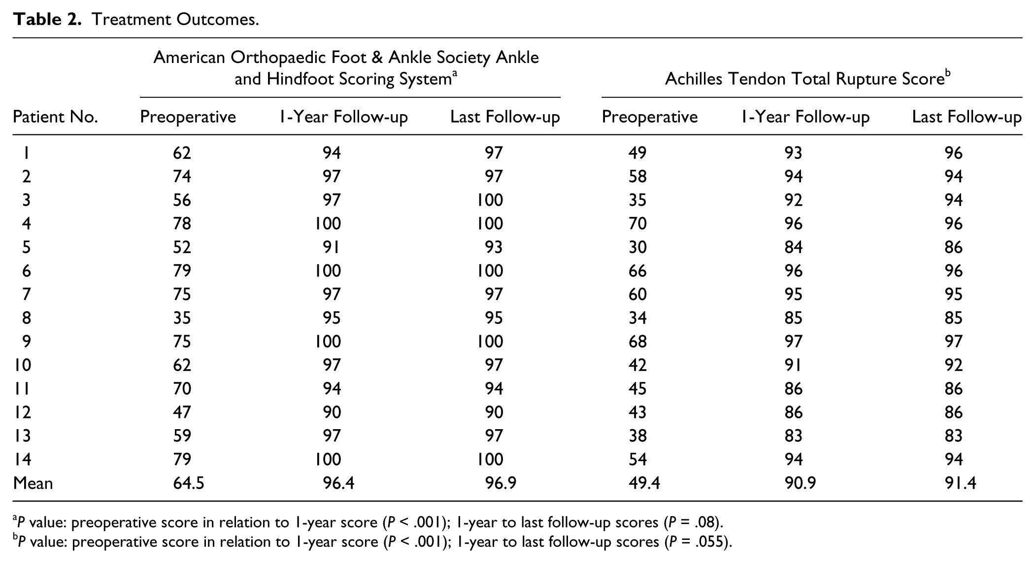
P value: preoperative score in relation to 1-year score (P < .001); 1-year to last follow-up scores (P = .08).
P value: preoperative score in relation to 1-year score (P < .001); 1-year to last follow-up scores (P = .055).
The mean ATRS score improved from 49.4 (range, 30-70) preoperatively to 90.9 (range, 83-97) at the 1-year follow-up (P < .001). At the latest follow-up, the mean ATRS score was 91.4 points (range, 83-97). The improvement in the score was not statistically significant between the 1-year and latest follow-up (Table 2).
Weight bearing was begun at 7 weeks in all patients. All patients were able to stand on their toes. The mean time to return to the preinjury activity level (daily activities except sports) was 9.3 weeks (range, 8-12). One patient who was engaged in recreational sports returned to sporting activity in 16 weeks. The preoperative degree of active dorsiflexion (mean, 20.5) was not significant compared with either the postoperative (mean, 19) or the healthy side (mean, 21.6). However, the postoperative value showed a statistically significant reduction compared with the healthy side (P = .004). The preoperative degree of active plantarflexion (mean, 23.9) showed a significant difference from both the postoperative (mean, 43.7) and the healthy side (mean, 46.1) values (P < .001 in both). The postoperative values showed a significant reduction in comparison to the healthy side (P = .021). None of the patients developed any complications. All patients had good wound healing (Figure 4). None developed skin necrosis or wound dehiscence. No rerupture or sural nerve damage was noted by the latest follow-up.
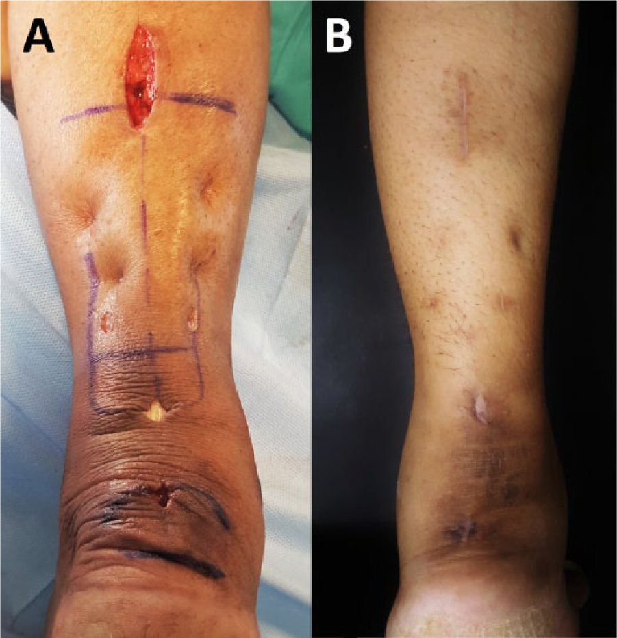
(A) Incisions at time of surgery. (B) Wound healing at 3 weeks.
Discussion
A chronic Achilles tendon rupture poses a unique set of challenges in management compared with an acute rupture. It is known that a longstanding rupture results in a gap between the tendon ends due to the continued contraction of the gastroc-soleus muscle complex. 46 As the muscle fibers shorten, the tension that they can produce decreases and finally becomes zero when the fibers are at 60% of their resting length. 40 Depending on the magnitude of the gap, end-to-end apposition may not be possible. Thus, one of the main objectives of treating a chronic rupture is restoring the original length and the tension of the tendon.32,55 Compounding the above problem is the poor blood supply in this area. 29 The tendon end may become irregular and dull in appearance. 58 The tendon sheath may not be clearly visible, and the retracted tendon may adhere to the posterior fascia. 65 Debriding the tendon ends is usually required, which further increases the gap. Thus, the repair of chronic ruptures requires augmentation and reinforcement by using a turndown flap, a tendon transfer, a tendon graft, or synthetic materials. 29
Operative management gives the best results for a chronic Achilles tendon rupture. In a series of 57 patients, Christensen 5 reported that patients who were managed operatively showed 90% improvement compared with only 70% with those managed nonoperatively. A nonoperative approach may be used in cases of comorbidities such as peripheral vascular disease, anesthetic contraindications, and in patients who do not have any significant functional defects and are able to continue their activities of daily living.29,48
Smaller gaps ranging from 2 to 3 cm can often be repaired end-to-end. Myerson 46 recommended end-to-end repair for defects smaller than 2 cm and advised a V-Y lengthening or a flexor hallucis longus (FHL) transfer for larger gaps. Kuwada 20 recommended an end-to-end repair for gaps less than 3 cm, a turndown flap with or without a synthetic graft for 3- to 6-cm gaps, and a gastrocnemius recession with free tendon graft for gaps larger than 6 cm. Similar recommendations were given by Den Hartog 7 and Maffulli and Ajis. 30 Thus, with the increase in the gap, greater reinforcement is required to obtain a strong repair to withstand the high functional requirement of the tendon. Also, the quality of the turndown flap created from the proximal stump may be suboptimal, thus requiring additional reinforcement with other means.32,33 All the above guidelines recommend the use of a V-Y lengthening or a turndown flap with additional augmentation using a tendon transfer, free tendon graft, or a synthetic graft material for larger defects.
A turndown flap and its modifications have been described by various authors with satisfactory results.29,48 Christensen 5 and Gerhardt 12 separately described a single turndown from the proximal gastrocnemius aponeurosis. The flap could be twisted 180 degrees as described by Silfverskiold. 56 Two flaps, one from the medial side and one from the lateral side, could be raised as described by Toygar. 59 Alternatively, 2 flaps, one from the proximal stump and one from the distal stump, could be raised as described by Weisbach. 62 Rush 51 created a tube from the flap, which was sutured to the distal stump. Recently, Guclu and colleagues 13 reported good long-term functional results of a central turndown flap with a V-Y tendon plasty.
Several studies have augmented a turndown flap with a tendon graft. Mann and colleagues 40 described a flexor digitorum longus (FDL) transfer with central turndown flap augmentation with good to excellent results in 6 of 7 patients. Studies have also described FHL tendon augmentation with turndown flaps with excellent results.2,57 However, 1 study expressed concern that FHL harvesting could reduce the push-off during stance phase. 6 Another study described a central turndown flap combined with a triple-loop plantaris tendon augmentation in Myerson type 3 ruptures. 52 However, the authors reported a 16.7% incidence of wound-healing problems or superficial wound infections. Mao and colleagues 41 managed chronic ruptures in 10 patients with FHL transfer and 2 gastrocnemius turndown flaps reinforced with the plantaris tendon.
The semitendinosus tendon has been employed by a number of authors to reconstruct a chronic Achilles tendon rupture.8,11,16,33,53 There are several advantages of using the semitendinosus graft. It is easily harvestable by a small incision at the pes anserinus insertion, and it is consistently thicker and firmer than the gracilis tendon. 10 It is long enough to bridge the gap between the tendon ends sufficiently.10,33 Harvesting it is safe, and a free graft does not deprive the foot of motor power or strength. 33 Ji and colleagues 16 used the semitendinosus tendon to augment a V-Y advancement, while others have used it in isolation.8,10,53 All the procedures employing a turndown flap with augmentation have used an open approach. But a minimally invasive central turndown flap with tendon augmentation has not been described in the literature.
Achieving a strong and stable repair is only half a battle won. The other half lies in countering the wound complications associated with these repairs. Wong and colleagues 64 in their review found the highest rate of minor wound complications with open repair and immobilization (12.3%) compared with percutaneous (4.9%) and conservative repairs (0.5%). Another study reviewed 60 patients who underwent open tendoachilles repair at their institute and reported a 16.7% incidence of postoperative wound infections despite prophylactic antibiotic therapy. 42 One study comparing an open and a percutaneous repair reported a wound infection rate of 21%, while another similar study reported deep infection rate of 5.7%, both in open repairs, but 0% each for percutaneous repairs.14,23 To counter the problem of wound complications, various minimally invasive techniques have been employed by authors. Since the first description of a percutaneous technique by Ma and Griffith 26 in 1977, various authors have described a minimally invasive approach by percutaneous stab incisions, mini-open techniques, or endoscopic techniques.11,25,31,33,35,44 A meta-analysis of randomized controlled trials comparing conventional to minimally invasive techniques for repair of Achilles tendon rupture concluded that patients were nearly 3 times more likely to report a good or excellent outcome following minimally invasive surgery (MIS) repair compared with open surgery at a minimum of 6 months follow-up.24,43
In our technique, we used a combination of a mini-open technique assisted by endoscopy. The role of endoscopy was to debride the tendon ends and to clear the retrocalcaneal area, which is not possible by a purely percutaneous or a mini-open technique. A central turndown flap augmented with a free semitendinosus tendon graft provided the necessary strength and thickness required to achieve a stable repair. We added a percutaneous suture similar to the Ma and Griffith 26 technique, which served to close the gap in the proximal gastrocnemius aponerosis (created by the turndown flap harvest) and to create a single strong tendon mass to be inserted into the calcaneus. We inserted this tendon mass into the calcaneus with the help of interference screw fixation (Figure 5). This created an onlay effect on the existing distal stump when present.
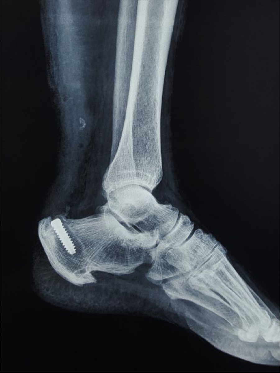
Postoperative radiograph.
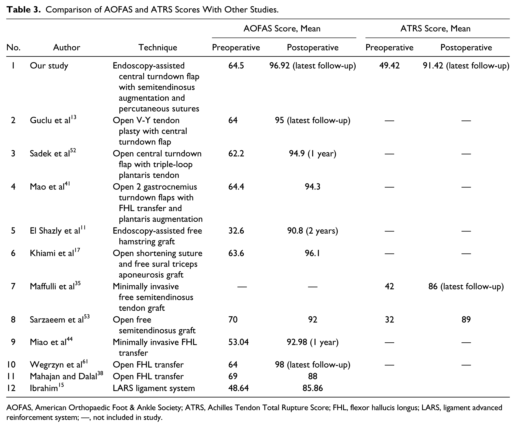
Many techniques require the presence of a distal stump for the turndown flap to be sutured to.5,12,56,59,62 But in most cases, the distal stump is too short to obtain a strong repair.48,49 Besides, in cases of degenerative tears, the distal stump tissue is unhealthy and poor in quality. An interference screw fixation has been shown to be stronger than tendon-to-tendon fixation and reduces the dependence on the quality of the distal stump.3,9 MRI studies have shown that insertion of a graft into the calcaneus leads to incorporation of the graft into the original stump. 11 Although sural nerve damage has been reported in various studies employing percutaneous, mini-open, or open techniques, 45 we did not have single case of sural nerve damage at the latest follow-up. The functional results of our study assessed by the AOFAS and the ATRS scores have been compared with other studies with several different techniques in Table 3.
Comparison of AOFAS and ATRS Scores With Other Studies.
AOFAS, American Orthopaedic Foot & Ankle Society; ATRS, Achilles Tendon Total Rupture Score; FHL, flexor hallucis longus; LARS, ligament advanced reinforcement system; —, not included in study.
In the current study, we debrided the unhealthy tendon tissue, inflamed peritendinous tissue, and released the adhesions via endoscopy. Along with the above, we believe drilling of the tunnel in the calcaneus brought about significant pain relief and healing of the lesion. A bony Haglund’s deformity if present can be debrided but not completely excised with a minimally invasive technique. However, we believe that drilling the calcaneal tunnel in proximity to the bony deformity would prevent any potential impingement on the reconstructed tendon.
This study has its limitations in being a retrospective case review study with a small number of cases. However, we were able to obtain excellent outcomes in all patients with our procedure, with the longest follow-up being 78 months. More prospective studies comparing this technique with others would enlighten us in our quest for the ideal reconstruction technique.
Conclusion
Our study presents a new technique for the reconstruction of chronic Achilles tendon ruptures by augmentation with a central turndown flap with a free semitendinosus tendon graft and percutaneous sutures in a minimally invasive approach and assisted by endoscopy. All patients in our study had a favorable outcome with no complications through the latest follow-up. We believe that with this triple-repair technique, one can achieve as strong and robust a repair as in an open surgery while at the same time reducing the incidence of complications by being minimally invasive.
Footnotes
Declaration of Conflicting Interests
The author(s) declared no potential conflicts of interest with respect to the research, authorship, and/or publication of this article.
Funding
The author(s) received no financial support for the research, authorship, and/or publication of this article.