
Abstract
Background:
The Trabecular Metal Total Ankle Implant differs from other newer-generation implants in the transfibular approach, multiplanar external frame for alignment, tantalum trabecular metal interfaces, curved geometry, and shallow resection depths. The primary aim of this study was to report midterm clinical and radiographic results, as well as survivorship and adverse events at a minimum of 5-year follow-up.
Methods:
A total of 83 ankles (81 patients, average age 60.6 years old, 50.6% females) with average 6.3 years’ (range, 5.0-10.1) follow-up were included. Postoperative patient-reported outcome measures (PROMs) included SF-12 physical (PCS) and mental component summary (MCS) scores, Ankle Osteoarthritis Scale (AOS), pain visual analog scale (VAS). Radiographic outcomes included postoperative range of motion (ROM) and coronal/sagittal alignment. Adverse events were reported using the Canadian Orthopaedic Foot and Ankle Society Reoperation Coding System (CROCS).
Results:
Preoperative tibiotalar coronal deformity included 27 valgus (10 degrees, range 2-20 degrees) and 25 varus ankles (−9 degrees, range −2 to −25 degrees), corrected to neutral postoperatively. Postoperative tibiopedal ROM was 17.8 degrees dorsiflexion and 21.8 degrees plantarflexion. Adverse events occurred at average 28.7 months, most commonly gutter debridement (n = 17, 16.7%) and subsequent operative treatment unrelated to metal components (n = 10, 12.0%). There were 2 cases (2.4%) of acute deep infection treated with irrigation and debridement, polyethylene exchange, and retention of metal components without recurrence of infection. There were no cases of fibular nonunion, septic or aseptic loosening, or implant subsidence. Postoperative PROMS included SF-12 PCS: 40.4; SF-12 MCS: 56.0; VAS: 2.3; AOS Pain: 17.0; and AOS Disability: 24.9. Overall implant survival, defined by retention of the metal components, was 100% at final follow-up.
Conclusion:
At a minimum of 5 years, patients who underwent TM TAA reported minimal ankle pain and regained neutral ankle alignment and mobility, without septic or aseptic implant loosening. Although having certain limitations, this study suggests that TM TAA is a viable option for the treatment of end-stage ankle arthritis.
Introduction
Utilization of total ankle arthroplasty (TAA) in the surgical treatment of end-stage ankle arthritis continues to garner increased interest. As these procedures are performed more often over the years, it becomes crucial to evaluate the long-term outcomes and survivorships of these implants. Most common causes of implant failure described in the literature requiring revision surgery include aseptic loosening, peri-implant osteolysis, subsidence, and infection. 11 Studies of 5-year minimum follow-up of modern TAA designs yield an implant survivorship ranging from 78% to 98%.10,11,14,16,25
Currently, TAA is available in the United States with a variety of implant designs, most of which use a traditional anterior approach to the ankle. In contrast, the Trabecular Metal (TM) (Zimmer Biomet, Warsaw, IN) prosthesis uses a lateral transfibular approach. 30 Although this does require creating and subsequently fixing a fibular osteotomy—which carries its own risks for malunion and nonunion—the benefits of the lateral exposure is that it provides superior visualization of the ankle joint and allows for curved cuts made by a router on a fixed radius of the tibia and talus. This can potentially minimize bone resection and allow for a more anatomic implant positioning along the natural trabecular lines of the talus.15,24
Although early to midterm results of the Zimmer prosthesis reported in the literature have been promising,1,4,12,27,28,31 there remains a need to determine longer-term survivorship, including clinical and radiographic outcomes of this implant. We present our experience of the TM TAA with minimum 5-year follow-up by a single-surgeon designer. The primary aim of this study was to report the survivorship and adverse events related to the TM implant, with a secondary aim of describing functional and radiographic outcomes.
Methods
A retrospective review was performed on patients who underwent primary transfibular TAA by the senior author, who is a designer and patent holder of the device. Surgeries were performed from October 2012 to January 2018. Inclusion criteria included a minimum of 5 years of follow-up, with complete postoperative patient-reported outcome measures (PROMs) and radiographs. Exclusion criteria included patients with incomplete follow-up, prior ipsilateral TAA (revision TAA), ankle arthrodesis (fusion takedown), and/or previously infected ankle. Institutional Review Board approval was obtained to report patient outcomes. This study was designed and reported in accordance with the Strengthening the Reporting of Observational Studies in Epidemiology (STROBE) statement on strengthening the reporting of observational studies. 32
Patient demographic, perioperative, comorbidity, and radiographic data were retrospectively collected via review of the electronic medical record including pre- and postoperative clinical notes, anesthesia notes, operative notes, discharge summaries, emergency department visit notes, and additional operative notes. Demographic variables collected included age at the time of index procedure, sex, and body mass index (BMI). Perioperative data collected included the length of follow-up, laterality, etiology of the ankle arthritis (posttraumatic arthritis, primary osteoarthritis, inflammatory arthritis, or other), and history of prior ankle or hindfoot surgery. Comorbidity data included the American Society of Anesthesiologists (ASA) score, tobacco use, diagnosis of diabetes, or a history of malignancy. The length of clinical and radiographic follow-up were recorded from time of surgery to the last postoperative visit where PROMs and weightbearing ankle radiographs were collected, respectively.
Patient-Reported Outcomes
PROMS included 12-item Short Form Health Survey (SF-12) physical (PCS) and mental component summary (MCS) scores 34 ; Ankle Osteoarthritis Scale (AOS) 13 ; and visual analog scale (VAS) for pain. 26 All PROMs included are validated measures that have been applied to numerous foot and ankle pathologies. 21 The SF-12, AOS, and VAS are all scored ranging from 0 to 100. A higher score indicates impairment for VAS and AOS; however, a lower score indicates impairment for SF-12. Patients without complete PROM data at a minimum of 5 years postoperation were excluded from the study.
Radiographic Outcomes
For all patients, standardized radiographic assessment included weightbearing anteroposterior (AP), mortise, and lateral view radiographs of the ankle obtained preoperatively and postoperatively at 3, 6, and 12 months and annually. Nonweightbearing radiographs were obtained at 2 weeks and 6 weeks postoperation. The radiographs were analyzed by a foot and ankle fellow (A.N.F.) on the preoperative and most recent radiographs taken at a minimum of 5 years postoperation. The observer did not participate in any of the operations.
Postoperative tibiotalar and tibiopedal range of motion (ROM) were measured on weightbearing radiographs as previously described (Figure 1).8,19 Standing lateral films of the ankle with the patient in maximal plantarflexion and dorsiflexion were obtained; tibiotalar ROM was defined as the angle between the articulating surfaces of the tibial and talar components, whereas tibiopedal ROM was defined as the angle between the anatomic axis of the tibia and a line drawn perpendicular to the floor. Ankle alignment was measured using well-accepted and reliable measurements as previously described. Preoperatively, the coronal tibiotalar alignment angle was measured on the AP radiograph and reported as the tibiotalar tilt1,5,9,33 and lateral distal tibia angle (LDTA).1,2,5,27 The sagittal tibiotalar alignment was measured on the lateral radiographs and reported as the anterior distal tibia angle (ADTA).5,27 Postoperatively, angular assessment of the tibia and talar component alignment was performed. The AP radiograph was used to measure the tibial component coronal alignment angle (alpha angle [α]) whereas the lateral radiograph was used to measure the tibial sagittal/slope alignment angle (beta angle [ß]) and talar sagittal/slope alignment angle (gamma angle [γ]).1,4,18,20,22,31,35 Intracomponent tibiotalar coronal alignment was measured as the angle between the tibial and talar surfaces. 2
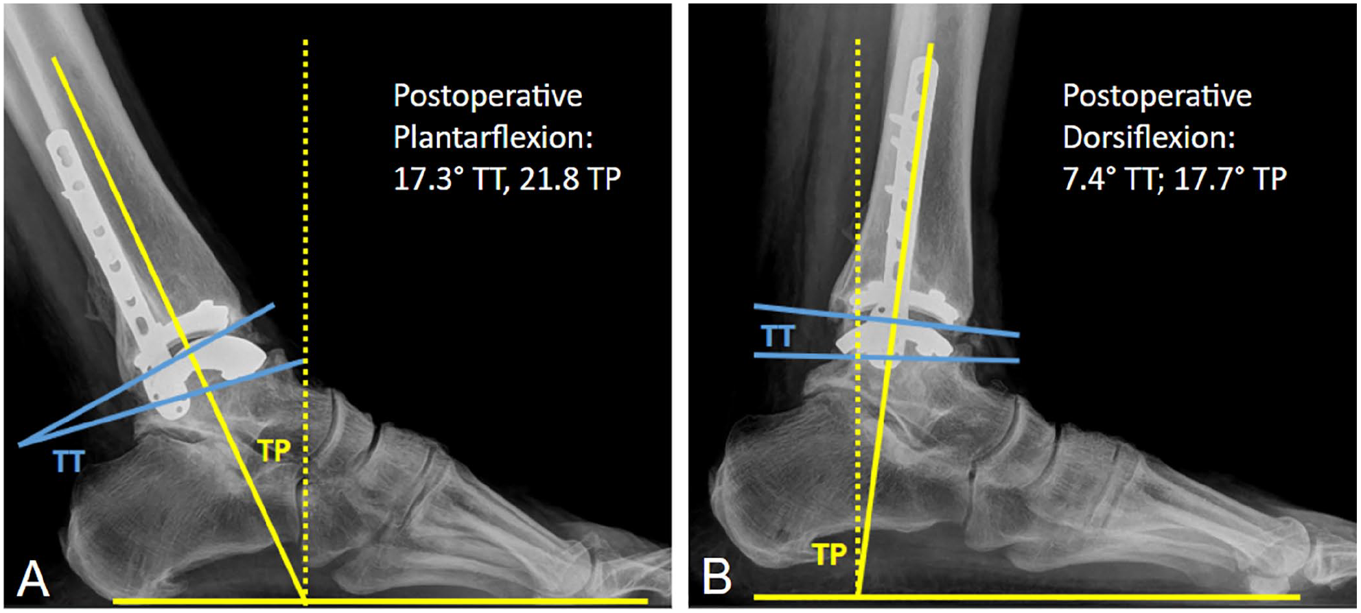
Postoperative (A) plantarflexion and (B) dorsiflexion range of motion (ROM) were measured radiographically on lateral weightbearing plain films of the ankle. Tibiopedal (TP) ROM was defined as the angle between the anatomic axis of the tibia relative to a line drawn perpendicular to the floor. Tibiotalar (TT) ROM was defined as the angle between the articulating surfaces of the tibial and talar components.
Complications
Adverse events and reoperations were reported using the Canadian Orthopaedic Foot and Ankle Society (COFAS) Reoperation Coding System (CROCS). 36 For the purpose of this study, adverse events were defined as nonoperative perioperative treatments, as well as surgical treatments unrelated to TAA components (COFAS codes 2, 3, 4, 5, 6, 7); revisions were defined as postoperative surgical treatments related to TAA metal component (COFAS codes 9-11). As previously defined in the literature, 3 radiographic healing of the fibular osteotomy was defined as satisfying clinical criteria (no pain, no warmth, improvement in swelling, and stability to stress) and radiographic criteria (at least 85% visible trabecular bridging across the osteotomy site and no lucency around the hardware on the AP and lateral weightbearing views of the ankle).
Surgical Technique and Postoperative Protocol
The TM TAA was used in all patients. The implant differs from other newer-generation implants in the transfibular approach, non–mobile-bearing prosthesis, semiconstrained design, curved surface at every interface in the sagittal plane, coronal orientation of the rails, shallow resection depth, coupled cuts, highly cross-linked polyethylene inset into the tibial (titanium) tray, and articulating with the talar (cobalt chrome) surfaces. The titanium tibial tray and the talar cobalt chrome components are bonded with the trabecular metal (porous tantalum) at the bone-implant interface.
The surgical technique was previously described in detail. 27 Regional anesthesia, most commonly either high ankle block or popliteal and adductor canal nerve blocks, was administered by the anesthesia team. Patients were positioned 15 degrees lateral off the horizontal supine plane with a bean bag under the ipsilateral hip and operative extremity elevated on a bone foam. A thigh tourniquet was applied but not routinely inflated. All patients received a TM TAA implanted based on the manufacturers’ technique guides. An external fixator was used to facilitate radiographically guided corrections of the calcaneus, talus, and tibia in the coronal and sagittal planes. Curved resections of the tibia and talus were created using an attached router. Trials were inserted with the frame unlocked to determine final polyethylene thickness. Final implants were then inserted from lateral to medial. The fibular osteotomy was reduced and fixed with a fibular plate. Iliac crest bone graft and marrow were harvested with a 2-mm trephine in a minimally invasive fashion to supplement the fibular osteotomy and to fill in preoperatively identified cysts around the arthroplasty. Prophylactic medial malleolar fixation with a 4.0-mm screw was indicated for patients at risk for subsequent fracture. Concomitant bony and soft tissue procedures were performed as needed to achieve appropriate foot and ankle alignment and stability.
For the first 2 weeks postoperatively, patients were immobilized in a well-padded short leg splint with the ankle in neutral position and maintained nonweightbearing. Sutures were removed at postoperative week 2 and patients were transitioned to a removable night splint to hold the ankle in neutral position and a controlled ankle motion (CAM) walking boot. At this time, weightbearing deep knee bends and Achilles stretching for early ROM and preservation of gastrocnemius tone was initiated. Patients were instructed to perform knee bends for 100 minutes a day (20-minute sessions 5 times daily) out of the boot with full weightbearing to tolerance in standing and bending. Deep knee bends were initiated later at 6 weeks in patients who underwent concomitant tendon Achilles lengthening, foot osteotomy, or arthrodesis. No weightbearing ambulation was permitted in the first 6 weeks.
At 6 weeks postoperation, patients began weightbearing with ambulation, with progression to full weightbearing in a walking boot over the course of 4 weeks. At 12 weeks, patients transitioned out of the boot to strengthening exercises, proprioceptive training, water activities, and use of an exercise bike. Importantly, patients were instructed to avoid any twisting motion at the ankle while performing weightbearing exercises.
Statistics
Descriptive statistics were tabulated for patient, operative, and outcome data. Continuous variables were summarized with means, SDs, and 95% CIs. Categorical variables were summarized with frequency and percentage. A Kaplan-Meier analysis was used to evaluate implant survival and rate of reoperation with 95% Hall-Wellner CIs at a minimum of 5 years postoperatively. All statistical analyses were performed using SAS, version 9.2 (SAS Institute, Cary, NC).
Results
Of the consecutive 151 TAAs performed by the senior surgeon during the study period (between October 6, 2012, and January 26, 2018), 83 (55.0%) were successfully contacted for enrollment and consented with a minimum of 5-year clinical and radiographic follow-up. Demographic and clinical data are outlined in Table 1. The patients were evaluated at a mean of 5.8 years clinical and 6.3 years radiographic follow-up. The average age was 60.6 years (range, 28-80), and about half of the patients were female (n = 42; 50.6%). Most patients were nonsmokers (n = 55; 66.3%), whereas 27.7% patients were former smokers (n = 23) and 3.6% patients were current smokers (n = 3). In addition, 13.3% patients had diabetes (n = 11) and 10.8% patients had a history of malignancy (n = 9). The majority of patients had an ASA score of 2 (n = 49; 59.03%), followed by ASA scores of 3 (n = 25; 30.1%) and 1 (n = 8; 9.6%). Most patients had a primary diagnosis of posttraumatic arthritis (n = 61; 73.5%) and had previously undergone ankle and/or hindfoot surgery (n = 57; 68.7%). The concomitant surgical procedures performed besides the routine fibular osteotomies and ligament reconstructions were iliac crest bone graft (n = 83; 100%), removal of hardware (n = 21; 25.3%), Achilles tendon lengthening (n = 10; 12%), and prophylactic stabilization of the medial malleolus (n = 6; 7.2%).
Patient Demographics and Perioperative Data (N = 83).
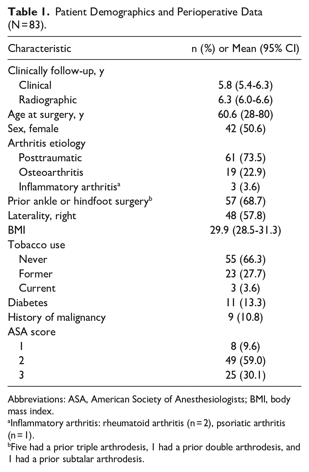
Abbreviations: ASA, American Society of Anesthesiologists; BMI, body mass index.
Inflammatory arthritis: rheumatoid arthritis (n = 2), psoriatic arthritis (n = 1).
Five had a prior triple arthrodesis, 1 had a prior double arthrodesis, and 1 had a prior subtalar arthrodesis.
The postoperative PROMs were SF-12 PCS: 40.4, SF-12 MCS: 56.0, VAS: 2.3, AOS Pain: 17.0, and AOS Disability: 24.9 (Table 2). Pre- and postoperative radiographic data are outlined in Table 3. Preoperative tibiotalar coronal deformity included 27 valgus (10 degrees, range 2-20 degrees) and 25 varus (−9 degrees, range −2 to 25 degrees) ankles. Postoperative radiographic tibiotalar dorsiflexion and plantarflexion averaged 6.7 and 17.5 degrees, respectively, and tibiopedal ROM averaged 17.8 degrees dorsiflexion and 21.8 degrees plantarflexion.
Postoperative Patient-Reported Outcomes. a
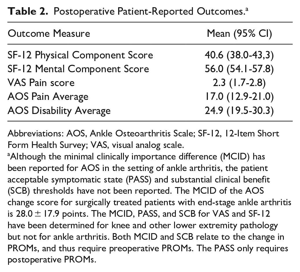
Abbreviations: AOS, Ankle Osteoarthritis Scale; SF-12, 12-Item Short Form Health Survey; VAS, visual analog scale.
Although the minimal clinically importance difference (MCID) has been reported for AOS in the setting of ankle arthritis, the patient acceptable symptomatic state (PASS) and substantial clinical benefit (SCB) thresholds have not been reported. The MCID of the AOS change score for surgically treated patients with end-stage ankle arthritis is 28.0 ± 17.9 points. The MCID, PASS, and SCB for VAS and SF-12 have been determined for knee and other lower extremity pathology but not for ankle arthritis. Both MCID and SCB relate to the change in PROMs, and thus require preoperative PROMs. The PASS only requires postoperative PROMs.
Preoperative and Postoperative Radiographic Assessment Including Alignment and Range of Motion. a
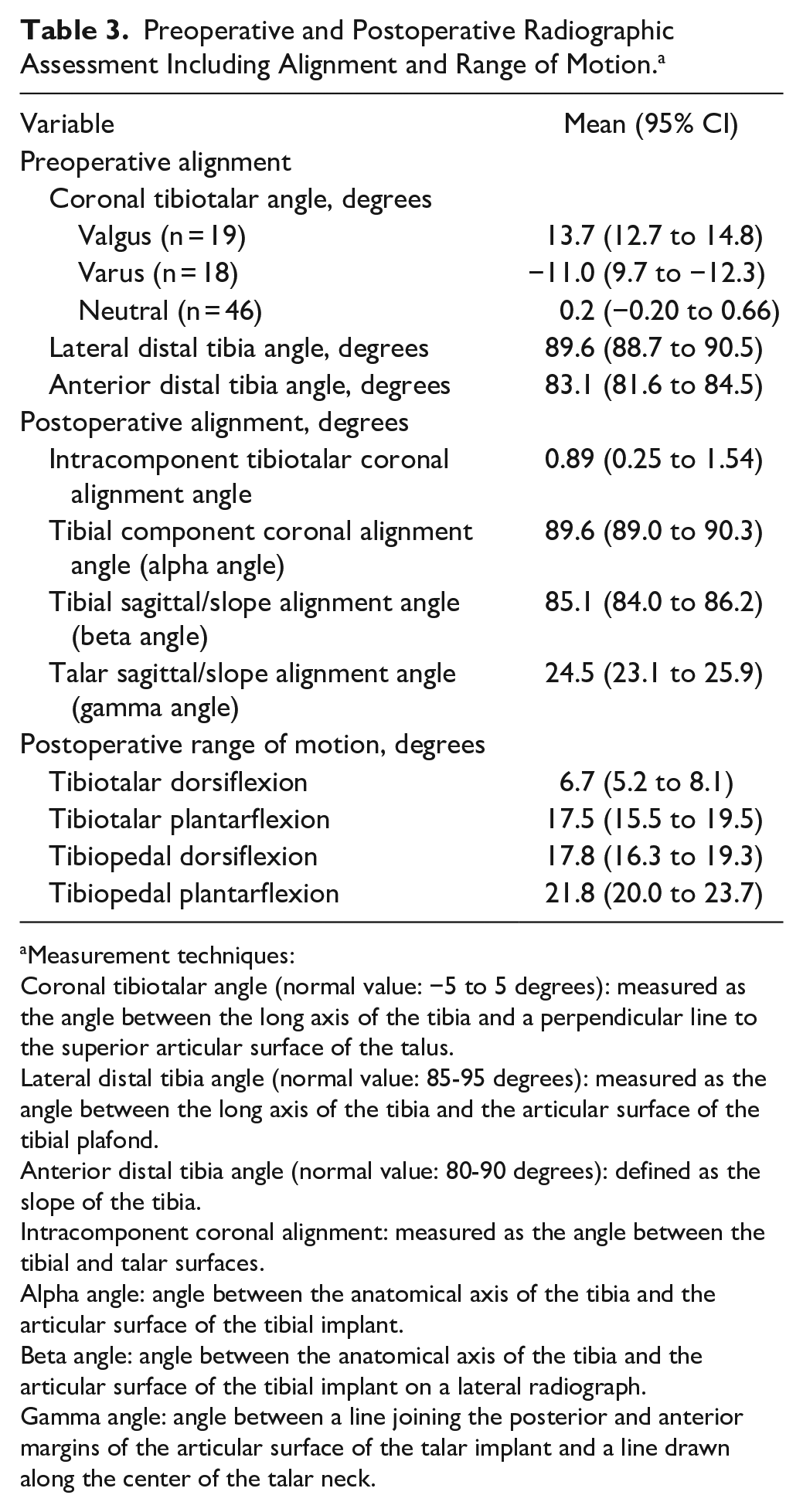
Measurement techniques:
Coronal tibiotalar angle (normal value: −5 to 5 degrees): measured as the angle between the long axis of the tibia and a perpendicular line to the superior articular surface of the talus.
Lateral distal tibia angle (normal value: 85-95 degrees): measured as the angle between the long axis of the tibia and the articular surface of the tibial plafond.
Anterior distal tibia angle (normal value: 80-90 degrees): defined as the slope of the tibia.
Intracomponent coronal alignment: measured as the angle between the tibial and talar surfaces.
Alpha angle: angle between the anatomical axis of the tibia and the articular surface of the tibial implant.
Beta angle: angle between the anatomical axis of the tibia and the articular surface of the tibial implant on a lateral radiograph.
Gamma angle: angle between a line joining the posterior and anterior margins of the articular surface of the talar implant and a line drawn along the center of the talar neck.
A total of 32 (38.6%) adverse events occurred throughout the study period, at an average of 28.7 months postoperative (Table 4). Most common were gutter debridement (n = 17, 16.7%), subsequent operative treatment unrelated to metal components (n = 10, 12.0%), and isolated removal of fibula hardware (n = 10, 7.2%). There were 2 cases (2.4%) of acute postoperative deep infection (<3 months) treated with operative irrigation and debridement, with retention of the metal components. There were no cases of septic or aseptic loosening or subsidence. There were no cases of polyethylene wear due to failure. There were no cases of implant subsidence, or fibular nonunion, and all implants maintained neutral alignment without evidence of loosening due to lack of osseous growth. The overall implant survival, defined by retention of the metal components, was 100%.
Reoperations by COFAS Reoperation Coding System (CROCS) in Patients with a Documented Adverse Event
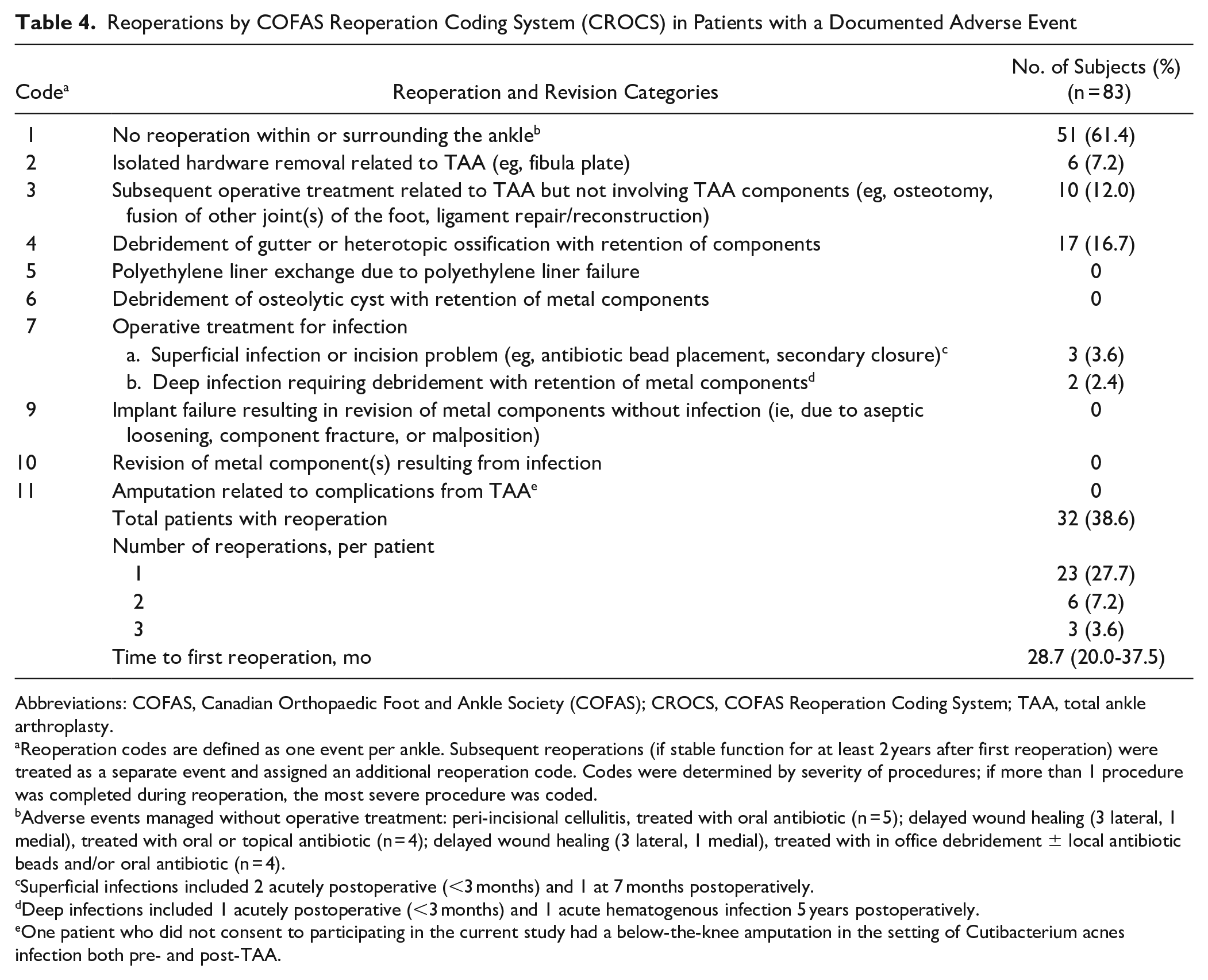
Abbreviations: COFAS, Canadian Orthopaedic Foot and Ankle Society (COFAS); CROCS, COFAS Reoperation Coding System; TAA, total ankle arthroplasty.
Reoperation codes are defined as one event per ankle. Subsequent reoperations (if stable function for at least 2 years after first reoperation) were treated as a separate event and assigned an additional reoperation code. Codes were determined by severity of procedures; if more than 1 procedure was completed during reoperation, the most severe procedure was coded.
Adverse events managed without operative treatment: peri-incisional cellulitis, treated with oral antibiotic (n = 5); delayed wound healing (3 lateral, 1 medial), treated with oral or topical antibiotic (n = 4); delayed wound healing (3 lateral, 1 medial), treated with in office debridement ± local antibiotic beads and/or oral antibiotic (n = 4).
Superficial infections included 2 acutely postoperative (<3 months) and 1 at 7 months postoperatively.
Deep infections included 1 acutely postoperative (<3 months) and 1 acute hematogenous infection 5 years postoperatively.
One patient who did not consent to participating in the current study had a below-the-knee amputation in the setting of Cutibacterium acnes infection both pre- and post-TAA.
Discussion
This minimum 5-year follow-up of the Zimmer transfibular TAA demonstrated 100% implant survivorship with retention of metal components, and an overall adverse event rate of 38.6%. Patients demonstrated good clinical outcomes including PROMs and ROM at final follow-up, while maintaining radiographic implant alignment. Overall, the results of this study support transfibular TAA as a durable approach and treatment option for end-stage ankle arthritis.
Similar to other joint replacements, TAA implant survivorship is a primary criterion of clinical success. Patient selection, expectations, and revision options are all guided by the durability of the implant. Previous short-term studies for TAA through the lateral approach have demonstrated consistently high survivorship. Barg et al 1 reported 55 ankles with a survival probability of 100% and 93% after 1 year and 2 years, respectively. Similarly, Tan et al 27 reported on 85 patients with a 100% survival rate at 24 months, and Usuelli et al 31 reported a 98.9% survival rate at 24 months in 89 patients. Henricson et al 17 recently reported the largest series of the trabecular metal TAA implanted in 239 consecutive cases from a Swedish ankle registry. The overall prosthesis survival probability was reported at 95% after 3 years, with a 3-year risk of revision of 3%. 17
In contrast, there have been fewer studies evaluating midterm follow-up for the transfibular TAA. The 100% implant survivorship observed in this study corroborates previous midterm reports of trabecular metal TAA and other TAA designs. Maccario et al 37 reported on 85 patients at 5-year follow-up and found that 97.7% of implants were retained, with 2 cases (2.3%) of failure due to septic loosening. In a recent 5-year follow-up of 38 patients, Kim et al 20 reported a survivorship of 92.1% and a reoperation rate of 23.7% with 1 case (2.6%) of amputation. The longevity of other modern TAA designs is variable, with studies of 5-year minimum follow-up yielding an implant survivorship ranging from 78% to 98%.10,11,14,16,25
In terms of overall surgical reintervention, the average time to first reoperation reported was 28.7 months. After stratifying reoperations using the COFAS Reoperation Coding System, the most common indications for return to OR were for gutter debridement (COFAS code 4), subsequent operative treatment unrelated to metal components (COFAS code 3), and isolated removal of fibular hardware (COFAS code 2). Although removal of lateral hardware is inherently unique to the surgical design of this implant, the rate of removal was within the range of previous studies, including Kim et al 20 who reported fibular hardware removal in 23% of their cohort. Of note, other authors have reported success in minimizing rates of fibular hardware removal using a longer oblique osteotomy fixed with screws only. 29 In our case, the senior author decided to switch to a lower profile plate to address painful prominent hardware. On the other hand, medial gutter impingement requiring debridement is not unique to the lateral trabecular TAA, and is a common indication for reoperation following TAA. We observed a similar rate of gutter debridement compared with Kim et al, 20 who reported a 13.2% reoperation rate for medial gutter debridement in their midterm study of 38 patients. We believe this is likely due to the surgical preference of the senior author, who only performed an index medial gutter debridement for preoperative gutter impingement and varus ankles. Of note, none of the 9 cases with postoperative gutter impingement had undergone medial gutter debridement at the index procedure. We did not observe any cases of septic or aseptic loosening, or implant subsidence at final follow-up, which is consistent with previous reports of lateral trabecular TAA. 20
Unique to this study was the assessment of postoperative ROM following TM TAA using standing lateral plain films. Postoperative tibiotalar dorsiflexion and plantarflexion was 6.7 degrees (5.2-8.1) and 17.5 degrees (15.5-19.5), respectively. Tibiopedal dorsiflexion and plantarflexion postoperatively was 17.8 degrees (16.3-19.3) and 21.8 degrees (20.0-23.7), respectively. Mosca et al 23 evaluated both pre- and postoperative clinical ROM in 73 patients who underwent lateral transfibular TAA, reporting improvements in dorsiflexion from 6.8 ± 5.8 degrees to 17.2 ± 3.1 degrees postoperatively, and plantarflexion from 9.9 ± 4.2 degrees to 18.4 ± 6.3 degrees postoperatively. Despite earlier arthroplasty reports of preoperative ROM being the primary determinant of postoperative motion, most TAA studies have demonstrated successful improvement in ROM similar to this study as well as gait function.6,7
Although the current study reports favorable postoperative PROM, baseline preoperative PROMs were not available for analysis. However, significant improvements in multiple tested PROMs have been consistently demonstrated in the literature. This includes the American Orthopaedic Foot & Ankle (AOFAS) score, the Foot Function Index (FFI), Short Form-12 and 36, SEFAS and EQ-5D, and EQ-VAS, Foot and Ankle Outcome Score (FAOS), and the EuroQol–5 Dimensions (EQ5D).
We acknowledge several limitations of this study. Although 151 were initially identified during the study period, only 55% of these patients had available minimum 5-year clinical and radiographic outcomes. We attributed this loss to follow-up to several factors, mainly change of practice location of the primary surgeon. Other factors include geographic distance as many of the patients live beyond a 300-mile radius. Finally, the COVID pandemic limited our ability to have patients return during the time frame of this cohort. Although this may introduce a potential source of bias, we did not find any significant differences in patient demographics or operative details between patients included in the final analysis and those lost to follow-up. Furthermore, unique to this study is our analysis of postoperative ROM using radiographic parameters. Although this may differ from previously reported clinical ROM values, it is also subjected to variability due to patient effort and other extrinsic factors. Moreover, preoperative ROM and PROMs were not available for this study given that they were not routinely collected as standard of practice. This baseline functional data would be useful in assessing for significant functional improvements following TM TAA. However, multiple postoperative PROMs at minimum 5-year follow-up were available for each patient, and these values corroborate postoperative outcomes reflected in the literature.
Conclusion
Transfibular TM TAA appears to be an effective treatment for end-stage ankle arthritis at 5-year midterm follow-up. This study confirms good postoperative clinical results including PROMs and ROM as well as radiographic alignment correction. The survival rate was 100% for retention of the trabecular metal components, with an overall adverse event rate of 38.6%. There were no cases of implant subsidence, or fibular nonunion, and all implants maintained neutral alignment without evidence of loosening. Patients should be counseled on the possibility of reoperation for postoperative expectation management.
Supplemental Material
sj-pdf-1-fai-10.1177_10711007241290222 – Supplemental material for Transfibular Total Ankle Arthroplasty: Clinical, Functional, and Radiographic Outcomes and Complications at a Minimum of 5-Year Follow-up
Supplemental material, sj-pdf-1-fai-10.1177_10711007241290222 for Transfibular Total Ankle Arthroplasty: Clinical, Functional, and Radiographic Outcomes and Complications at a Minimum of 5-Year Follow-up by Amanda N. Fletcher, Jonathan Day, Morgan Motsay, Maggie Manchester, Zijun Zhang and Lew C. Schon in Foot & Ankle International
Footnotes
Ethical Approval
Ethical approval for this study was obtained from Mercy Medical Center Institutional Review Board (Study 1648405).
Declaration of Conflicting Interests
The author(s) declared the following potential conflicts of interest with respect to the research, authorship, and/or publication of this article: Lew C. Schon, MD, reports general disclosures from Zimmer: co-inventor, IP royalties; paid consultant; paid presenter or speaker; research support. Disclosure forms for all authors are available online.
Funding
The author(s) received no financial support for the research, authorship, and/or publication of this article.
References
Supplementary Material
Please find the following supplemental material available below.
For Open Access articles published under a Creative Commons License, all supplemental material carries the same license as the article it is associated with.
For non-Open Access articles published, all supplemental material carries a non-exclusive license, and permission requests for re-use of supplemental material or any part of supplemental material shall be sent directly to the copyright owner as specified in the copyright notice associated with the article.