






Abstract
Introduction
Deep learning has rapidly reshaped breast cancer imaging, but the evolution of segmentation, detection, and diagnostic research remains insufficiently characterized. This bibliometric review mapped global trends, collaboration patterns, thematic evolution, and emerging frontiers from 2006 to 2025.
Methods
This bibliometric study retrieved publications on deep learning in breast cancer imaging from the Web of Science Core Collection and Scopus. English-language articles and reviews published between 2006 and 2025 were included. After deduplication, Bibliometrix, VOSviewer, and CiteSpace analyzed publication trends, country contributions, collaboration patterns, keyword co-occurrence, temporal topic evolution, and citation bursts.
Results
A total of 3,568 publications were included. Annual output remained limited before 2016 but increased markedly thereafter, with especially rapid growth after 2020, indicating the transition of this field from an exploratory stage to accelerated development. China ranked first in corresponding-author publications, whereas the USA and the United Kingdom showed stronger citation impact, reflecting differences between publication scale and academic influence. Keyword analysis showed that the field was primarily structured around deep learning, breast cancer, mammography, segmentation, detection, and computer-aided diagnosis. Temporal analyses further indicated a shift from early computer-aided diagnosis frameworks and conventional neural-network approaches toward more advanced and clinically relevant themes, including explainable artificial intelligence, nomogram, self-attention, transformers, neoadjuvant therapy, and axillary lymph node metastasis.
Conclusion
Deep learning in breast cancer imaging has evolved into a rapidly expanding and increasingly sophisticated field centered on segmentation, detection, and clinically relevant diagnostic research. Future progress will depend on improved interpretability, robust validation, and stronger clinical integration.
Plain Language Summary
Breast cancer is one of the most common cancers worldwide, and early and accurate detection is critical for improving patient outcomes. In recent years, a type of artificial intelligence called deep learning has been increasingly used to help analyze medical images, such as mammograms, to find and diagnose breast cancer. In this study, we reviewed and analyzed thousands of scientific papers published between 2006 and 2025 to understand how research in this field has developed over time. We found that before 2016, there were relatively few studies, but research activity increased rapidly after that, especially in the past five years. This shows that deep learning has become an important and fast-growing area in breast cancer imaging. We also found that early studies mainly focused on basic computer programs to assist doctors, while more recent research is exploring advanced methods that can not only detect cancer but also help explain how decisions are made and support treatment planning. New topics such as improving model transparency, using more advanced algorithms, and linking imaging results with clinical outcomes are becoming increasingly important. Overall, our findings show that deep learning is playing a growing role in breast cancer imaging and has the potential to improve diagnosis and patient care. However, further work is needed to ensure that these technologies are reliable, understandable, and widely applicable in real clinical settings.
1. Introduction
Breast cancer remains one of the leading malignancies worldwide, particularly affecting women. 1 Data indicated that, in 2022, there were approximately 20 million new cancer cases globally, with breast cancer accounting for 11.6% of all new cases and contributing to 6.9% of cancer-related deaths. 2 Despite continuous advances in early screening and treatment methods in recent years, breast cancer remains one of the primary malignancies facing women globally, 3 highlighting its persistent threat to long-term female health. Furthermore, the distribution of breast cancer is significantly uneven across different regions, with these disparities closely linked to the accessibility of healthcare resources. 2 With the aging population and the accumulation of various risk factors, the incidence of breast cancer is expected to rise, with over 3 million new cases annually projected. 4 This underscores the importance of early screening and standardized treatment for breast cancer to reduce the associated risks.
Currently, breast cancer screening and diagnostic approaches rely on complementary imaging techniques, which collaborate and serve distinct roles in various clinical settings. 5 Conventional screening still relies on mammography as the cornerstone, 6 while digital breast tomosynthesis helps alleviate the issue of missed diagnoses due to tissue overlap and improves detection efficiency in real-world screening. 7 For women with extremely dense breast tissue, the European Society of Breast Imaging recommended MRI screening every 2 to 4 years for middle-aged and older women, with ultrasound as a supplementary option when MRI was not available. 8 Additionally, recent studies have shown that contrast-enhanced mammography can enhance the visibility of breast lesions and, in specific contexts, provide diagnostic value comparable to or complementary with MRI, offering more options for stratified screening and diagnostic decision-making. 9 In resource-limited environments or among dense breast populations, breast ultrasound remains an essential supplementary method. 10
Deep learning has reshaped the automation of these imaging modalities, shifting towards convolutional and transformer-enhanced architectures for lesion detection, segmentation, and diagnosis. In recent years, deep learning has made rapid advancements in automated breast imaging segmentation and diagnosis across various imaging modalities, including mammography, digital breast tomosynthesis, MRI, and ultrasound. 11 For example, Rahman utilized a ResNet-50 deep convolutional neural network to achieve efficient breast cancer diagnosis from mammography images, achieving 93% class accuracy. 12 Qasem developed the AMS-U-Net model for fully automated breast tumor segmentation in digital breast tomosynthesis, achieving high accuracy in tumor segmentation. 13 Carvalho proposed a method based on MRI for automatic breast cancer segmentation and classification, achieving a Dice score of 0.93 in segmentation and 100% accuracy in malignant tumor classification. 14 In the ultrasound domain, Li’s research demonstrated that the deep learning model based on ultrasound had an AUC of approximately 0.73, with sensitivity and specificity of 0.93 and 0.90, respectively. Additionally, multimodal ultrasound fusion outperformed conventional B-mode ultrasound, supporting the use of multi-source sonographic information to enhance diagnostic performance. 15 Furthermore, deep learning research across modalities and tasks for breast cancer has emerged rapidly.16,17 These advancements indicate that deep learning-based breast cancer segmentation and diagnosis are now progressing towards more complex and realistic clinical tasks. More importantly, current research is no longer confined to proof-of-concept classification models, but is increasingly extending to clinically meaningful tasks such as lesion detection, automated segmentation, diagnostic decision support, treatment-response assessment, and outcome prediction.
Although research on deep learning for breast cancer is growing, several related bibliometric studies have examined artificial intelligence or deep learning applications in breast cancer diagnosis and imaging. Ekinci et al. investigated artificial intelligence applications in breast cancer diagnosis, 18 Wajid et al. focused on transfer learning techniques for breast cancer detection. 19 However, task-oriented bibliometric characterization of deep learning research across segmentation, detection, and diagnostic applications in breast cancer imaging remains limited. There is a lack of systematic characterization of collaboration networks, knowledge themes, and the evolution of technological pathways. Additionally, there is a shortage of structured mappings comparing segmentation, diagnosis and detection as three major task domains in multimodal scenarios. Bibliometric analysis can depict the collaborative structure, co-citation networks, and thematic evolution of research at the level of large-scale literature, identifying hotspot shifts and potential frontiers based on temporal and geographical dimensions. 20 Therefore, this study systematically analyzes the research landscape and evolutionary trajectory of deep learning-based segmentation, detection, and diagnosis of breast cancer imaging from 2006 to 2025, identifying key collaboration clusters and high-impact themes. The goal is to provide a quantifiable evidence base and a practical roadmap for technological iterations and clinical translation.
2. Methods
This study was designed as a bibliometric study of published literature on deep learning in breast cancer imaging. The reporting of this study conforms to the BIBLIO guidelines for bibliometric studies of biomedical literature. 21 This bibliometric analysis used the Web of Science Core Collection (WoSCC) and Scopus as the data sources because they are two major multidisciplinary citation databases with broad and complementary journal coverage and structured citation metadata suitable for bibliometric analysis.22,23 Their combined use can improve dataset completeness and reduce single-database bias. Other databases were not included because of limitations in reproducibility, citation metadata completeness, standardized bulk export, or compatibility with citation-network analyses. The literature search was conducted on January 30, 2026. The detailed search strategies for WoSCC and Scopus are provided in Supplementary Table S1.
This study limited eligibility to publications published from 2006 through 2025, to documents labeled Article or Review, and to records written in English. Although the search was designed to cover the early development of this field, no eligible records were identified in either WoSCC or Scopus before 2006. Therefore, 2006 was defined as the starting year of the analysis. Records retrieved from WoSCC and Scopus were merged and deduplicated in Python. Duplicate records were removed by sequential matching based on DOI and standardized title information, and the final deduplicated dataset comprised 3,568 records (Figure 1). Because all eligible records retrieved from the predefined databases were included after deduplication, no formal sample-size calculation was required. Flowchart of this study
We used Bibliometrix R package (version 4.4.2), VOSviewer (version 1.6.20), and CiteSpace (version 6.3.1) to derive annual publication trajectories, country-level collaboration and co-authorship networks, keyword co-occurrence structures, and co-citation clusters that characterize the intellectual and thematic landscape of deep learning for segmentation, detection, and diagnosis in breast cancer imaging from 2006 to 2025. The analyses were based on standard descriptive bibliometric and network-based indicators, including publication trends, collaboration patterns, keyword co-occurrence, co-citation structures, temporal topic evolution, and citation bursts.
Because this bibliometric study was based exclusively on publicly available bibliographic metadata and did not involve human participants, human tissue, clinical interventions, individual patient data, or identifiable personal information, formal ethics committee approval and informed consent were not required.
3. Results
A total of 3,568 publications on deep learning in breast cancer imaging from 2006 to 2025 were included in the final bibliometric dataset. As shown in Figure 2A, these studies were published in 826 sources, involved 8,857 authors, and contained 5,406 author keywords and 321,152 cited references. The annual growth rate reached 43.4%, with an average of 3.47 co-authors per document and an international co-authorship rate of 16.62%, indicating a rapidly expanding and increasingly collaborative field. The average citations per document were 27.15, and the average document age was 3.12 years. As shown in Figure 2B, no eligible records were identified before 2006, and annual publication output remained minimal until 2015. Since 2016, the number of publications has increased markedly, rising from 11 in 2016 to 943 in 2025, with particularly rapid growth after 2020. The cumulative publication curve likewise showed a steep upward trend, suggesting that deep learning in breast cancer imaging has progressed from an early exploratory stage to a period of accelerated development. This trend indicates a clear transition from early methodological exploration to rapid field expansion. Overall bibliometric characteristics and annual publication trends of research on deep learning in breast cancer imaging from 2006 to 2025. (A) Summary of the main bibliometric indicators of the final dataset. (B) Annual and cumulative numbers of publications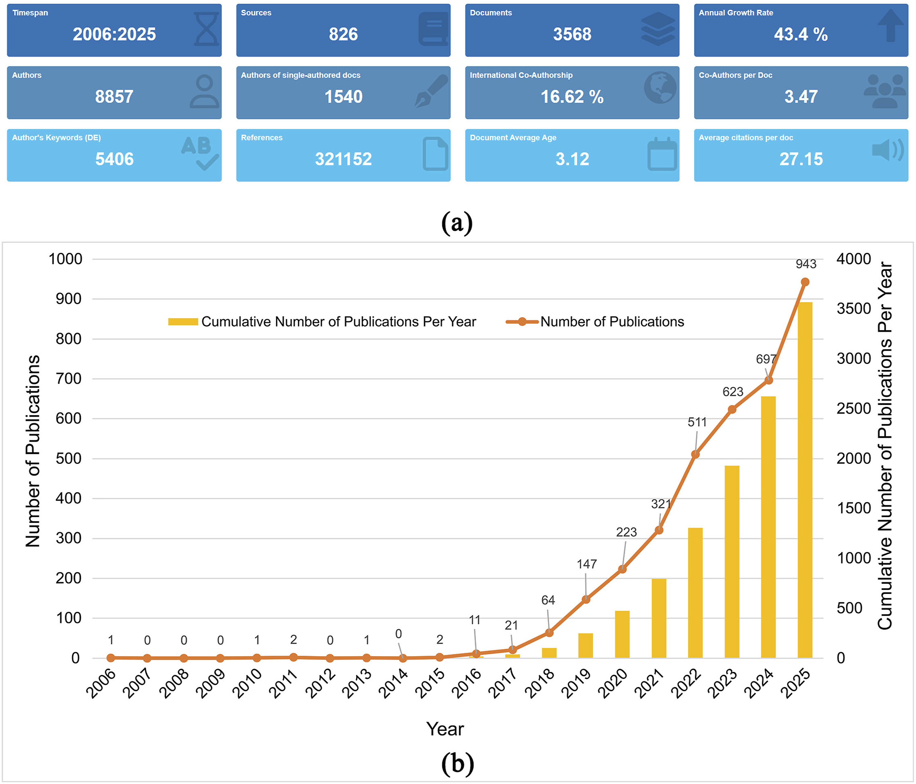
Top 10 Countries by Corresponding-Author Publications in Research on Deep Learning in Breast Cancer Imaging From 2006 to 2025
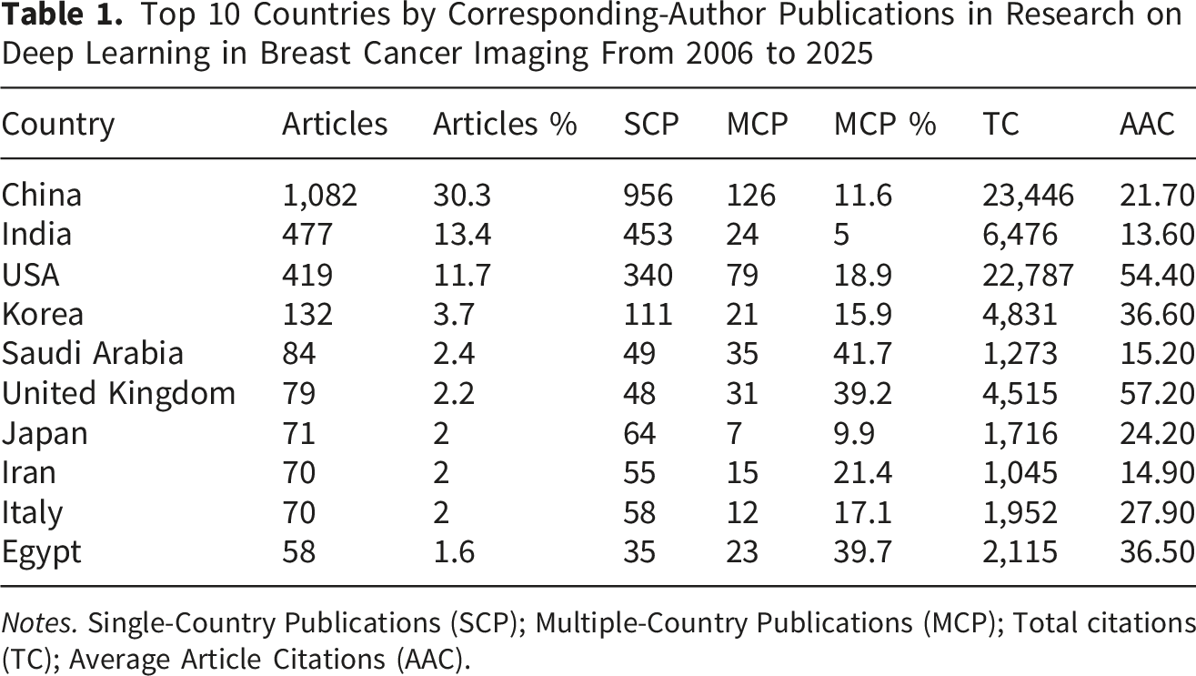
Notes. Single-Country Publications (SCP); Multiple-Country Publications (MCP); Total citations (TC); Average Article Citations (AAC).
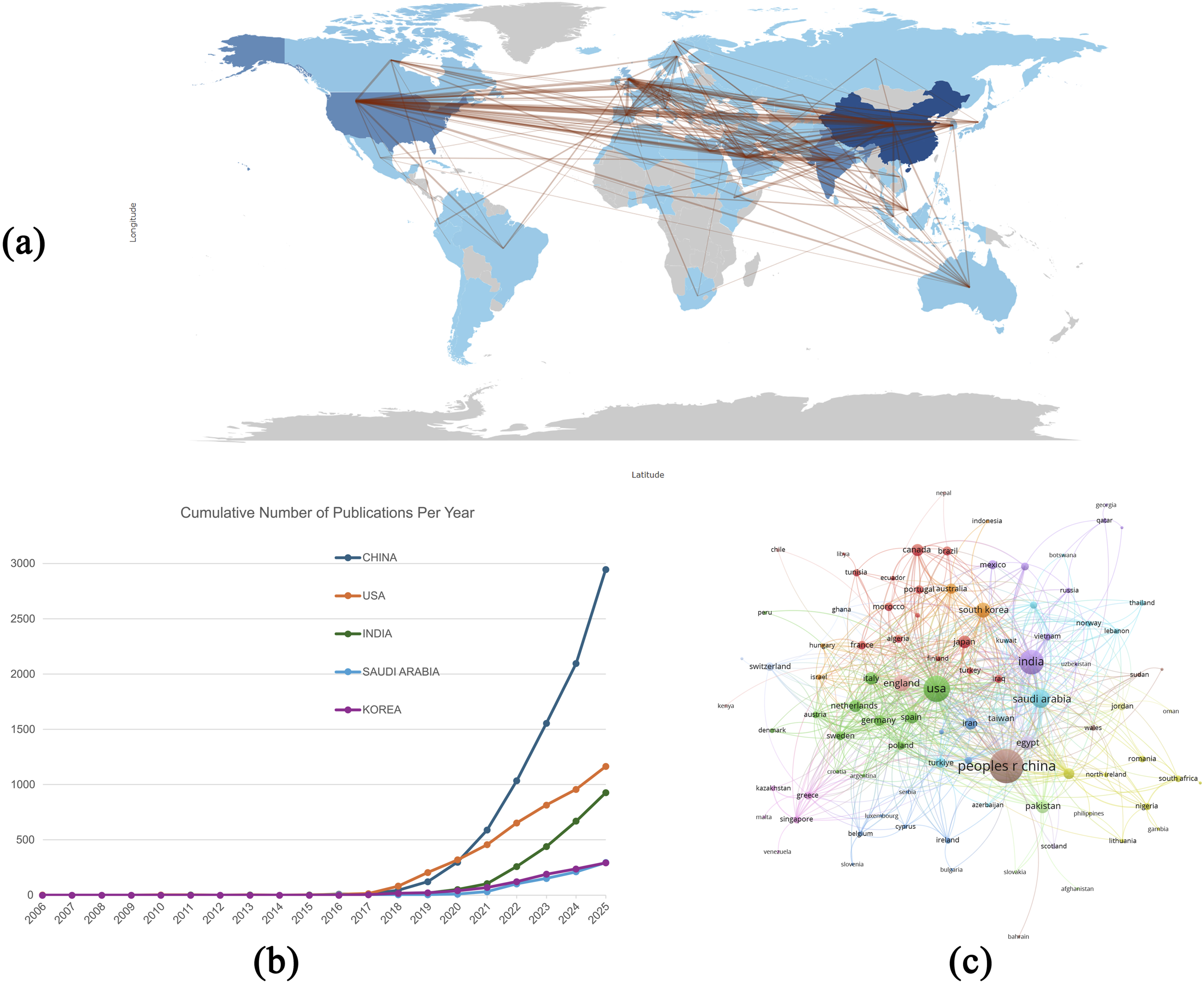
Country-level distribution, publication trends, and collaboration patterns in research on deep learning in breast cancer imaging from 2006 to 2025. (A) Geographic collaboration map. (B) Cumulative annual publication trends of the top five most productive countries. (C) International collaboration network among countries based on co-authorship analysis
A total of more than 3,000 institutions contributed to research on deep learning in breast cancer imaging. As shown in Supplementary Figure S1, Sun Yat-sen University was the most productive institution, with 44 publications, followed by Fudan University with 38 publications and the Chinese Academy of Sciences with 32 publications. In terms of collaboration intensity, Sun Yat-sen University also had the highest total link strength (TLS = 107), followed by Fudan University (TLS = 86) and Radboud University Nijmegen (TLS = 67). These findings indicate that leading Chinese institutions not only contributed substantially to publication output but also occupied central positions in the institutional collaboration network. In particular, Sun Yat-sen University and Fudan University combined high productivity with strong collaborative connectivity, suggesting an important role in promoting inter-institutional cooperation and knowledge exchange in this field. This pattern suggests that institutional leadership in this field is shaped by both productivity and collaborative connectivity.
The analysis of authors further revealed the major contributors and collaboration patterns in this field. As shown in Supplementary Figure S2, according to the publication output, Ha, Richard ranked first with 14 publications, followed by Zhang, Lei with 13 publications and Chang, Cai with 12 publications. In terms of collaboration intensity, Ciritsis, Alexander and Rossi, Cristina had the highest total link strength (TLS = 51), followed by Boss, Andreas (TLS = 50). These findings suggest that the most productive authors were not entirely identical to the most collaborative ones, indicating a certain distinction between publication leadership and network connectivity in this research area. The author collaboration network exhibited several interconnected clusters, reflecting active cooperation among researchers from different institutions and regions. Thus, author-level contributions appear to be organized around both productive individuals and collaborative research clusters.
Top 10 Most Productive Journals in Research on Deep Learning in Breast Cancer Imaging From 2006 to 2025
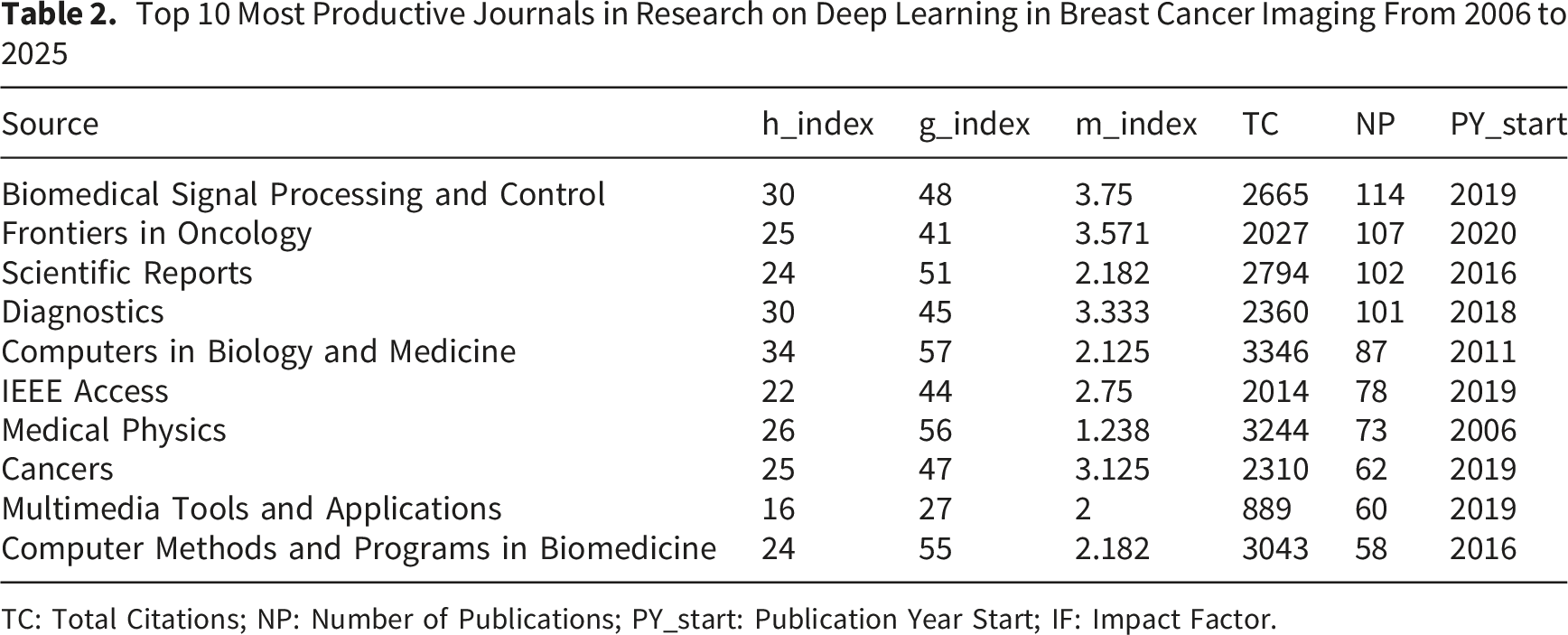
TC: Total Citations; NP: Number of Publications; PY_start: Publication Year Start; IF: Impact Factor.
A VOSviewer-based co-occurrence analysis identified 392 author keywords that appeared at least five times in the final dataset. As shown in Figure 4A, the keyword network was centered on “deep learning,” “breast cancer,” “artificial intelligence,” “mammography,” and “machine learning,” indicating that these terms form the core knowledge structure of the field. Closely connected keywords such as “segmentation,” “breast cancer detection,” “computer-aided diagnosis,” “classification,” “radiomics,” “ultrasound,” and “magnetic resonance imaging” further suggest that the field has been primarily organized around three task domains: segmentation, detection, and diagnostic research across multiple imaging modalities. The dense interconnections among these terms reflect a strongly interdisciplinary landscape spanning radiology, oncology, and computational medicine. The overlay visualization in Figure 4B shows that more recent research has shifted toward advanced and clinically relevant themes, particularly “explainable artificial intelligence,” “nomogram,” “self-attention,” “transformers,” “neoadjuvant therapy,” and “axillary lymph node metastasis.” This pattern indicates that the field is evolving beyond conventional classification and lesion analysis toward improved model interpretability, more sophisticated network architectures, and increasingly clinically oriented applications. Thus, the keyword structure supports the task-oriented framework of this study, showing that segmentation, detection, and diagnosis represent the central application domains while interpretability and advanced architectures are emerging frontiers. Co-occurrence network and temporal evolution of author keywords in research on deep learning in breast cancer imaging from 2006 to 2025. (A) Keyword co-occurrence network generated by VOSviewer based on 392 author keywords occurring at least five times. (B) Overlay visualization of the same keyword network, showing the temporal evolution of research topics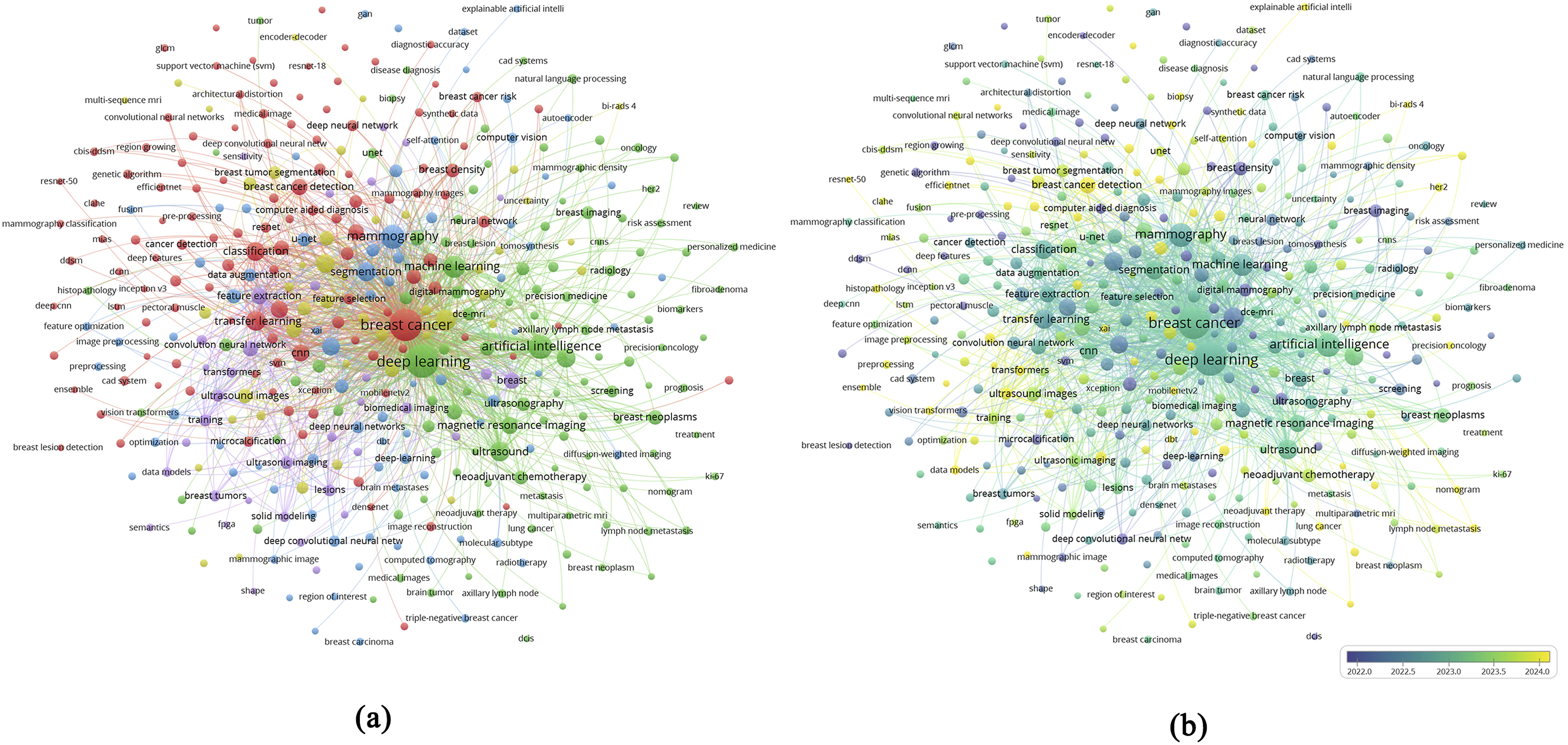
The temporal evolution of representative topics is further illustrated in Figure 5A. Early research was mainly characterized by terms such as “computer-aided detection (cad),” “artificial neural network,” “encoder-decoder,” “fully convolutional network,” “breast mass classification,” and “automated breast ultrasound (abus),” reflecting the initial emphasis on computer-aided diagnostic frameworks, early neural-network methods, and modality-specific lesion analysis. From around 2021 onward, the thematic focus expanded toward broader terms including “breast imaging,” “breast density,” “artificial intelligence,” “breast cancer,” and “deep learning,” indicating the rapid consolidation of the field. In the most recent period, keywords such as “radiomics,” “segmentation,” “breast lesion segmentation,” “vision transformer,” and “accuracy” became increasingly prominent, highlighting continued methodological refinement and performance-oriented development. Consistent with these trends, the burst detection analysis in Figure 5B showed that earlier citation bursts were dominated by “artificial neural network,” “computer assisted diagnosis,” and “computer aided detection,” whereas more recent ongoing burst terms included “learning algorithm,” “positron emission tomography,” “cancer prognosis,” “mammograms,” “prospective study,” and “lymph node metastasis.” Taken together, these findings indicate that the field has evolved from early computer-aided diagnosis paradigms into a more integrated research landscape centered on segmentation, detection, and increasingly clinically relevant diagnostic applications in breast cancer imaging. The temporal pattern further suggests a recent shift toward clinically oriented and translational research themes. Temporal evolution and citation bursts of keywords in research on deep learning in breast cancer imaging from 2006 to 2025. (A) Trend topics of representative keywords over time. (B) Top 25 keywords with the strongest citation bursts
4. Discussion
This study analyzed 3,568 publications from the WoSCC and Scopus, providing a comprehensive overview of research activity from 2006 to 2025 on deep learning for segmentation, detection, and diagnostic research in breast cancer imaging. Compared with previous bibliometric studies that mainly focused on artificial intelligence applications in breast cancer diagnosis, transfer learning techniques for breast cancer detection,18,19 the present study provides a more task-oriented bibliometric perspective. Specifically, it characterizes segmentation, detection, and diagnostic research as three interconnected application domains across breast imaging modalities. Since 2016, publications in this field have grown markedly, reflecting the widespread adoption of artificial intelligence in oncology, a trend also confirmed by global bibliometric analyses. 24 China has been a leading contributor in terms of publication volume, while the USA demonstrated the highest citation impact, jointly promoting cross-regional exchange and rapid dissemination of research. Keyword analysis revealed several thematic clusters centered on “breast cancer” and “deep learning,” such as segmentation, classification, mammography, diagnosis, and neural networks, which together define the current research landscape. More importantly, the combined evidence from keyword co-occurrence, trend topics, and citation bursts indicates that the field has gradually evolved from early computer-aided diagnosis frameworks toward a more integrated landscape centered on segmentation, detection, and clinically relevant diagnostic applications across multiple imaging modalities.
The development of deep learning in breast imaging can be summarized into three overlapping phases. Early studies largely followed radiomics and classical machine-learning pipelines, extracting handcrafted texture features from relatively small, single-center datasets, thereby revealing the limitations of the initial exploratory stage.25,26 Around 2015, convolutional neural networks and encoder–decoder architectures rapidly emerged as the predominant methods for segmentation and lesion classification in medical imaging, 27 laying the algorithmic foundations for subsequent translational work. This transition coincided with the increase in publications observed in Figure 2, as methods were quickly adapted to breast-specific tasks such as lesion detection and classification. 28 More recently, especially after 2020, research has shifted toward multimodality integration, clinically oriented diagnostic modeling, and more advanced architectures, as reflected by the emergence of keywords such as “self-attention,” “transformers,” and “explainable artificial intelligence.” This temporal pattern suggests that the field is no longer driven solely by algorithmic novelty, but increasingly by the need to improve interpretability, clinical relevance, and workflow integration.29,30
Despite these advances, important gaps remain between algorithmic performance and clinical application. For instance, Abdullah’s meta-analysis demonstrated that substantial heterogeneity persists due to variation in imaging protocols, dataset sizes, and validation strategies, emphasizing the continued need for refinement and methodological standardization in deep learning models. 31 Moreover, many models achieved high accuracy in internal test sets but exhibited notable performance drops in prospective, multi-institutional studies. 32 Heterogeneity of data, limited external validation, lack of integration with non-imaging clinical variables, and algorithmic bias further restrict the generalizability of current models and hinder their wider adoption in routine clinical classification and diagnosis. In addition, recent reviews have emphasized that reproducibility, bias control, and explainability remain unresolved barriers to clinical translation, even in studies reporting promising diagnostic performance.33,34 Therefore, the main challenge facing the field is no longer simply whether deep learning can achieve high accuracy under controlled conditions, but whether segmentation, detection, and diagnostic systems can remain reliable, transparent, and clinically useful across institutions, populations, and imaging platforms.
To bridge these gaps and to reflect the evolution of recent keywords, several future research directions should be prioritized. Multimodal architectures are particularly critical, as deep learning models capable of jointly analyzing mammography, MRI sequences, and ultrasound are likely to outperform single-modality networks. Mammography remains a highly active research modality, as indicated by keyword bursts in Figure 5, highlighting its ongoing centrality in clinical practice. For example, Chen et al. developed a multimodal model combining mammography and ultrasound, which significantly improved breast cancer screening accuracy. 35 Similarly, Atrey et al. reported that multimodal classification achieved an accuracy of 99.22%, substantially higher than single-modality approaches. 36 Such fused models can capture complementary information about breast lesions and achieve superior segmentation and diagnostic performance when paired with appropriate architectures. However, future development should move beyond modality stacking alone and place greater emphasis on clinically grounded multimodal integration, transparent reporting, and robust external validation. The sustained appearance of keywords such as “network” and “artificial intelligence” underscores the continuous innovation in model architectures. Taifi et al. demonstrated that optimized architectures achieved 99% accuracy in breast cancer classification, 37 while Singh showed that combining transfer learning with classifier ensembles improved accuracy by over 8 percentage points. 38 These examples illustrate that architectural optimization and hybrid frameworks provide promising directions for future segmentation and diagnostic applications. At the same time, the recent emergence of “transformers,” “self-attention,” and “explainable artificial intelligence” suggests that future progress will depend not only on performance gains, but also on model interpretability and deployability in real clinical settings.
In addition, the emergence of “lymph node metastasis” and “neoadjuvant chemotherapy” as recent keywords highlights the growing interest in prognostic applications. Accurate prediction of axillary lymph node status and systemic treatment response is essential for guiding therapeutic planning. 39 Lee et al. reported that deep learning models based on breast MRI showed encouraging diagnostic potential for predicting axillary lymph node metastasis. 40 Similarly, Guo et al. demonstrated that cross-modality deep learning frameworks could predict pathological responses to neoadjuvant chemotherapy in breast cancer. 41 These findings indicate that future research is likely to place stronger emphasis on predictive modeling for metastasis and systemic treatment outcomes, ultimately advancing precision oncology and individualized treatment strategies. Importantly, these directions should be understood as clinically oriented extensions of diagnostic research rather than a departure from the core task framework of segmentation, detection, and diagnosis defined in the present study.
Although deep learning models are increasingly being explored for prognostic assessment and treatment-response prediction, several limitations should be acknowledged before their routine implementation in breast cancer care. Recent studies have suggested that deep learning models based on longitudinal breast ultrasound and MRI data may enable early and noninvasive prediction of response to neoadjuvant therapy. 42 In another clinically relevant application, a meta-analysis by Lee et al. showed that MRI-based deep learning models had promising diagnostic performance for predicting axillary lymph node metastasis; however, the available evidence also revealed heterogeneity and underscored the need for more robust validation. 40 More broadly, deep learning studies in breast MRI remain affected by variations in imaging protocols, datasets, and validation strategies, which may compromise model generalizability across different institutions and clinical environments. 31 These limitations are particularly important because prediction of treatment efficacy and prognosis depends not only on imaging features but also on treatment regimens, molecular subtype, pathological characteristics, and longitudinal follow-up. Therefore, imaging-only deep learning models may provide incomplete risk assessment unless they are integrated with relevant clinical and biological variables. From the perspective of clinical translation, deep learning should be regarded as a decision-support tool rather than an independent decision-maker. Real-world implementation remains challenging because model performance may vary across clinical settings, and limited generalizability may reduce confidence among healthcare providers. 43 Additional barriers include data privacy, algorithm transparency, potential bias, ethical considerations, regulatory requirements, and workflow integration in breast imaging practice. 44 Future studies should therefore prioritize multicenter prospective validation, transparent reporting, clinically meaningful endpoints, and human–AI collaborative workflows to improve the reliability, interpretability, and practical utility of deep learning models in breast cancer imaging.
This study has several limitations. Although combining WoSCC and Scopus improved coverage compared with a single-database search, the analysis was restricted to English-language publications indexed in these two databases; therefore, relevant studies indexed only in other databases, regional databases, preprint servers, or non-English sources may not have been captured. Furthermore, bibliometric approaches can identify publication trends and thematic evolution but cannot directly assess the methodological rigor or clinical validity of deep learning models for segmentation and diagnosis. Because bibliometric analyses rely on indexed metadata, residual inconsistencies in author names, institutional affiliations, and keyword assignment may also influence the network structure despite data cleaning and deduplication. Finally, citation-based metrics are inherently time-dependent, and recently published high-quality studies may not yet be adequately reflected in our analysis. This limitation is particularly relevant for publications from 2025, which had limited time to accumulate citations. Despite these limitations, this study provides a comprehensive overview of research trends in deep learning for breast cancer imaging and offers valuable insights to guide future methodological development and clinical translation.
5. Conclusion
This study demonstrates that research on deep learning in breast cancer imaging has evolved into a rapidly expanding and increasingly sophisticated field from 2006 to 2025. More recent keyword patterns further suggest a shift from conventional computer-aided diagnosis and basic classification tasks toward advanced architectures, improved interpretability, and clinically relevant applications, as reflected by the emergence of terms such as explainable artificial intelligence, self-attention, transformers, nomogram, neoadjuvant therapy, and axillary lymph node metastasis. Although methodological innovation has accelerated substantially, future progress will depend on whether these models can achieve greater robustness, transparency, external validity, and clinical utility across real-world imaging settings. Overall, this study provides a task-oriented bibliometric framework for understanding how deep learning in breast cancer imaging has developed and where its next translational priorities are likely to lie.
Supplemental Material
Supplemental Material - The Evolving Landscape of Deep Learning in Breast Cancer Imaging: A Bibliometric Study of Segmentation, Detection, and Diagnostic Research From 2006 to 2025
Supplemental Material for The Evolving Landscape of Deep Learning in Breast Cancer Imaging: A Bibliometric Study of Segmentation, Detection, and Diagnostic Research From 2006 to 2025 by Shengxiong Zeng, Lei Wang, Yuqin Zhang, Yue Deng, Xiaocheng Shen, Yong Li in Cancer Control
Footnotes

Ethical Considerations
This bibliometric review was based exclusively on publicly available bibliographic records and did not involve human participants, human tissue or samples, clinical interventions, individual patient data, or identifiable personal information. Therefore, formal ethics committee approval was not required.
Consent to Participate
This study did not involve human participants or identifiable individual-level data.
Author Contributions
Conceptualization, S.Z. and L.W.; Methodology, S.Z. and L.W.; Software, S.Z.; Validation, L.W., Y.Z. and Y.D.; Formal Analysis, X.S.; Investigation, S.Z.; Resources, Y.L.; Data Curation, S.Z.; Writing – Original Draft Preparation, S.Z. and L.W.; Writing – Review & Editing, S.Z. L.W. and Y.L.; Visualization, S.Z.; Supervision, Y.L. All authors have read and agreed to the published version of the manuscript.
Funding
The authors declare that no financial support was received for the research, authorship, and/or publication of this article.
Declaration of Conflicting Interests
The authors declared no potential conflicts of interest with respect to the research, authorship, and/or publication of this article.
Data Availability Statement
The original contributions presented in this study are included in the article, further inquiries can be directed to the corresponding authors.
Supplemental Material
Supplemental material for this article is available online.
References
Supplementary Material
Please find the following supplemental material available below.
For Open Access articles published under a Creative Commons License, all supplemental material carries the same license as the article it is associated with.
For non-Open Access articles published, all supplemental material carries a non-exclusive license, and permission requests for re-use of supplemental material or any part of supplemental material shall be sent directly to the copyright owner as specified in the copyright notice associated with the article.
