






Abstract
Scientists and philosophers have long appreciated that active somatosensation requires the sensory and motor systems to exchange information about body the body’s movements as well as touch in order to accurately interpret incoming somatosensory information and plan future movements. However, the circuitry underlying this sensory and motor integration is complicated and is difficult to study without tools to label specific cellular components in the various brain regions involved. Here, I review the general pathways that convey ascending sensory and descending motor information, using the rodent whisker system as a model to take advantage of the cell type specificity possible in this model. I then detail the circuits in motor cortex in which incoming information from somatosensory cortex and thalamus is integrated. I close with a brief description of changes in these circuits during motor learning.
Keywords
Introduction
A beautiful display of exotic birds-of-paradise might capture the eye at a natural history museum. For me, the real excitement in encountering animals comes when, at an aviary, I see a burrowing owl and impatiently wait for them to fly! Curiosity about how the brain produces the broad behavioral range in animals fascinates and motivates motor systems neuroscientists as well as animal lovers worldwide. Coordination of these tasks, such as taking a short flight and accurately landing on a branch or catching food in mid-air, relies not only on a motor plan but also on the integration of incoming sensory information conveying the animal’s current body position as well as the location of relevant objects in the outside world. The full range of circuits for control of movement is not yet understood, so our studies and this review will address a subset: the motor cortex, and in particular the circuits by which somatosensory information is integrated into motor circuitry.
Why Do the Sensory and Motor Systems Interact in the Brain?
It is said that “If you know your enemies and know yourself, you will not be imperiled in a hundred battles” (Wee 2003). It has long been appreciated that, in order to make sense of incoming sensations, and especially active touch, it is necessary that the motor and sensory system exchange information, including the limb and muscle positions, as well as incoming sensory information and future planned movements (Gibson 1962). Similarly, in order to generate accurate movements, the brain must ideally know sensory information not only about the external world but also about the current state of the body. Thus, one main sensory input to motor areas must include such proprioceptive information. This includes information from muscle spindles to supply information on the current state of the musculoskeletal system, such as joint angles and muscle tension. Proprioception might be used for planning the initial movement, but peripheral feedback is too slow to provide continuous control of motor output (Kawato 1999; Scott 2008). In a forward model of motor control, the estimated position and state of the body can be updated using an efferent copy of the transmitted motor command, which could be used to more rapidly update the motor system’s estimate of the body, and thus generate revised motor commands. Such information may also be used to estimate the intended sensory consequences of an ongoing movement. In the specific case of whiskers, which may lack muscle spindles (Bosman and others 2011; Bowden and Mahran 1956; Moore and others 2015), some of the information regarding whisker position likely originates in an efferent copy of the outgoing motor command. This might include motor cortex, which provides an accurate signal corresponding to whisking phase (Hill and others 2011).
To complete the internal model of the body state and real world, the somatosensory system needs to bring in information from sensory receptors. A great variety of mechanoreceptors in the skin bring in somatosensory information including those in whisker follicles. Incoming sensation allows objects in the outside world, either as targets or obstacles, to be integrated into the motor plan, as well as to reach central cortical regions where they might inform the thoughts and future desires or goals of the animal. This incoming sensation can be active or passive. Active somatosensation involves the animal actively moving the body to activate mechanoreceptors, such as a child handling a toy or a mouse whisking against a wall. Passive somatosensation occurs when movement in the external world actives mechanoreceptors, including poking or tickling someone else, or for rodents, when an experimenter moves a rod across the whiskers at rest. These sensations are interpreted differently, at least in part because the animal is aware of its own movement during active touch. To assist in describing this incoming sensation and the uses of sensory information in interpreting and controlling motor output, Figure 1 includes a functional diagram for motor control. The motor controller (in blue) needs input representing both the desired plan, as well as information about the body and the world. This information comes in the form of proprioceptive inputs (black—potentially a form of servo control), efferent copy signals from outgoing motor control (red—a forward model of motor control), and somatosensory input (gold), which generate an internal representation of the body and the real world. Several brain regions have been proposed to perform these computations (Scott 2012; Shadmehr and Krakauer 2008). However, there are many more cell types and connections within the sensorimotor pathway (Fig. 2) than needed to implement this system (Fig. 1). It is not clear whether S1 to M1 projections represent strictly somatosensory information or both somatosensory and proprioceptive information. However, inactivation of S1 results in loss of finger coordination in a precision grip task prior to movement onset (Brochier and others 1999), suggesting a role for somatosensory (cutaneous) information in the S1 to M1 projection (Witney and others 2004). Thus, the sensory projections to M1 likely include representations of incoming sensory information and of the current state of the physical plant, though it is also possible that M1 has more complex circuitry to build these state estimates. In the following sections, I will describe what we know about M1 inputs and how they project to specific M1 cell types.
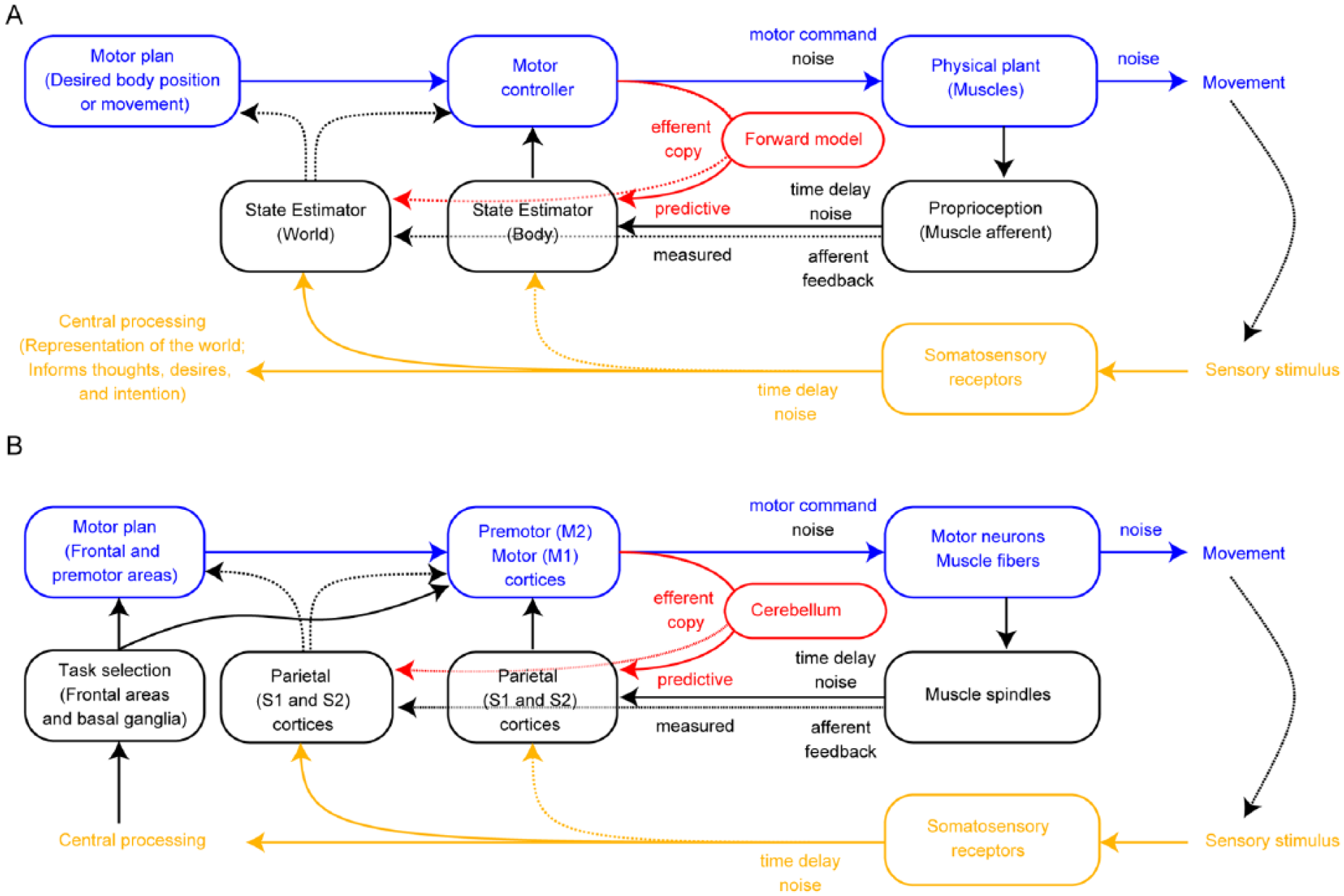
A block diagram for motor control. (A) A scheme for how motor control including a forward model might be implemented. Boxes note the functional role of each component, with motor outputs shown in blue, sensory inputs in gold, and the forward model added in red. Computations within each circuit include, for example, output from the motor controller to make the state estimator match the motor plan. A second state estimator is added for the internal representation of the world, which may directly influence motor planning and control. (B) Motor control scheme, with boxes indicating brain regions proposed to be involved (Scott 2008; Shadmehr and Krakauer 2008) in the computations of (A). As complex as these schemes are, motor areas contain vastly more cell types and connections than are represented here.
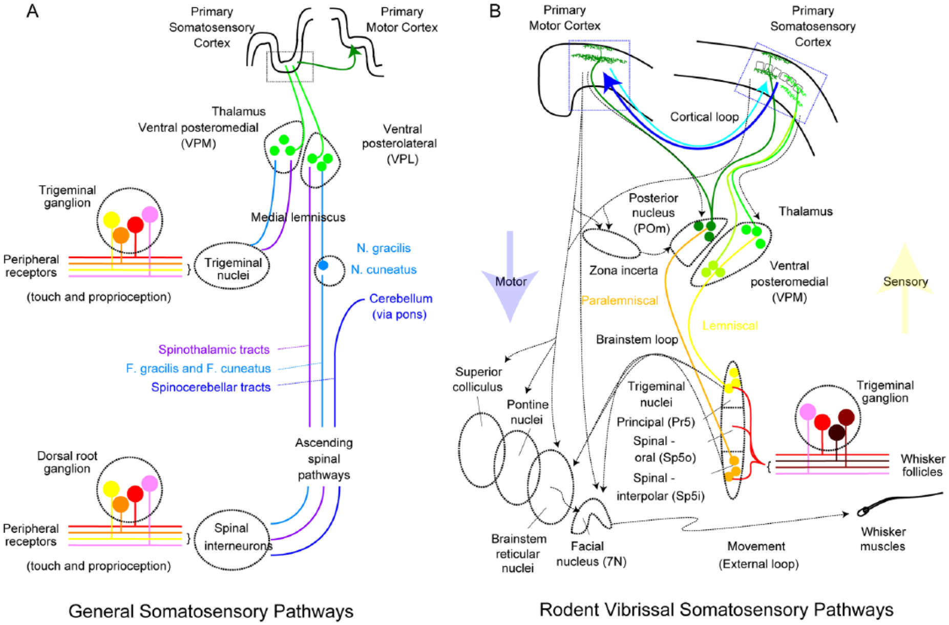
Somatosensory pathways. (A) General somatosensory pathways. Peripheral sensation enters the nervous system via mechanoreceptors whose cell bodies lie in dorsal root ganglia (body) and the trigeminal ganglion (face), ascending through several tracts. These reach somatosensory cortex via principal sensory thalamic nuclei, the ventral posteromedial (VPM) and ventral posterolateral (VPL) for face and body, respectively. (B) Rodent vibrissal somatosensation is presented in greater detail. Mechanosensation from the whisker follicles enters via the trigeminal ganglion, whose outputs target both the principal (Pr5) and spinal-interpolar (Sp5i) divisions of the trigeminal nuclei. A somatotopic representation of the whiskers is present in both brainstem areas, as well as their targets in thalamus and cortex. The most rapid sensorimotor loop relays this information to brainstem circuitry and the facial motor nucleus (7N), which controls the musculature underlying whisker movement. Ascending paths reach thalamus via lemniscal and paralemniscal pathways, with Pr5 reaching the principal thalamic nucleus VPM. Sp5i reaches the higher order posterior (POm) thalamic nucleus. VPM and POm both project to primary somatosensory cortex (S1), targeting distinct layers. VPM particularly targets the layer 4 barrel structures (boxes in [B]), which each topographically represent a single whisker. POm also projects directly to primary motor cortex (M1). Sensorimotor projections to M1 originate in S1, as well as secondary somatosensory cortex (S2). The sensorimotor loop is closed by feedback projections to S1, as well as projections to thalamus (including recruitment of feedforward inhibition) and to brainstem, including the facial motor nucleus.
Sensory and Motor Pathways for Active and Passive Somatosensation
With the development of genetically engineered mouse lines to label a variety of cell types in neocortex (Gerfen and others 2013; Taniguchi and others 2011), mouse models have drawn great interest in the hope of correlating the connectivity, excitability, and functional roles of cortical cell types, and thus elucidating how cortical circuits work. Since active whisker somatosensation is a major mouse sensory modality, I will focus on the rodent whisker system as a model of somatosensory integration, where great effort has been expended to understand the circuitry involved in detail. My sensory systems colleagues have an inherent advantage in understanding the seemingly rational organization of most sensory systems: the attributes of the physical world represented in each primary sensory region seem straightforward, and from the sensory receptors themselves to neocortex, the visual, auditory, and somatosensory systems retain topographic organization corresponding to their spatial, tonotopic, and somatotopic input. Somatosensory representation can be as fine as a single rodent whisker (Woolsey and Van der Loos 1970). Whereas motor systems, especially cortex, are generally somatotopic (Penfield and Jasper 1954), these maps also show overlap between nearby muscles (Rathelot and Strick 2009). In the mouse, these is a whisker motor cortex that is reciprocally connected to the corresponding somatosensory representation and whisker movement can be evoked by microstimulation (Ayling and others 2009; Hooks and others 2011; Tennant and others 2011), but these maps are coarse. Thus, defining primary motor cortex (M1) and its subregions, such as forelimb and vibrissal divisions, can be ambiguous. Furthermore, what exactly is represented in motor cortex is not nearly as clear as in primary sensory areas. While not necessarily mutually exclusive, hypotheses range from kinematic parameters such as muscle position and force (Evarts 1968; Todorov 2000), to movement direction (Georgopoulos and others 1982), or complex behaviors (Graziano and others 2002).
Figure 2A describes the general ascending pathways for somatosensory input in mammals, following touch from fingers or whiskers to central processing areas of cerebral cortex. This picture is simplified, as including all brain regions and cell types is immensely complex (Bosman and others 2011). A variety of peripheral mechanoreceptors in the skin, specialized for different types of touch, send information to the dorsal horn, where many classes of mechanoreceptors impinge on local interneurons and projection neurons, in turn conveying the signal up the spinal cord (Abraira and Ginty 2013). Sensory information from facial regions enters instead via the trigeminal nucleus. Sensory information then reaches primary somatosensory cortex by way of the ventral posterior nuclei in thalamus (medial and lateral, VPM and VPL), where VPM is specialized for facial inputs, such as whiskers, while VPL is specialized for inputs from the body, such as the fingers and limbs. This review will focus on the rodent vibrissal system, which many investigators have traced the circuitry from brainstem to cortex (Fig. 2B). As a result, some differences with forelimb regions may not be treated in detail. In rodents, incoming whisker touch information via the trigeminal ganglion reaches both the principal trigeminal (PrV, yellow) and spinal trigeminal-oral subdivision (orange) and ascends to two distinct thalamic nuclei (Veinante and Deschenes 1999; Veinante and others 2000), but even this description is simplified (Pierret and others 2000). The lemniscal pathway via VPM conveys rapid, single whisker touch information to the cortex, while the paralemniscal pathway via a higher order thalamic nucleus, posterior nucleus (POm), conveys multiwhisker information, responds more slowly, and includes information such as pain and temperature as well. VPM is the principal thalamic nucleus targeting primary somatosensory cortex (S1), called barrel cortex, while afferents from POm target the cortex more broadly (Fox 2008; Ohno and others 2012). Of note, trigeminal output can project directly to brainstem nuclei and regulate motor output (“Brainstem Loop”; Nguyen and Kleinfeld 2005), which may mediate rapid feedback in a reflex-like manner. Sensory information thus reaches primary motor cortex (M1) via connections from S1 (“Cortical Loop”), as well as direct input from POm and less directly via secondary somatosensory cortex (S2). Subsequent figures will address these interconnections in cell-type specific detail. Motor output in turn interacts with ascending sensory inputs at the levels of corticocortical feedback connections to S1, direct corticothalamic projections to POm, as well as indirect connections via zona incerta (Urbain and Deschenes 2007). Brainstem-projecting neurons (Matyas and others 2010) then influence whisking by targeting reticular nuclei (Hattox and others 2002) as well as the facial motor nucleus controlling whisker muscles directly (Grinevich and others 2005). Whisker sensation is generally an active sensation, with movement required to generate contact with objects. Rodents are seen to actively palpate objects during exploration. Passive responses to deflection occur, but differ from active touches (Crochet and Petersen 2006), suggesting that planned movements influence incoming sensory responses.
Why do multiple sensorimotor loops exist at the brainstem and cortical levels? In order to be an effective predator or to avoid becoming prey, animals must be able to process incoming sensory information to formulate and execute an appropriate response. Time is of the essence in this sensorimotor loop: systems that can respond faster have the advantage of catching their competitors addressing circumstances that have already changed (Boyd, 1976). Thus, it makes sense that sensory and motor pathways interact on many levels—shorter loops in the brainstem might mediate situations requiring more rapid responses, whereas subtle stimuli that require additional processing and response may reach the cerebral cortex. In the case of whisking, for example, the short latency retraction on touch to generate gentle contact (Deutsch and others 2012; Mitchinson and others 2007) may be mediated by brainstem circuits, whereas whisking to actively investigate objects can be controlled by long latency loops.
Circuitry Underlying Sensorimotor Integration in Primary Motor Cortex
Why has the rodent somatosensory system become a prevalent model for studying the neural circuitry of sensorimotor integration? Circuits in primary visual and somatosensory areas have received substantial attention as models for local connectivity (Douglas and Martin 2004; Markram and others 2015). The large size, dorsal location, and somatotopic whisker representation of barrel cortex (Woolsey and Van der Loos 1970) in rodents make it an accessible model for experiments ranging from in vivo recording and imaging to developmental manipulation (Trachtenberg and others 2002). Studies of local circuit connectivity has been extended into primary motor and secondary somatosensory areas, the other principal whisking regions in rodents (Hooks and others 2011). The ability to rapidly develop and test new methodologies, especially viruses for regionally targeted gene expression (Atasoy and others 2008) and opsins for optogenetic circuit mapping (Boyden and others 2005), coupled with mouse genetics to label and manipulate specific cell types (Gerfen and others 2013; Taniguchi and others 2011), gives mice a strong advantage over primates as a mammalian model system. Anatomically, topographically corresponding primary somatosensory and motor areas are strongly interconnected by direct corticocortical projections (Zingg and others 2014). Thus, this projection has become an effective model for studying corticocortical connectivity (Mao and others 2011; Petreanu and others 2009; Rocco-Donovan and others 2011).
It is important to note that rodent motor cortical areas are inherently smaller than primate motor areas. The cortical surface of primates is greatly expanded, with topographic areas representing distinct regions of the limb, and in some primates includes new and old subdivisions characterized by presence and absence of corticomotoneuronal neurons, respectively (Rathelot and Strick 2009). There are a number of different locations within primary motor cortex that have been studied in primates, but it is not clear if the circuit connectivity between M1 and S1 will be the same for all areas. Prior studies have focused on which cortical areas are interconnected (Leichnetz 1986), though some have examined the laminar origin of corticocortical projections (Jones and Wise 1977), with layers of origin similar to that found in rodent (Mao and others 2011). The circuit concepts uncovered by studies in mice will be more valuable if similar circuits exist and are accessible to study in primates. This assumption merits rigorous testing once the basic circuitry is understood, and experimenters can use tools and methods to study comparable cell types in primates.
How Sensorimotor Integration Is Instantiated in Motor Cortex
Since the elucidation of the sensory and motor homunculus by Wilder Penfield (Penfield and Jasper 1954), projections between the somatotopically corresponding regions of M1 and S1 have been studied anatomically and functionally in mammalian models including cat (Jones and Powell 1968) and monkey (Jones and Powell 1969). However, the literature, based on extracellular recording and microstimulation studies, disagrees on the laminae of pyramidal neurons targeted by S1 input. Asanuma and others have found that S1 inputs strongly target neurons in layer 3 in the cat (Kaneko and others 1994; Porter and others 1990). In contrast, others find S1 inputs to pyramidal neurons across all layers, including brainstem targeting L5 pyramidal neurons, in both the cat (Zarzecki 1989) and macaque (Ghosh and Porter 1988).
More recently, S1 to M1 cortical projections have been heavily studied in rodents (Fig. 3). These two distinct cortical areas show significant regional specialization. S1 is a superb example of six-layered mouse neocortex, with a cell sparse layer 1 (L1), a dense granular layer 4 (L4) that divides pyramidal cell containing layers into supragranular (layer 2/3) and subgranular (layers 5A, 5B, and 6). M1, in contrast, is in the lateral and medial agranular areas of mouse cortex, with the whisker region more medial (based on microstimulation mapping; Ayling and others 2009; Brecht and others 2004; Hooks and others 2011; Tennant and others 2011). L4 is traditionally regarded as absent in frontal areas (Shipp 2005), though some molecular markers of L4 are present in rodent M1 (Schaeren-Wiemers and others 1997) and some L4-like neurons exist in the local circuit of primates and rodents (Garcia-Cabezas and Barbas 2014; Yamawaki and others 2014). Expression of these markers, such as ROR-beta, tapers from high in granular regions such as S1 to low near the midline. This gradient from lateral to medial agranular areas is one of the main differences in the circuitry of rodent forelimb and whisker areas. The relative thicknesses of the layers also varies, with L5B wider in medial areas. Vibrissal S1 projections to M1 are elongated in the anterior/posterior axis, and they vary in location from medial to lateral depending on the S1 injection location (Mao and others 2011). Anatomically, fluorescence from these projections is present across layers (Oh and others 2014; Zingg and others 2014), though perhaps strongest in the upper layers of M1. However, what specific cell types in M1 are targeted by these S1 afferents?
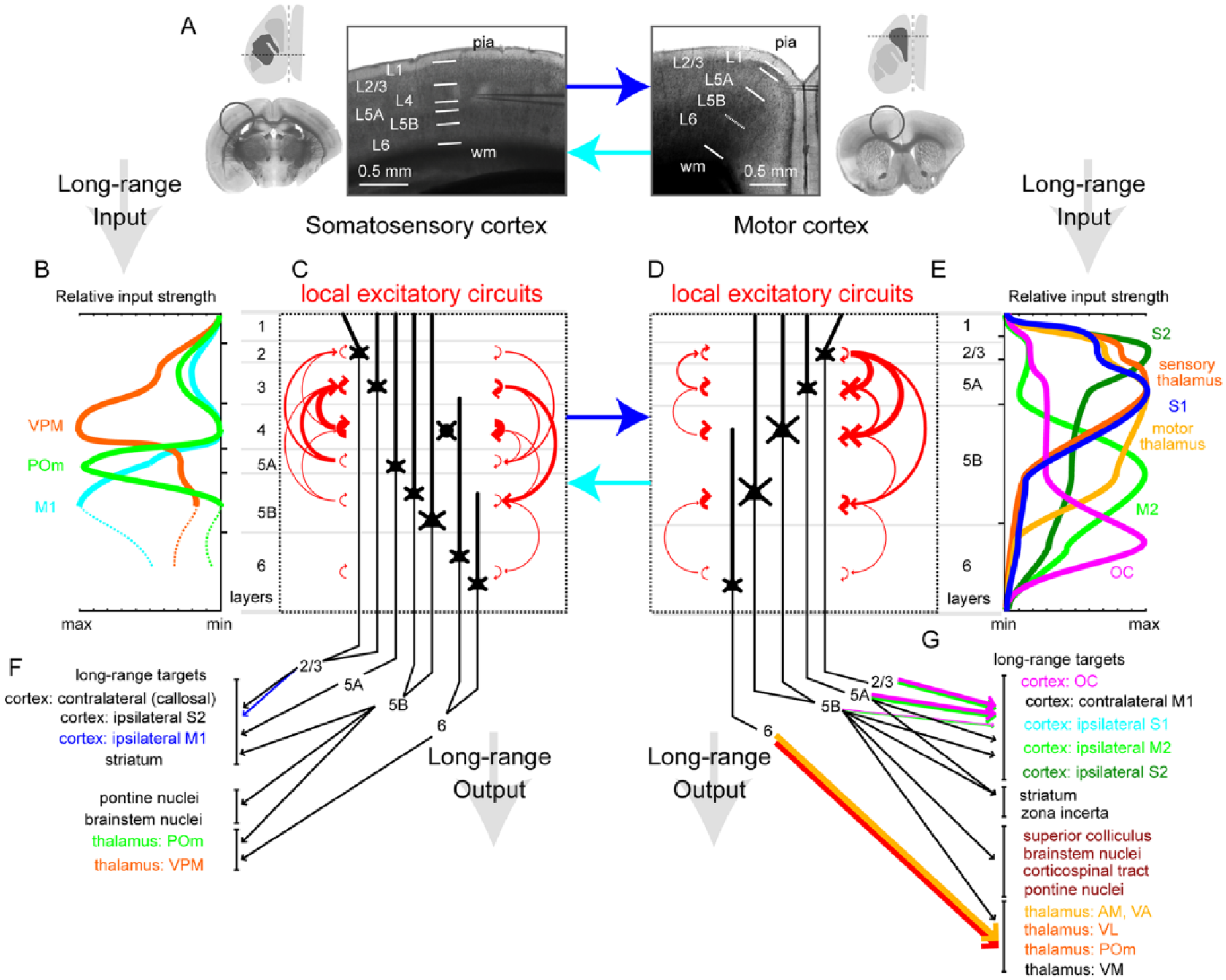
Corticocortical sensorimotor connections. (A) Overview of rodent sensory and motor cortex. Cartoon includes a dorsal view of the mouse brain, with the cortical area (S1, left; M1, right) highlighted in gray. Dashed line shows the plane of coronal section. Low magnification inset of coronal section shows S1 (left) and M1 (right), with gray circle indicating approximate position of the cortical region. High magnification microscope image shows laminar structure of S1 and M1. White bars give approximate position of laminar borders, with Layers (L) 1, 2/3, 4, 5A, 5B, and 6 marked. Note absence of granular L4 in vibrissal M1. Relative layer thickness are used for vibrissal regions. Pia (surface) and white matter (wm, ironically black in brightfield image) are noted as the top and bottom margins of cortex. (B and E) Plot of the relative magnitude of excitatory input to cortex from different thalamic and cortical inputs. Input to S1 is at left (VPM, POm, M1), with maximal input targeting distinct layers for each pathway. Input for M1 (S1, S2, M2, OC, and thalamic inputs) is at right with min/max scale reversed. (C and D) The local excitatory circuits of these regions are shown, with thickness of red arrows proportional to the connectivity across layers. Ascending pathways (to more shallow layers) are on the left of each panel, and descending pathways (to deeper layers) are at right of each panel. The S1-M1 interconnections (blue arrows) are addressed in subsequent figures. (F and G) The long range outputs are shown with the layer of origin of the projection noted. Many corticocortical pathways are reciprocal, and thus color selected to correspond to input in panel B and E.
Channelrhodopsin-2-assisted circuit mapping (CRACM; Mao and others 2011; Petreanu and others 2007; Petreanu and others 2009) suggests S1 afferents form the strongest synaptic connections in the upper layers of mouse cortex, including pyramidal neurons of layers 2/3 and 5A (L2/3 and L5A). Retrograde labeling indicated that pyramidal tract-type (PT-type) neurons, a subset of L5B neurons that project to the thalamus and brainstem (Shepherd 2013), did not receive especially strong input. However, M1 neurons projecting to S1, which reside particularly in L2/3 and L5A and are intratelencephalic type (IT-type) neurons (Shepherd 2013), did receive particularly strong S1 input (Mao and others 2011).
The S1 input to M1 does not originate from all layers homogeneously. Retrograde label in M1 has revealed the laminar specificity of the S1 to M1 projection (Hooks and others 2011; Mao and others 2011). Following injection of fluorescent LumaFluor Retrobeads into M1 (Fig. 4), the most strongly labeled band of neurons is L5A, with contributions from L2/3 as well as L5B. There is also a substantial number of cells labeled deep in white matter (the mysterious fringe in Fig. 4A) that contribute to this projection, though little is known about them. In similar studies in the rat, where differences between cortical columns associated with and between barrels are more pronounced, inputs to M1 are more likely to originate from the septa-related columns between cortical barrels as opposed to the barrel column itself (Alloway and others 2004). It is not yet clear how widespread the output from a single S1 barrel column is to M1 due to the limitations of labeling and studying single neurons or making small injections in a single barrel, though methods for reconstruction of single neurons are of growing interest (Economo and others 2016) and would permit direct evaluation. Consistent with broad S1 output, however, two-channel optogenetic approaches suggest M1 neurons integrate input from across widely spaced areas of S1 (Hooks and others 2015).
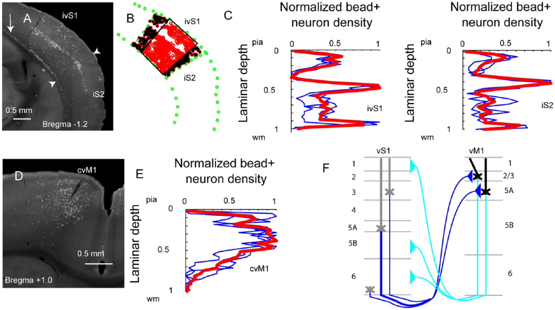
The origin of corticocortical projections. (A) Injection of retrograde label (LumaFluor Retrobeads) marks the somata of corticocortical neurons projecting to M1. A coronal section shows label in ipsilateral S1 (ivS1) and S2 (iS2). Arrow: fringe of cells deep in white matter. Arrowheads: boundary of S1/S2. (B) The location of retrogradely labeled neurons (red), as well as the margins of cortex (green), was marked manually, and neurons inside the black quadrilateral were quantified for relative cortical depth. (C) Quantification of laminar depth of M1 projections from ivS1 (left) and iS2 (right), with individual sections (blue) and average (red) shown. Relative laminar depth is measured from pia (0) to white matter (1), and cell counts are normalized to the densest projection (L5A in these cases). The absence of L4 projections from both S1 and S2 is evident. (D, E) Contralateral M1 (cvM1) is also shown, with a distinct pattern compared to S1 and S2. (F) Cartoon showing the laminar origin of reciprocal projections between M1 and S1.
What aspects of sensory input are represented in each layer of M1? Combining calcium imaging of neuronal responses in mice with simultaneous monitoring of incoming sensory stimuli (such as whisker contact with object) and motor outputs (such as whisking and licking) demonstrated that superficial layers of M1 accessible to this imaging approach contain individual neurons that responded during motor outputs as well as other cells that were sensory (whisker touch) responsive, consistent with receipt of S1 or other sensory input (Huber and others 2012). These same types of signals are present in the M1 feedback to S1 (Petreanu and others 2012), derived predominantly from L2/3 and L5A pyramidal neurons. However, this method did not address responses of layers 5 and 6 neurons. In single unit studies in the rat, mechanical whisker stimulation also produced responses in deeper (>1 mm below cortical surface) layer neurons, and not perfectly consistent with local feedforward signals from S1 to L2/3, the response latency to this stimulation in lower layers was not slower than that in the layers closer to the pia (Chakrabarti and others 2008). Thus, there is no consensus on how sensory information propagates through M1.
Determining the Location of M1
Defining whisker M1 as the region receiving input from whisker S1 is not without flaws. Rodent M1 may contain functionally distinct subdivisions at the border between medial and lateral agranular cortex. In rats, for example, the S1 projection zone in M1 may be separate from and lateral to the whisker M1 region as determined by microstimulation (Smith and Alloway 2013). Microstimulation maps have not found single regions corresponding to a specific whisker, such as a single C2 whisker column in M1 corresponding to the C2 barrel in S1 (Lefort and others 2009), but instead tend to evoke movement of multiple whiskers (Brecht and others 2004), though adjacent sites for opposing abduction and adduction could be distinguished in the mouse (Harrison and others 2012). Penfield himself (Penfield and Jasper 1954) noted the topographic map’s imprecision in mankind (“one representation is not sharply demarcated from adjacent representations”), and stimulation experiments in mouse (Hooks and others 2011) show overlap with the S1 projection zone in mice (Huber and others 2012; Mao and others 2011). However, individual variation exists (Tennant and others 2011) and the size of the map is intensity- and anesthesia-dependent. The imprecision of these maps may be due to the fact that spatial limitations of the microstimulation technique (Histed and others 2009) or the fact that corticomotoneuronal projections originate from partially overlapping areas (Rathelot and Strick 2009). Although whisker movement can be evoked by stimulation in both S1 and M1 (Matyas and others 2010), the separation of the whisker S1 and M1 does make these areas easier to distinguish, as opposed to forelimb regions, where these regions are adjacent or, possibly, overlapping. Forelimb movement in response to microstimulation falls off as a gradient as one moves from anteromedial (fM1) to posterolateral (fS1), for example (Ayling and others 2009). Indeed, different mammals show different degrees of separation in motor and somatosensory cortical representations (Frost and others 2000; Nudo and Frost 2009).
The Host of Corticocortical and Thalamocortical Inputs to M1
Although S1 provides a major corticocortical input to M1, it is not the only sensory input to M1. While primary sensory areas, such as S1 and V1, are characterized by a principal sensory nucleus that provides feedforward excitation to L4 and thus characterizes the major representation of that cortical area, the single major input to primary motor areas is not as well defined. Instead, mouse M1 receives input from several cortical and thalamic areas (Fig. 3), including higher order sensory areas such as secondary somatosensory cortex (S2) as well as frontal areas including secondary motor cortex (M2) and orbital cortex (OC) and posterior parietal areas such as retrosplenial cortex (Hooks and others 2013; Suter and Shepherd 2015). While these inputs have received less study than S1 projections, they do illustrate several rules for corticocortical connections. S2 projections preferentially innervate L2/3 pyramidal neurons, and these connections appear twice as strong as those with L5A. S2 projections also were horizontally restricted to a narrow range (Suter and Shepherd 2015). Input to L5B, including corticospinal neurons, is less than to these shallow layers, though this difference is not as steep as the seven-fold reduction reported for S1 input to L5B compared to L5A (Mao and others 2011). Thus, it seems that sensory information from S1 and S2 preferentially enters primary motor areas in the upper layers (L2/3 and L5A) and is consistent with observations in cat (Kaneko and others 1994; Porter and others 1990). L5B and L6 dendrites may theoretically sample these inputs at dendritic locations distal to the recording site at the cell body. This targeting is in contrast to frontal areas, such as dorsal premotor regions (M2) and OC, whose axons preferentially arborize and provide synaptic input directly to corticospinal and corticothalamic output neurons of L5B and L6 (Hooks and others 2015). Making an accurate comparison of corticocortical inputs to primary motor areas in primates becomes quite complex, as the expansion of the frontal cortex in primates results in a large number of areas not present in mice. Primate M1 connections with frontal premotor areas include six premotor regions with direct projections to the spinal cord (Dum and Strick 2002).
Thalamic inputs include classical motor thalamic nuclei consisting of anteromedial (AM), ventral anterior (VA), and ventrolateral (VL) thalamic nuclei, as well as higher order sensory nuclei such as the posterior nucleus (POm; Hooks and others 2013; Kuramoto and others 2009; Kuramoto and others 2015; Ohno and others 2012). POm carries somatosensory information of a qualitatively different nature than the principal somatosensory nuclei: for rodents, responses in the vibrissal pathway tend to be slower and integrate across multiple whiskers (Diamond and others 1992). Individual POm axons project not only to S1 but also to M1 (Ohno and others 2012), and the axons arborize densely in L1 and L5A of both regions (Herkenham 1980; Hooks and others 2013). Of note, the broad arbors of these axons disregard “the columnar structure that is the basis of information processing in sensory areas,” suggesting the circuit organization of motor areas differs fundamentally from sensory areas (Kaneko 2013). Functionally, these connections also excite L5A neurons most strongly in S1 (Bureau and others 2006; Petreanu and others 2009) and M1 (Hooks and others 2013), while also exciting L2/3 neurons of M1 in a similar pattern to corticocortical input from S1. This similarity suggests that somatosensory information is integrated in the upper layers (L2/3 and L5A) of primary motor cortex instead of being sent directly to the corticofugal neurons in L5B and L6. This pattern of arborization and afferent connectivity was similar across thalamic injections in adjacent posterior thalamic nuclei, including VL, and is thus marked sensory thalamus in Figure 3 (Hooks and others 2013). More anterior thalamic injections into traditionally motor thalamic nuclei target both PT- and IT-type cells of L5 as well as L2/3 (Hooks and others 2013; Yamawaki and Shepherd 2015). The corticothalamic input pattern in cat VL also includes broad arborizations (Asanuma and others 1974), and the afferents preferentially target layers 1 and 3 (Strick and Sterling 1974). Because of the convergence of this cortical and thalamic input in M1, Asanuma and colleagues have suggested that this convergence and the associative LTP it produces may be a cortical signature of sensory feedback during learning (Iriki and others 1989; Kaneko and others 1994). As in cat, S1 and POm input converges on the same neurons in mouse (Hooks and others 2015). The more anterior thalamic nuclei have distinct patterns of arborization (Herkenham 1980; Hooks and others 2013; Kuramoto and others 2009), and these tend to include direct input to some of the deeper neurons of cortex. As the motor thalamus is defined by input from cerebellum and basal ganglia, two circuits involved in motor learning, it is perhaps consistent with the division seen in corticocortical input to M1 where frontal and motor-related signals, not sensory input, flows directly to corticofugal output neurons.
What happens to these sensory signals from S1, S2, and thalamus that enter M1? How does this information flow through local circuits in motor cortex? In the cat, it has been suggested for some time that S1 input impinges on L2/3 neurons and is connected by local excitatory connections to output cells in L5 (Kaneko and others 1994). Similarly, L3 connects directly to PT-type neurons in rats (Kaneko and others 2000). Using glutamate uncaging to study local connectivity quantitatively in mice has determined that the main translaminar projection of M1 is a feedforward excitation from L2/3 to L5A and L5B (Hooks and others 2011; Weiler and others 2008), consistent with the idea that incoming somatosensory information may be processed in the upper layers of M1 and then transmitted to lower layers by local feedforward excitation (Fig. 3C, D). Further studies that examined the local connectivity of these neurons in a subtype-specific manner have elucidated a hierarchical circuit (Fig. 5) in which local excitation is fed forward from L2/3 corticocortical neurons and L5 IT-type neurons to excite the upper half of L5B PT-type neurons (Anderson and others 2010; Hooks and others 2011; Kiritani and others 2012). Thus, some local computation is performed on incoming sensory information prior to its transmission in descending corticospinal pathways.
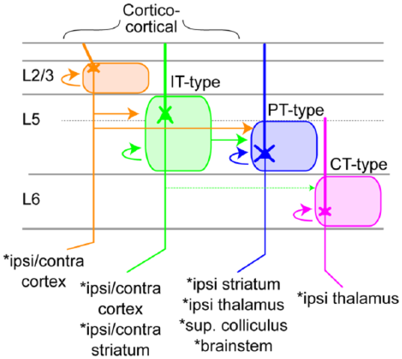
Cell-type-specific local excitatory connections of M1. L2/3 pyramids excite L5A IT-type pyramids as well as upper L5B IT- and PT-type L5 pyramids. IT-type cells of L5A and L5B excite one another and funnel input forward to L5B PT-type neurons. CT-type neurons in L6 receive some L5 IT input but predominantly excite one another. At bottom, the principal long-range targets of each cortical pyramidal cell type are shown. Based on several studies (Anderson and others 2010; Gerfen and others 2013; Hooks and others 2011; Kiritani and others 2012; Mao and others 2011; Yamawaki and Shepherd 2015).
A Role for Sensorimotor Inputs to M1 in Motor Skill Learning
It is not only interesting what circuits are involved in control of movement, but also how these circuits change when we learn a new task. Baby birds and mammals move in an uncoordinated way, but this quickly improves. Similarly, practicing scientists and other humans learn some complex skills such as typing—if not playing the violin—during adulthood. The clinical relevance of understanding M1 circuitry derives in part from the observation that plasticity in M1, especially plasticity of projections from S1, in humans is believed to play a role in learning new motor skills (Karni and others 1995; Karni and others 1998). Understanding the mechanisms of this plasticity might help in the treatment of a variety of movement disorders, such as recovery of function following stroke or treatment of neurological disorders such as dystonia.
As sensory information plays a role in developing the motor plan, one straightforward hypothesis is that plasticity at corticocortical synapses from S1 to M1 is a necessary component of motor skill learning, though this may depend on the complexity of a specific task. Gross changes in M1 representations can be induced by early developmental manipulations of sensory experience, such as whisker trimming (Huntley 1997). Connections from S1 to M1 facilitate learning, as monkeys could not learn a new skilled task if S1 were ablated, though retention of previously learned skills were not abolished (Pavlides and others 1993). Based on cortical lesions of M1 projecting regions of S1, Sakamoto and Asanuma found that the S1 → M1 connection was necessary for motor learning in a reach task in cats (Sakamoto and others 1989). Similarly, rats require M1 to learn but not perform a lever press task that involved only basic movements, as ablation of M1 blocked new skill learning, not the execution of previously learned skills (Kawai and others 2015). This suggests that M1 is only needed to tutor subcortical areas during skill learning, and may be dispensable for execution. In contrast to this, silencing of M1 seemed to prevent execution of a more complex grasping movement in mice (Guo and others 2015).
At the circuit level, many lines of evidence suggest there are changes in M1 during motor learning. Asanuma and colleagues demonstrated that tetanic stimulation could cause long-lasting potentiation in the strength of S1 to M1 projections in cat (Sakamoto and others 1987). Furthermore, though thalamic projections from VL could not be potentiated when stimulated alone, this pathway could be potentiated when coactivated with S1 (Iriki and others 1989), suggesting a role for corticothalamic inputs in motor learning. In mice, multiple changes are described during learning. Dexterous task learning induces persistent new spines on the apical dendrites of L5 pyramidal neurons, and these new spines are likely to be spatially clustered with others (Fu and others 2012; Xu and others 2009). Spine turnover in distal dendrites as well as changes in the population activity of L2/3 neurons also occurs during learning (Peters and others 2014), and inhibition by somatostatin-positive interneuron of these dendrites is one possible circuit mechanism involved in this reorganization (Chen and others 2015). The reorganization of firing patterns during learning demonstrated here is consistent with distinct cell types firing in a stereotyped pattern during different phases of preparation and execution (Isomura and others 2009). However, it is not yet known how all inputs change strength and connectivity (e.g., S1 input to L2/3 during learning of the reach task). Does cortical plasticity then reflect storage of the learned skill? Would task execution remain unaffected if potentiation were reversed? These studies imply this plasticity is a necessary part of learning and one locale where the motor skill is stored. Because behavior is preserved for some tasks after M1 ablation (Kawai and others 2015), this raises the question of the significance of this long-term plasticity at M1 synapses. This is not a trivial question to resolve.
Conclusion
This review is an overview of the specific synaptic circuits underlying sensorimotor integration in primary somatosensory and motor cortex, focused on mouse models, which are currently well equipped to dissect the circuitry in a cell-type-specific way. As transgenic mice allow reliable identification of the same circuit elements from animal to animal in a way that is not currently straightforward in primate models, we hope that such studies in mice are useful for understanding general rules of mammalian motor cortex circuitry. However, as the diversity of primate cortical areas vastly exceeds that of the mouse, rodent studies may result in oversimplification and thus difficulty in making a more direct comparison to the findings of primate and human literature. Nevertheless, this simplified view suggests that the upper layers of M1, especially the corticocortical and corticostriatal pyramids of L2/3 and L5A, receive S1 input and M1 then transmits output via deeper pyramidal tract-type neurons. However, there is much work to be done to determine whether the same circuitry is present in primates and humans, and if plasticity in this projection is also relevant to motor skill learning.
Footnotes
Acknowledgements
I thank Joshua C. Brumberg, Yi Zuo, Brendan P. Lenhert, Sarah E. Ross, Susan C. Hooks, and YingXin Zhang-Hooks for comments and suggestions on the figures and article.
Declaration of Conflicting Interests
The author declared no potential conflicts of interest with respect to the research, authorship, and/or publication of this article.
Funding
The author received no financial support for the research, authorship, and/or publication of this article.