
Abstract
Michelangelo’s unparalleled frescoes in the Sistine Chapel have been traditionally construed to represent the traditional, Roman Catholic interpretation of the Seven Days of Creation in the Book of Genesis and the Last Judgement. Indeed, in September 2018, Pope Francis I offered a benediction for the Vatican’s art treasures and for “those who contributed to the Church’s history through art.” A number of studies have suggested that Michelangelo concealed anatomic information about the human brain in his renderings of God and biblical prophets and saints. Was the anatomic content of these images purposeful or coincidental? Was the anatomy intentionally concealed? Is there a common pattern with symbolic connotations in the anatomic representations? Was Michelangelo sending an encoded message? In this study, we analyze the imagery in the Sistine frescoes in light of the literature surrounding Michelangelo’s artistry with a particular interest in the relationship between the certain purported anatomic depictions and details demonstrable using scientific and anatomic techniques available during the High Renaissance.
Introduction
The Renaissance was a pivotal period in the evolution of Western culture. It embodied the beginnings of what would later become modernity. New ideas blossomed in a bridge between the medieval period and the Enlightenment. While faith remained strong, empirical evidence began to supplant scholasticism. Pioneering ideas that drew upon philosophy, art, and science were catalyzed. Man moved to center stage, revitalizing the teachings of Protogaras of Abdera (485–415 BCE), who wrote, “Of all things the measure is Man.” Concomitantly, a movement that came to be known as Renaissance humanism took root. Humanism was deeply anthropocentric. It emphasized the importance of man and terrestrial existence rather than the divine or the supernatural. In addition, as a movement, humanism both resulted from and contributed to a restitution of interest in classical Greek and Roman thought and expression.
The bubonic plague of 1348 laid waste to more than 20 million souls, representing one-third of Europe’s population. As it waned, an interest in science and medicine slowly emerged. This interest accelerated in the mid-to-late 15th century as forgotten classical texts arrived in translation after the fall of Constantinople in 1453 and after Spanish Jews exiled by the Inquisition arrived in Tuscany from 1492 onward. Classical tropes derived from Greco-Roman pantheism permeated Europe by the 16th century, together with literary and scientific works in Hebrew and Arabic that came from the Iberian Peninsula.
This was an era of patronage. Italian banks dominated Europe and European trade. The early Italian Renaissance was centered in Tuscany. In the words of the Florentine statesman Leonardo Bruni (c. 1370–1444), Florence became the “new Athens on the Arno.” Wealthy families exhibited their success not only by building and decorating palaces and churches but also by supporting the sciences, the arts, music, and literature in their courts and through private collections of objects and artifacts. The great banking dynasties, including the Medicis, the Baldesis, the Baroncellis, the Rucellais, and the Tornaquinci, encouraged and supported artists, sculptors, scientists, poets, musicians, and philosophers, including Francesco Petrarca (1304–1374), a poet who tried to reconcile secularism with an intimate relationship with God; Marsilio Ficino (1433–1499), who reintroduced Platonism into Western philosophy and theology; and Pico della Mirandola (1463–1494), who articulated many sentinel ideas about the dignity of man. Petrarca is remembered for his poetry, Ficino for working to combine Christian morality with classical philosophy, and della Mirandola for becoming the spokesman for humanism.
Members of wealthy families also became powerful in the Catholic Church, and their patronage as popes and cardinals was transformative once again. The scale of creativity during the mid-15th century was truly extraordinary.
Together with the humanism and anthropomorphism that characterized the Renaissance, ideas about God, faith, and belief began to change and reform. The intellectuals of the Renaissance were deeply invested in man’s freedom to know and choose; however, they never rejected the moral purpose of theological ethics. Indeed, della Mirandola explicitly espoused the idea that unselfish man (in the Christian moral sense) could evolve into the highest attainable angelic form and achieve a mystical union with God. While the ultimate authority of God was not explicitly challenged, the authority of the Church did come into question, and its corruption was certainly acknowledged by the mid-14th century, as shown in Giovanni Boccaccio’s Decameron (1353) (Boccaccio 1995).
A secondary movement called the High Renaissance began approximately 1490 and lasted until 1527. The High Renaissance became a beacon of illumination fueled with a hybrid of pagan philosophy wedded to Christian theology. Like the ancient Pharos of Alexandria, it convened and encouraged the most fertile of thinkers, artists, and human explorers. Unsurprisingly, it continues to inspire scholars, art historians, literary researchers, and classicists into our own time. This was the period in which Michelangelo and da Vinci thrived. It emerged as a “perfect moment” of wealth, knowledge, relative comfort, and widespread peace harnessing a summa of intellectuality and knowledge. At the same time, however, the teachings of Martin Luther were spreading across Europe. It is often argued that the Reformation was less of a religious revolution than a political one. It was fueled not by religious zeal but by the endorsement of the noble classes whose dependence on the pope could be severed and whose consolidation of power and wealth could continue unrestrained. The Reformation was also accelerated by the proliferation of a totally disruptive technology that democratized the promulgation of ideas: Johann Gutenberg’s printing press, invented in 1440.
Rome was sacked in 1527 by Hapsburgian Emperor Charles V. His mercenaries, the Landsknechts, reiterated the depradations of the vandals before. In the manner of the Athenians who rededicated the sacred Parthenon on the Acropolis and entered a “golden age” after the Persians ruined it in the mid-fifth century BCE, the learned Pope Julius II commissioned the rebuilding of St. Peter’s Basilica. Three artistic masters flourished at this time: Leonardo da Vinci (1452–1519), a sculptor, painter, architect, inventor, and engineer, as well as an anatomist and a naturalist; Michelangelo di Lodovico Buonarroti Simoni (1475–1564), also a polymath: sculptor, painter, architect, urbanist, anatomist and poet; and Raphael Sanzio da Urbino (1483–1520), arguably the apotheosis of the classical artist and the one who most faithfully embodied the neo-Platonist ideal. Each of them qualified as a uomo universale, a universal man.
Between 1508 and 1512, Michelangelo embellished the ceiling of the Sistine Chapel in the papal palace with scenes of the Creation as described in Genesis. Between 1536 and 1541, in the wake of the response of the Council of Trent to the Protestant apostasy to the north, he was commissioned by Pope Paul III to complete a 45′ × 40′ mural above the high altar depicting The Last Judgment. This work marks the pivotal transition from the unifying themes and realism of the High Renaissance to the exaggerated, decorative, and sometimes tortuous ambiguities of the Mannerist period.
Michelangelo’s Faith
From the time of his first successes to the end of his life, Michelangelo was a demonstrably religious Catholic and seemed to have accepted uncritically the Catholic liturgy of his day. He deployed his faith as a creative tool for deciphering the Divine and the Divine will. Michelangelo’s art is open to analysis and interpretation through many lenses, including poetic metaphor, allegory, parable, and open symbolism. In this study, we even see in the imagery of the Sistine Chapel a metamorphosis of biblical imagery into an exquisite anatomic map.
It is fair to inquire about Michelangelo’s religious beliefs. He lived in tumultuous times. Many theological ideas that had been solidly anchored by dogma for centuries were upended. What was his relationship to the Church? How did he manage express fealty to successive popes? How did he see and portray his own faith during the violent religious conflicts of the 16th century? How did he manage the battlefield of shattered schemastic that shattered Europe schismatically? Many persons of faith were shaken.
Michelangelo might well have agreed with Albert Einstein’s view that “it is the most important function of art and science to awaken a religious feeling and keep it alive in those who are receptive to it” (Einstein 1934). Did Michelangelo believe in God? It has been noted that Michelangelo lived twice the average life expectancy of the Renaissance. He is described as “hugely devout.” Wecker argued that Michelangelo spent “a lot of time thinking about death, which brough him closer to his faith” (Wecker 2017).
His career spanned several phases of the 16th century. As a triumphant 37-year-old genius, in 1512, he completed the arduous task of painting the frescoes in the Sistine Chapel. Michelangelo was then an exhausted but glorified servant of his papal patron, Julius II. Together with his pride, however, he expressed a plaint. In correspondence with Giovanni da Pistoia, he complained that “I’ve already grown a goiter from this torture, hunched up here like a cat in Lombardy. . . . My skin hangs loose below me, my spine’s all knotted from folding over itself. . . . My painting is dead” (Gilbert 1980). Nevertheless, his faith seemed unaffected and so his work.
Michelangelo was celebrated as an artistic prodigy after creating the sublime Pieta (1499) in St. Peter’s Cathedral and then for the completion of what is now considered the supreme painting project in the history of humankind. Notwithstanding his contentious exchanges with Julius II, Michelangelo’s ceiling literally placed him on the summit of a Catholic universe five years before its disruption by the heretic, Martin Luther.
In his later years, the aging master developed a nuanced relationship with Rome’s intellectual elite, which had become more pious as part of the counterreformation. For one example, Michelangelo carried out an exchange of sonnets with the aristocratic Marchesa Vittoria Colonna (1490–1547), whom he addressed as “My Ladyship.” Colonna was influential in expanding Michelangelo’s ideas to the reformational, crypto-Protestant circle known as “Nicodemites.” This offers insight into Michelangelo’s self-portrait as Nicodemus in the mutilated Florence Pieta (1547–1555). As aristocratic intellectuals, they showed an “interest in some of the ideas of the Protestant reformers but sought to act within the Catholic order, thus preventing schism in the church and remaining loyal to the papacy” (Shrimplin-Evangelidis 1989). Late in life, writing to his favorite nephew Lionardo Buonarroti, Michelangelo describes himself in Rome as writes of “esservi stato messo da Dio”—“having been put there by God” (Wallace 2011).
From the Fingertips of Yahweh to Modern Medical Imaging
In 1990, Meshberger startlingly suggested that Michelangelo used images of the human brain in his depiction of the Creation of Adam to represent God (Meshberger 1990). In 2000, Eknoyan proposed that Michelangelo concealed the shape of a bisected right kidney in the mantle of the Creator in the painting of the Separation of Land and Water in the Sistine ceiling, which is immediately adjacent to the Creation of Adam (Eknoyan 2000). In 2010, Suk and Tamargo proposed that Michelangelo concealed a ventral view of the brainstem in the Separation of Light from Darkness (Suk and Tamargo 2010). In 2020, Ashford proposed that in The Last Judgment, Jesus can be seen sitting in the center of an ellipse that can be perceived as a mid-coronal cross section of a human brain (Ashford 2020). Others envisioned a placentalized God in the Creation of Adam (di Bella and others 2015). Was the anatomic content of these images purposeful or coincidental? Had the anatomy been intentionally concealed? Is there a common pattern of inclusion of anatomic representations with symbolic connotations? Was Michelangelo sending a coded message?
In this study, we review the imagery in the Sistine frescoes in light of the literature surrounding Michelangelo’s artistry with a particular interest in the relationship between the certain purported anatomic depictions and the anatomic details demonstrable using techniques available during the High Renaissance.
Methods
High-resolution images and photographs of the entire cycle of images in the Sistine Chapel—intensively magnified in scale—were methodically examined. Contemporary and historical references were also reviewed using contemporary and modern lenses. Then, philosophical and brain function theories prevalent during the Renaissance were reviewed with a focus on Galen’s ventricular theory of brain function, which was the dominant theory linking the human soul to functional neuroanatomy during Michelangelo’s life.
The body of work related to neuroanatomy of artists and surgeons/anatomists that interacted with Michelangelo was reviewed and iconography extracted and analyzed. Based on this, 10 cadaveric human heads, 5 fixed in formalin and 5 fresh, were dissected using the techniques and mimicking the perspectives described in the 15th and 16th centuries. The dissected specimens were then photographed and served to generate medical illustrations that, contextualized with the neuroscientific knowledge of Michelangelo’s time and the anthropocentric philosophical ideas of the Renaissance humanism, were used to interpret the Sistine Chapel’s frescoes.
Results
Imagery and Early Paradigms of Functional Neuroanatomy
For centuries, scholars debated the functions of the brain, the nerves, and the heart. In the fourth century BCE, Hippocrates taught that the brain housed faculties of sensation and intelligence. Aristotle disagreed and situated intelligence in the heart. The brain, he argued, cooled the blood. Galen situated man’s thought and intelligence in the ventricles (Rocca 1997), where, he believed, psychic pneuma was first stored and then distributed throughout the nervous system. He postulated the existence of a spiritus animalis—animal spirit—distilled in the anterior (our lateral) ventricles, passing through the middle (third) ventricle to the posterior (fourth) ventricle, and then flowing into the nerves. In Christian times, attention shifted to the relationship of the brain and the soul. In the fourth century CE, bishop Nemesius of Emesa located the soul in the ventricles, then known as the “cells” of the brain. The cell doctrine of brain function held that the anterior (lateral) or first ventricles received sensations from the special senses and the rest of the body, which then flowed into the middle (third) ventricle. Judgment, thought, and reason were placed in the middle ventricle and memory in the posterior cell, or fourth ventricle.
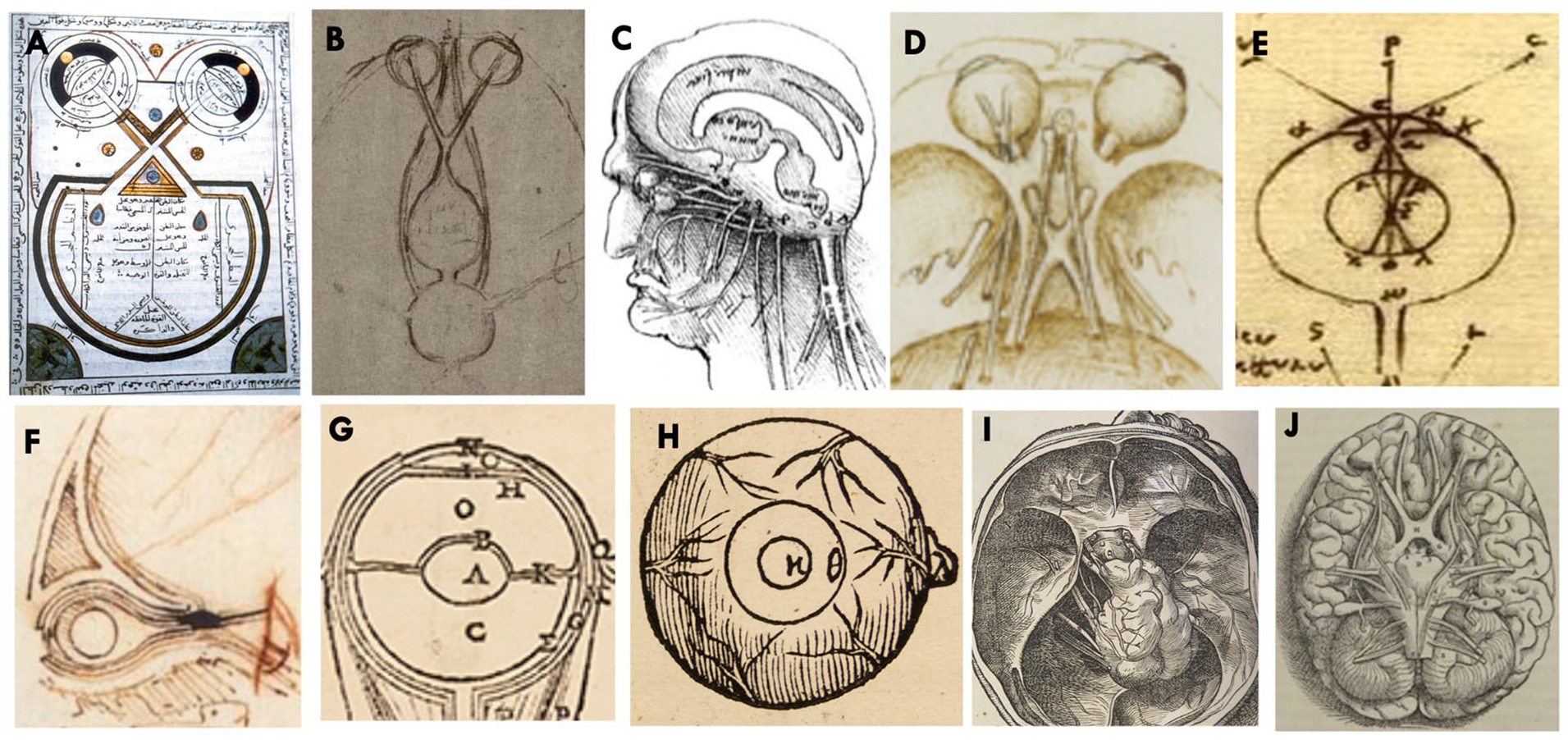
The association of neurologic function and the ventricles prevailed throughout the Middle Ages. It was reiterated by the Persian physician Avicenna (Ibn Sina) in his widely studied Canon of Medicine (Fig. 1A) (Licholai 1995). This association was reproduced in anatomic texts circulating in Italy during the High Renaissance (Fig. 1B–F), including Vesalius’s (Fig. 1H,I) masterwork published in 1543, De Humani Corporis Fabrica (Albertus Albertus 1496; Brunschwig 1512; Hundt 1501; Magnus 1506; Shotwell 2013). While not exactly “popularized,” the study of anatomy was hugely facilitated by the printing press and the introduction of moveable type and high-quality plates.
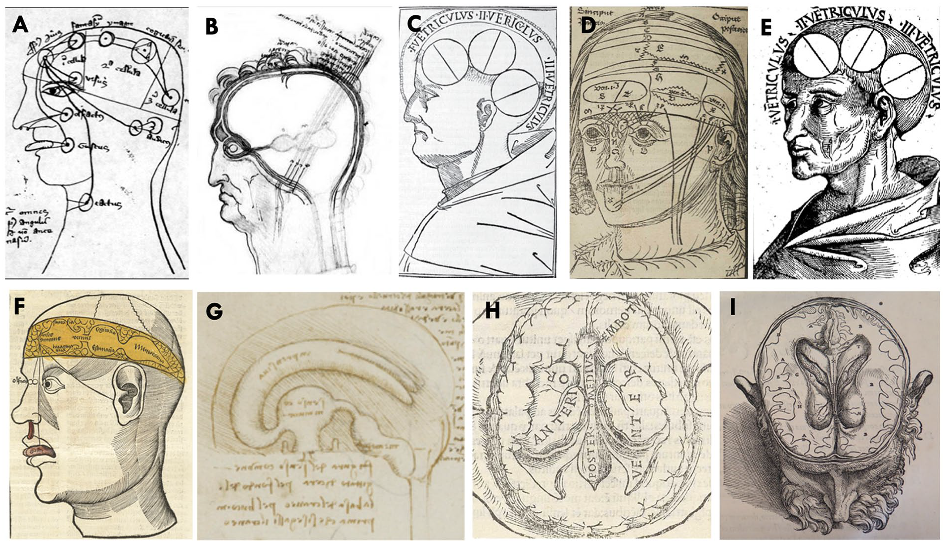
Iconographic evolution of cognitive neurosciences and functional neuroanatomy. (A) Ventricular theory of sensation and perception illustrated in a Latin edition of De generatione embryonis, edited in 1347 and written by the Persian philosopher Ibn Sina between 980 and 1037. (B) Ventricular theory of brain function by Leonardo Da Vinci showing the three cerebral ventricles, following the traditional medieval framework of Ibn Sina, circa 1490. (C) Ventricular theory illustration by Albertus of Orlamünde of 1496. (D) Ventricular theory by Magnus Hundt of 1501. (E) The ventricles from Philosophia Naturalis of Albertus Magnus (1506) with (I) common sense and basic imagination; (II) creative imagination, phantasy, rational thought, and evaluation; and (III) memory and reminiscence. (F) Ventricular theory per Hieronymus Brunschwig in 1512. (G) Representation of the three ventricles with reasonable accuracy by Leonardo Da Vinci circa 1508. (H) Three-dimensional form of the anterior, middle, and posterior ventricles in an axial anatomic cut of the brain by Jacopo Berengario da Carpi in 1523. (I) Axial transection of a human head demonstrating the cerebral hemisphere substance and a detailed anatomy of the surface of the lateral ventricles by Andreas Vesalius in De humani corporis fabrica.
Artists, Dissection, and Neuroanatomy during the High Renaissance
The traditional medieval neuroanatomic framework of the three cerebral ventricles remained dominant through the High Renaissance. By the turn of the 16th century, however, there was widespread interest in extending knowledge of the complex relationship between the human mind and the structure of the brain by direct observation. In 1435, Leon Battista Alberti urged artists to follow Seneca and draw the body as it existed in nature rather than projecting an ideal (Omnis arts imitatio naturae est—All art is [but] an imitation of nature) (Seneca 1917). He suggested an order of painting: first to build up the skeleton and musculature and then to cloak it in clothing (Alberti and Grayson 1972). Better at scholarship, perhaps, than at art, Alberti took holy orders, became a member of the curia, and came to hold great influence during the High Renaissance. In addition to the imitation of nature, he propounded other ideas around humanistic art, including formalisms that may have led to the incorporation of anatomy into paintings.
The role of anatomic dissections in furthering more accurate descriptions of the ventricular system and other brain structures is clearly demonstrated in Leonardo’s anatomy folios of the first decade of the 16th century (Fig. 1G) (Clayton and da Vinci 1992). Leonardo’s interest in anatomy is often linked to his association with artists who carried out dissections. In particular, he was said to have been influenced by Antonio del Pollaiuolo (~1429–1498), a sculptor with a reputation for conducting regular anatomic demonstrations during the time that Michelangelo apprenticed with the sculptor, goldsmith, and painter Andrea di Michele di Francesco de’ Cioni, better known as Verrocchio (1435–1488). Verrocchio’s workshop was adjacent to del Pollaiuolo’s. Leonardo’s practice of dissection was almost certainly a function of his interests in art rather than in science. He probably began dissecting on his own when he moved to Milan in 1483, which, coincidentally, was then a major center of medicine. Subsequently, Leonardo began collaborating with the anatomist Marcantonio della Torre in Pavia.
Michelangelo’s Anatomy
By his mid-teens, Michelangelo was almost certainly aware of public anatomic dissections at the princely court of Lorenzo de’ Medici (Hughes and Buonarroti 1997). By age 18, he had performed his own dissections in the Monastery of Santo Spirito, in the Oltrarno quarter of Florence. Michelangelo’s contemporary, the artist and biographer Giorgio Vasari (1511–1574), wrote [Michelangelo] “was constantly flaying dead bodies, in order to study the secrets of anatomy . . . dissecting men’s bodies in order to see the principles of their construction and the concatenation of the bones, muscles, veins, and nerves, the various movements and all the postures of the human body” (Vasari and others 1998). Ascanio Condivi (1525–1574), an artist best known for his biography of Michelangelo, echoed this report. “Michelangelo,” he wrote, “studied every known animal, and did so many human dissections that it outnumbers that of those who are professional in that field. This is a considerable influence that shows in his mastery in anatomy that is not matched by other painters” (Wohl and others 1976). Michelangelo seems to have wanted no less. In a letter of 1560, to Cardinal Rodolfo Pio of Carpi, he remarked, “Who has not been, or is not a good master of the figure, and especially of anatomy, cannot understand it” (Pearce 2019).
By the early 1500s, when he and Michelangelo both resided in Florence, Da Vinci’s anatomic studies were probably well known (Wilde 1953). Michelangelo also collaborated with Realdo Colombo (1515–1559), a surgeon and master anatomist who succeeded Vesalius as the chair of anatomy and surgery at the University of Padua. Colombo performed countless human dissections and autopsies and extensive vivisection in living animals, replicating (and correcting) some of Galen’s observations (Fye 2002; Taglialagamba 2010). Colombo and Michelangelo planned to collect their contemporary anatomic knowledge in an atlas. This collaboration did not take place because Colombo died in 1559 and Michelangelo died on February 18, 1564. The book, De Re Anatomica, completed just before Colombo’s death, has a single engraving that depicts Colombo dissecting a cadaver.
Over the course of his lifetime, Leonardo claimed to have dissected 30 corpses, generating over 700 drawings and using techniques such as intrathecal injection of beeswax to characterize the ventricles. Notably, he expanded the field of neuroanatomy beyond the ventricles to the brain itself and to the nerves (Clayton and da Vinci 1992). Even so, the extent of Michelangelo’s anatomic expertise has not been accurately established. Unfortunately, only a few of his anatomy studies survived (Eknoyan and de Santo 1997; Elkins 1984). Others may be kept in secret files in the Vatican. It is reasonable to speculate that he was familiar with contemporary anatomic (and to the extent the term is applicable) and physiologic thought and with the works of Leonardo and Colombo. An accurate representation of this knowledge, supported by unprecedently detailed anatomic illustrations, was to be found in Vesalius’s masterpiece De humani corporis fabrica. This book includes detailed illustrations of sequential neuroanatomic prosections representing the ventricles and related brain structures with unsurpassed precision. As best as can be determined, these illustrations accurately represent Michelangelo’s neuroanatomic knowledge and the dissection techniques he may have used. They have remained available to the present.
Neuroanatomic Dissection following Practices of Michelangelo’s Time
De humani corporis fabrica (Basel, 1543) describes a method of sequential dissection of the head (herein called dissection in situ) from the top downward without removing the brain from the skull. This method provides detailed visualization of the ventricular system and brain substance (Fig. 2A Elkins 1984G). The treatise also introduces a new dissecting method (herein called en bloc) in which the whole brain is removed from the skull base, allowing for a detailed visualization of the optic pathways and the ventral brainstem (Fig. 2H–H′).
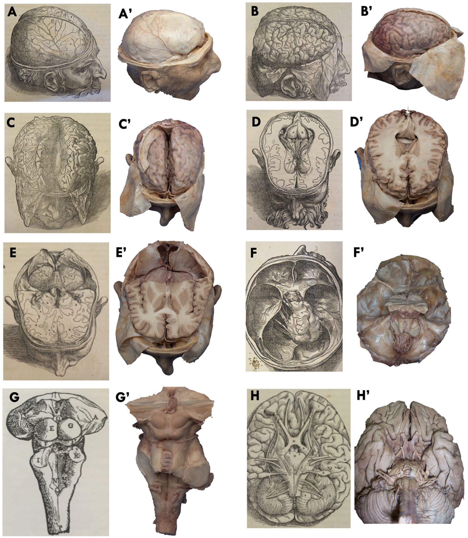
Neuroanatomic dissection reproducing techniques in Vesalius’s De humani corporis fabrica (1543). The in situ technique starts with the circumferential removal of scalp and skull (A–A′), cruciate sectioning and reflection of the dura (B–B′), and exposure of the brain hemispheres and interhemispheric fissure (C–C′). Then, at the top, the cerebral hemispheres are transected axially, revealing the lateral ventricles (D–D′), and the posterior one-third of the cerebral hemispheres are removed, exposing the top of the cerebellum (E–E′), which is also resected along the remaining anterior two-thirds of the cerebral hemisphere, exposing the emerging cranial nerves from the brainstem (F–F′). The dissection is then completed by sectioning the spinal cord and all the neurovascular structures at the skull base, allowing harvesting of the brainstem (G–G′). The en bloc technique described by Vesalius (H–H′) consists of harvesting the whole human brain by severing the cranial nerves from the skull base, including the eyes, followed by the removal of the vascular structures.
Visual analyses of Michelangelo’s work from an anatomic perspective
Separation of Light from Darkness
In Separation of Light from Darkness, the dorsal brainstem and the floor of the fourth ventricle appear to be concealed under the God’s vestments. With care, the superior and inferior colliculi, the pineal gland, the rhomboid fossa encircled by the superior and inferior cerebellar peduncles, and the trochlear nerves crossing below the inferior colliculi can be distinguished (Fig. 3). We think instead that the four bulges represent the tectum or quadrigeminal plate, with two superior and the two inferior colliculi.
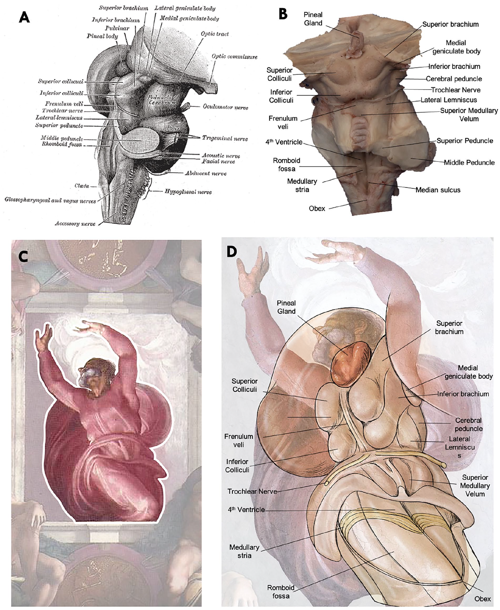
Contextual interpretation of the Separation of Light from Darkness. (A) Anatomic drawing of the dorsal brainstem by Henry Gray and Henry Vandyke Carter, from Anatomy of the Human Body (1918, lamina 681). (B) Photography of the dorsal brainstem in a specimen prepared following the in situ Vesalius dissection techniques with a detailed view of the dorsal brainstem. The most prominent features are the superior and inferior colliculi, the pineal gland, the rhomboid fossa encircled by the superior and inferior cerebellar peduncles, and the trochlear nerves emerging and crossing below the inferior colliculi. (C) Fresco Separation of Light from Darkness by Michelangelo with God’s representation highlighted. (D) Pictorial thesis of Michelangelo’s drawing of God with alleged anatomic structures observed in the dorsal surface of a human brainstem. It is possible to identify all the key and unique structures of the dorsal brainstem surface anatomy, including the pineal gland sitting on top of the brainstem, as well as bilateral superior and inferior colliculi with the cerebral peduncles laterally. Under the inferior colliculi, the trochlear nerves are clearly identified emerging in a crossed fashion and projecting around the midbrain ventrally. Caudal to this, the superior medullary velum partially covers the rhomboid fossa and floor of the fourth ventricle. The cerebral aqueduct can be seen as a conduit that runs vertically between the colliculi.
The dorsal brainstem and the fourth ventricle were of upmost importance in the Galenic doctrine of neurophysiology, which was supported by extensive vivisection experimentation. In this regard, Galen observed in De usu partium (On the usefulness of the part of the body), “And when one presses down upon that ventricle which is found in the part of the brain lying at the nape of the neck, then the animal falls into a very heavy and pronounced stupor” (Rocca 2008). As previously noted, Galen’s ventricular theory of brain function was popular during Michelangelo’s time, and therefore, he was likely familiar with the dorsal brainstem and the fourth ventricle’s function of arousal control that, when damaged, caused coma or even death.
Creation of the Sun and the Moon
Creation of the Sun and Moon is dominated by the figure of God, arms outstretched laterally, giving shape to the Sun and the Moon. The extended arms are suggestive of the optic nerves connecting both eyes to the brainstem, through the optic chiasm and tracts, and it is possible to infer the surface anatomy and internal structure of the eyes in the sun and in the moon (Fig. 4).
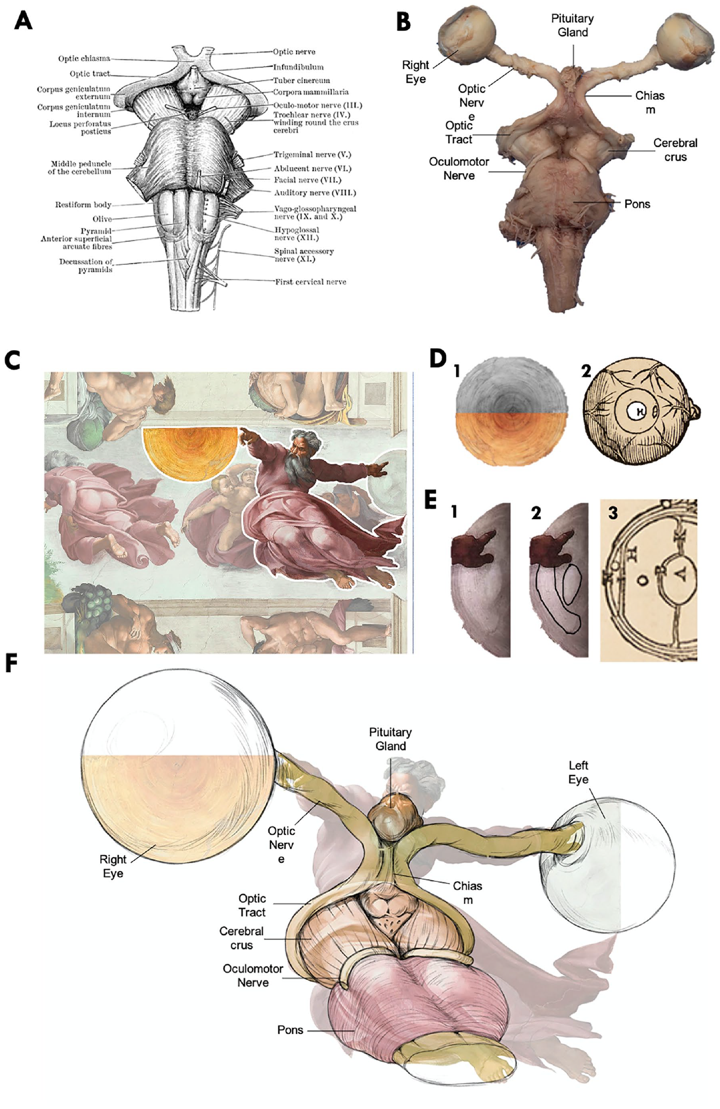
Contextual interpretation of the Creation of the Sun and Moon. (A) Anatomic drawing of the ventral brainstem by Henry Gray and Henry Vandyke Carter, from Anatomy of the Human Body (1918, lamina 681). (B) Photography of the ventral brainstem and optic pathway in a specimen prepared following Vesalius’s dissection techniques. (C) Fresco Creation of the Sun and Moon by Michelangelo with God’s representation creating the planets highlighted. (D) The reconstruction of the complete solar sphere through specular imaging replication (gray-scale) results in a circumferential pattern (1) that perfectly matches the “idealized depiction of the shapes and sizes of the parts of the eye” of Vesalius, which can be better appreciated in the specular image with gray-scale rendering (2). (E) On the surface of the moon (1), it is possible to identify a concave structure with an underlaying biconvex structure resembling the shape and positions of the cornea and the crystalline lens (2), as represented in Vesalius’s work (3). (F) Neuro-ophthalmologic interpretation of the fresco with God’s arms representing optic nerves connecting the eyes in the form of the sun and the moon and merging in the chiasm before entering the top of the brainstem represented by God’s body.
The association of the right eye with the sun and of the left eye with the moon goes back to Pharaonic Egypt (the Eyes of Ra and Horus). According to Galen, visual perception was achieved by the flow of “light-like” pneuma from the cerebral ventricles through hollow nerves connecting the eyes with the brain (Besser 2014; Ierodiakonou 2014). Galen taught that the organ of sight was made “luminous” to see objects and then by flowing back to the brain to have cognition and understanding of the surroundings. The iconography representing the eyes connected to the ventricles via hollow optic nerves crossing in the midline may date back at least 1000 CE (Fig. 5A).
Iconographic evolution of the organ of sight. (A) Islamic treaty of ophthalmology dated at least 1000 CE representing the optical pathway. (B) Leonardo da Vinci drawing from 1487 representing the optic nerves starting in the orbits and projecting to the anterior ventricular cavity. (C) Leonardo Da Vinci drawings of a more anatomically accurate ventricular system circa 1506–1507 with three ventricles are labeled imprensiva (anterior ventricle, corresponding to the paired lateral ventricles), senso comune (third ventricle), and memoria (posterior or fourth ventricle). Below the ventricles, seven pairs of cranial nerves are shown, with a representation of the optic pathway connecting the eyes with the base of the brain and ventricular system. (D) Da Vinci had an in-depth anatomic knowledge of the optical pathway and in 1508 accurately depicted a cross section of the skull, revealing the eyeballs, optic nerves, and the x-shaped optic chiasm formed by the optic nerves before they reach the brain. (E, F) Eyeball drawings of Da Vinci including the internal structures and highlighting the connection with the ventricular system. (G) Vesalius’s cross-sectional representation of the internal structure of the eye. (H) Vesalius also included in De humani corporis fabrica drawings of the superficial anatomy of the eyes with “idealized depiction of the shapes and sizes.” (I) Illustrations from Vesalius depicting the optic nerve arriving to the intracranial compartment from the orbits and entering into the top of the brainstem as visualized by a top-to-bottom dissection technique. (J) Anatomy of the visual pathway by Vesalius including the eyes, optic nerves joining in the chiasm, and the optic tract entering the brainstem at the ventral surface of the brain.
Eyesight had a special meaning for Leonardo and his contemporaries as visual perception enabled the observation of phenomena in the natural world. In 1508, he dedicated an entire manuscript to the study of eyesight and depicted with precision a cross section of the skull base, revealing the eyeballs, optic nerves, and the H-shaped optic chiasm formed by the optic nerves and tracts before they reach the brain (Fig. 5B–D). Leonardo also included several drawings of the eyeballs represented by two concentric circles and a hollow channel communicating with the anterior ventricle (Fig. 5E,F). This depiction was further expanded by Vesalius, who better characterized the internal structure of the eye through depictions of idealized shapes and sizes of the globe (Fig. 5G,H) and who also developed the en bloc techniques to improve the visualization of the visual pathway entering the top of the brainstem (Fig. 5I,J) (Reeves and Taylor 2004).
The “Sun—Right Eye” in the fresco was painted as an anteroposterior view, while the “Moon—Left Eye” was painted as a sagittal section, with the cornea, lens, and other intraocular anatomy visualized. In this fresco, the ventral brainstem, revealed by en bloc dissection, is hidden under God’s robes, with the mammillary bodies, the cerebral crus, and pons.
Creation of Adam
Creation of Adam inspired the first modern analysis of Michelangelo’s anatomic drawings. Meshberger concluded that Michelangelo introduced a mid-sagittal cross section of the brain (Meshberger 1990) in the image surrounding God and the angels. The large drapes resemble the contour of the human brain and a “cerebral mantle,” which in our interpretation encloses the ventricular cavities (Fig. 6). Vesalius demonstrates dissections of the cerebral hemispheres along the interhemispheric fissure followed by a combination of axial and sagittal sections revealing the cerebral anatomy (Fig. 2C,D). Leonardo’s folios of 1506–1507 also show a three-dimensional representation of the ventricles overlaying a human brain (Fig. 1G) (Clayton and da Vinci 1992). Michelangelo would likely have been familiar with these depictions.
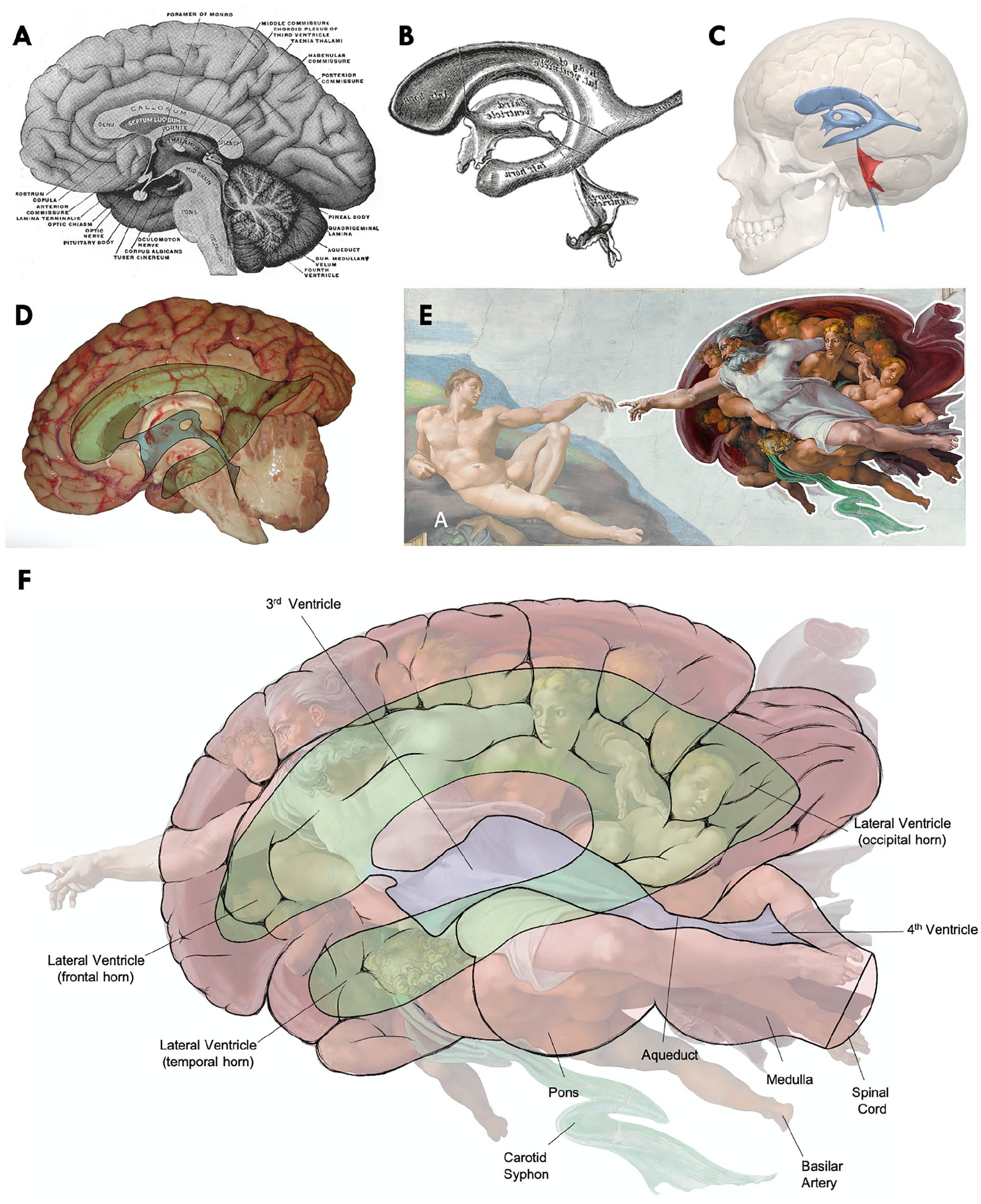
Contextual interpretation of the Creation of Adam. (A, B) Anatomic drawing of the mid-sagittal cross section of a human brain and a three-dimensional representation of the ventricular system by Henry Gray and Henry Vandyke Carter, from Anatomy of the Human Body (1918). (C) Three-dimensional depiction of the ventricular system overlaying a human brain. (D) Mid-sagittal cut of a fresh human brain with an overlay of the ventricular system. (E) Michelangelo’s representation of Adam and God surrounded by angels in the fresco Creation of Adam circa 1511. (F) Composite pictorial thesis suggesting that the image surrounding God and the angels is actually a depiction of the ventricular system and a mid-sagittal cross section of a human brain. In our interpretation, the large drapes resemble the human cerebral cortex, or mantle, and appear to enclose the lateral and third ventricular cavities.
The Last Judgment
The Last Judgment appears to project a coronal section of a human brain including the ventricular system by means of the fresco’s central image of Jesus surrounded by the Virgin Mary, saints, and apostles (Fig. 7). The blue spaces that highlight the central location of Jesus may actually represent the humor within the lateral and the fourth ventricle. The third ventricle, seemingly situated between the open legs of Jesus, is probably a blue loincloth added by Daniele da Volterra on papal insistence circa 1565 as an embarrassing example of painterly censorship (Fig. 1D) (Ashford 2020). A similar pattern was identified by de Bonis and others (2019) in the vault of San Giorgio’s Cathedral in Ferrara, Italy. That image is found in the main body of the Universal Judgment painted by Michelangelo’s pupil Bastianino (1528–1602), in which Jesus appears to have been situated in the third ventricle once again, only in a sagittal cross section of the brain. Bastianino later moved to Ferrara to paint for Canani (1515–1579), a renowned anatomist.
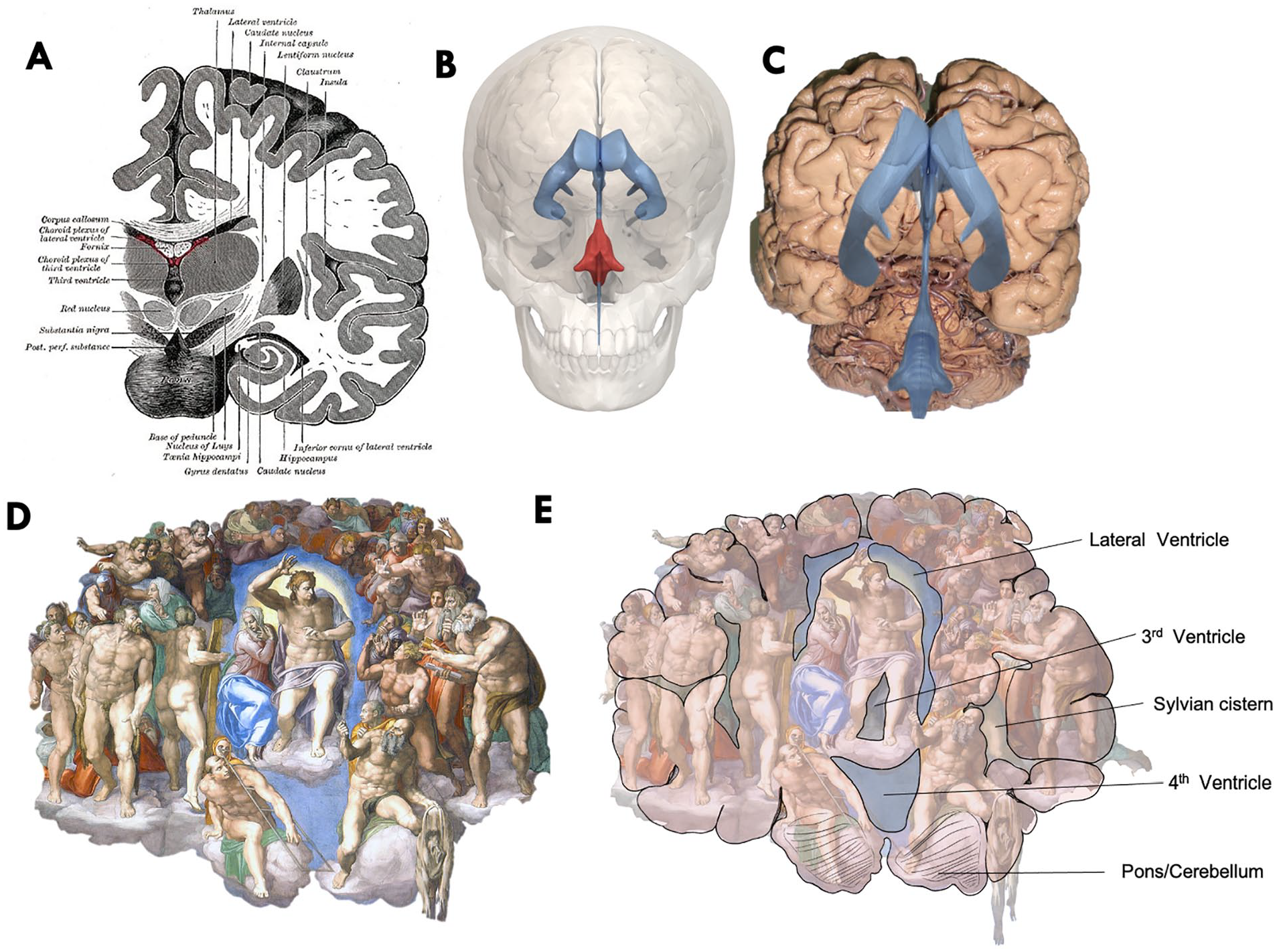
Contextual interpretation of The Last Judgment. (A) Anatomic drawing of a coronal section of a human brain by Henry Gray and Henry Vandyke Carter, from Anatomy of the Human Body (1918). (B) Three-dimensional depiction of the ventricular system of a human brain. (C) Anterior view a whole brain as removed by Vesalius’s en bloc technique with an overlay of the ventricular system. (D) Michelangelo’s representation of Jesus surrounded by the Virgin Mary, saints, and apostles in the fresco The Last Judgment (1536–1541). (E) Composite pictorial thesis suggesting that the image surrounding Jesus is actually a depiction of the ventricular system embedded within the brain. In our interpretation, the blue-colored spaces to highlight the central location of Jesus actually represent the lateral and the fourth ventricle as observed from a front view of the brain. The third ventricle, which we located in between Christ’s legs, is not a blue space but actually a blue loincloth. This was likely added by Daniele da Volterra shortly after the Council of Trent circa 1565 in an attempt to cover the genitals and backsides in this fresco as nudity in religious art had been condemned.
It is noteworthy that a bold and bearded man near the center of the image in Judgment holds a knife in his right hand and a flayed skin in his left. Unlike most of the characters in the scene, this man is contemplating Jesus’ left leg, as if thinking about a dissection. Jesus, in turn, seems to gaze at him in preference to the others (Fig. 8A–C). Traditionally, this figure was interpreted as representing Saint Bartholomew holding his own flayed skin as evidence of his martyrdom, but certain traditions in the iconography of Bartholomew, including the color and quality of his hair, seem to contravene this conclusion. Moreover, the flayed skin belongs to a person with different facial hair pattern than that of the bold man with a knife. A more plausible suggestion is that Michelangelo intended to depict anatomist Realdo Colombo, the anatomy professor at Padua, whose strikingly similar portrait is displayed on the front cover of De re anatomica (Fig. 8E–H) (Fye 2002).
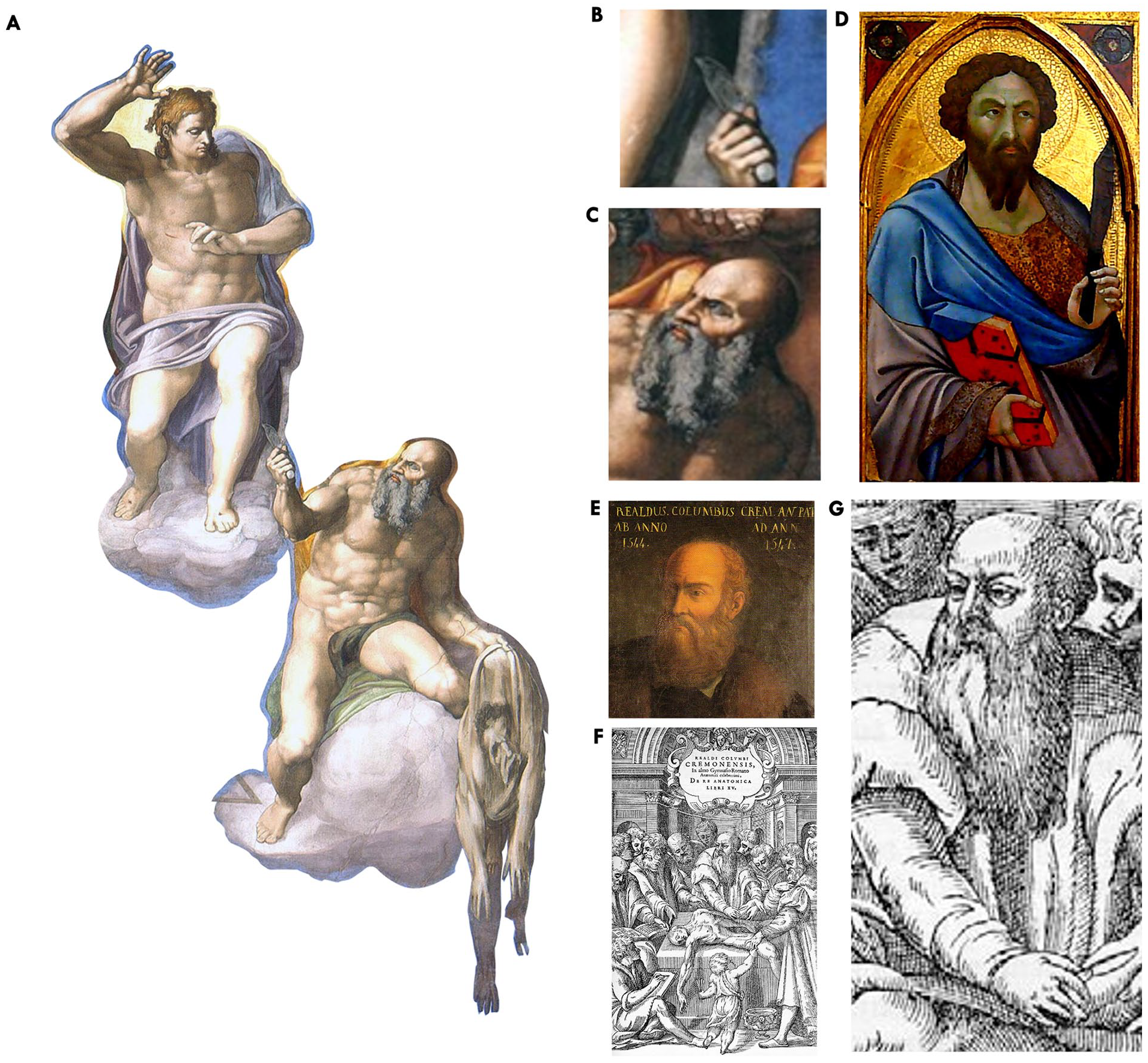
Michelangelo’s code of universal truth: religion and science. (A) Michelangelo’s representation of Jesus surrounded by the Virgin Mary, saints, and apostles in the fresco The Last Judgment (1536–1541). In this fresco, Jesus is staring at a man with a knife on his bottom left, and this man is staring at Christ’s leg. (B) Expanded view of the “flying knife” nearly touching the skin of Christ’s leg. (C) The man with a knife and holding his own skin was traditionally thought to represent Saint Bartholomew. (D) Tempera on wood by Bartolomeo Bulgarini (active 1337–1378) showing the phenotype of Saint Bartholomew at the time of his martyrdom as a middle-aged man with black and crisped hair and a thick beard. This facial phenotype is aligned with descriptions of Saint Bartholomew in the Golden Legend (collection of hagiographies widely read in the late medieval Europe that was compiled around the year 1260): “His hair is black and crisped, his skin fair, his eyes wide, his nose even and straight, his beard thick and with few grey hairs.” (E) Official portrait of Realdo Colombo during anatomy professorship in Padua (1544–1547). (F) Title page from Realdo Colombo’s De re anatomica (1559) designed by Paolo Veronese and showing Colombo performing a dissection. (G) Expanded view of Colombo’s face of the front cover of the book De re anatomica. Colombo’s knife, facial phenotype, and right-handedness are remarkably similarly to the man with the knife in The Last Judgment.
Interestingly, another flayed man is appreciated in the anatomy book Historia de la composición del cuerpo humano, published in Rome in 1556 by Juan Valverde de Amusco (Fig. 9). Amusco studied medicine in Padua under Colombo and commissioned Michelangelo’s pupil Gaspar Becerra (1520–1570) to provide the original illustration for this book, which again demonstrates how intertwined was Michelangelo’s life and work with the great humanistic artists and anatomists of his time.
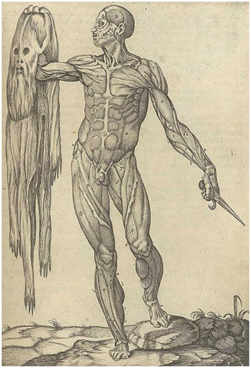
Amusco’s “flayed man.” In the book from Juan Valverde de Amusco’s Historia de la composición del cuerpo humano (1556), it is possible to identify a flayed man holding up his skin in one hand and a knife in the other as a symbolism of scientific experimentation of the human body.
Discussion
The purpose of this study is to review and reconsider certain examples of neuroanatomic imagery in the Sistine Chapel and provide contextual interpretations to support the presence of philosophical meanings that might have been expressed in encrypted neuroanatomic visual language. Michelangelo’s paintings most likely reflect his education and intellectual maturation in the neo-Platonist environment of the Florentine court of the de Medicis and northern Italy more broadly between 1489 and 1496.
Michelangelo’s symbolism has been the focus of intense study for many years. Several scholars have suggested that the frescoes in the Sistine Chapel symbolize more than simply biblical depictions alone. The possibility of encoded references to human anatomy has been repeatedly raised.
The acclaimed conservation project directed at the restoration of the vault and the altar wall of the Sistine Chapel around 2000 elucidated new details to support this possibility. While the risks of intentionality (the fallacy of attributing intention to the artist retrospectively on the basis of what can be seen currently) and anachronism (the error of interpreting a work of art in light of information unknown at the time the work was created) are all but unavoidable, the anatomic allusions and insinuations, once brought to light, are almost inescapable (Wimsatt and Beardsley 1946).
It is helpful to distinguish between the idea of a code as something hidden, which may be its purpose, and the idea of a code as a special way of transmitting information to those who know how to read it, akin to the way Morse Code was used. The term encoded does not necessarily mean “concealed” or “disguised.” Information or data might be encoded for emphasis, for efficiency of transmission, or for nuance. Thus, a reader may be instructed to pay particular attention to a word or a phrase by a difference in color, in size, in font, in emphasis (e.g., underlining), or in position. Each of these is an example of encoding. Indeed, by the middle of the 18th century, the Bible and eventually other books were printed or embellished by hand in two colors for purposes of emphasis as well as for decoration, thereby continuing the much older tradition of using illumination for distinguishing important letters and words and for encoding important ideas (Peddie 1914).
Which meaning of encoded, if either, applies to the putative but still convincingly anatomic codes of the Sistine Chapel? One explanation, for example, would dismiss the anatomic references in their entirety as nothing more than an exhibition of artistic virtuosity, which would not have been taken amiss at the time (ItalianRenaissance.org 2012). That seems overly severe an interpretation of the imagery. So long as it is understood that the attribution of codes to these frescoes must be deemed hypothetical from a strictly historical perspective in the absence of better corroborative evidence, it is fair to conclude that they are at least consistent with, if not exemplary of, the anatomic ideas and preoccupations that emerged during the High Renaissance and the artistic expression of the time.
But even so, and beyond speculation, apophenia, pareidolia and biologic reductionism, there is still compelling evidence of the anatomic symbolism in Michelangelo’s Sistine frescoes. Michelangelo strategically wove “double images” into the frescoes. Are the double images a code? The paintings are certainly replete with anatomic structures with subtle semiotic significance. In Separation of Light from Darkness, we see a dorsal brainstem and fourth ventricles under God’s robes. In Creation of the Sun and Moon, we identified the visual pathways and the ventral brainstem. And, as previously suggested, God and the surrounding angels in Creation of Adam seem to represent a mid-sagittal section of the brain, and Jesus surrounded by the Virgin Mary, saints, and apostles in The Last Judgment form the structures seen in a coronal brain section.
In each of these frescoes, large anatomic structures occupying the majority of the scene constitute a common stylistic pattern. Moreover, there seems to be a purposeful link between the anatomy and function of the structure (as it was understood at the time) and the theme of the painting.
Eknoyan, for example, argued that Michelangelo put a shape of a bisected right kidney concealed in the mantle of the Creator in the Separation of the Land from the Waters (Eknoyan 2000). Michelangelo likely suffered from nephrolithiasis. Whether as a function of his condition or not, his use of the renal outline in a scene representing the separation of solids (land) from liquid (water) could well have been purposeful and symbolic. Analogous allegories can be reasonably identified in Separation of Light from Darkness in the form of the inclusion of the dorsal brainstem and the floor of the fourth ventricle to symbolize the awakening of the human mind from darkness and in the depiction of the visual pathway in the Creation of the Sun and Moon to symbolize the capacity of the human mind to sense the environment.
Thus too, the coronal section of the brain highlighting the lateral and third ventricles in The Last Judgment may be a symbol of the capacity of human beings to interpret and judge the environment based on the ventricular theory of brain functioning. In addition, as previously proposed by Meshberger, the mid-sagittal depiction of the brain in the Creation of Adam may symbolize the ability of human mind of thought (Strauss and Marzo-Ortega 2002). The Last Judgment can be interpreted both as an allegory to the bestowal of Divine wisdom upon man but also as a coded inquiry directed at the question about who created whom. In the latter interpretation, Adam (who has an umbilicus) is creating God for humanity by using his intellect.
While Michelangelo was a devout and practicing Catholic, the fresco may belie the potentially heretical attempt to reconcile the centrality of humanity and the terrestrial immanence of the Christian god. It might also be interpreted as an illustration of man and God communicating directly without the intermediation of the Church. This very threatening idea was the essence of the allegation of Spiritualism leveled against Michelangelo toward the end of his life by Pope Paul IV and represented, for the time, a near heretical belief.
Finally, rather than a man’s martyrdom, the inclusion of Colombo, the anatomist, “at work” in The Last Judgment may symbolize the expansion of scientific knowledge to better interpret and judge the environment based on scientific experimentation and direct observation, in this case through anatomic dissections. The use of scientific in this context may be historically premature, but some experimentation and careful observation certainly took place. The dissection of Jesus has a powerful symbolic message, as it manifests the human nature of Christ with a body made of flesh and bone to be discovered. The search for the “universal truth” may be here encoded through the divine, the human, and the scientific all at once. Coincidentally or intentionally, few years after completing The Last Judgement Michelangelo removed the left leg of Jesus in the The Deposition (the “Florentine’s pieta”). Is this “missing leg” at Padua’s anatomical theater?
Michelangelo asked not to buried in the Vatican, and eventually his cadaver was spirited away for burial within Santa Croce by his Florentine friends. Was this because of his discomfiture with the pope? We cannot tell, but this part of the story may point to why that might have been his preference (Fields 2010).
Conclusions
A significant number of anatomic references and representations can be imputed to Michelangelo’s frescoes in the Sistine Chapel. These deft and potentially encoded visual allusions are not obvious. They must be sought out. Our analysis, undertaken from the perspective of neuroscientists, suggests that the iconographical encryption is neither incidental, nor mischievous, but rather carefully allegorical. It is largely recognizable to the trained neuroscientific eye, although whether this code is intended as encryptation is another question altogether.
We hope this anatomic interpretation of Michelangelo’s imagery will add a new and accessible scientific dimension to the glory of the Sistine Chapel. The hidden elements might just reflect a flourish of virtuosity and a mastery of anatomy, but they almost certainly no less allude to a chain of significant tropes. Michelangelo’s genius combined references complex relationships with Catholic faith, a Church wounded by the Reformation, and ideas about religious orthodoxy that could not be easily navigated in his times. While we cannot fully fathom all if his intentions, the application of 21st-century imaging techniques to the sublime figures that float across the Sistine’s vault bestow a sharper focus on Michelangelo’s masterpiece and the mind of the artist.
Footnotes
Declaration of Conflicting Interests
The author(s) declared no potential conflicts of interest with respect to the research, authorship, and/or publication of this article.
Funding
The author(s) received no financial support for the research, authorship, and/or publication of this article.