
Abstract
Although intracerebral hemorrhage (ICH) and cerebral small vessel disease (cSVD) have long been considered distinct clinical entities, emerging evidence reveals significant overlap in their etiologies and imaging markers. This review aims to explore the relationship between ICH and cSVD, suggesting that ICH may represent an acute manifestation of small vessel disease. ICH is primarily caused by cerebral amyloid angiopathy and hypertension, while cSVD is mainly attributed to cerebral amyloid angiopathy and arteriolosclerosis. Hypertension-induced arteriolosclerosis is one of the most common pathologic changes in cSVD. This overlap in etiology suggests a close relationship between ICH and cSVD. In patients with ICH, multiple imaging markers of cSVD are often observed. Recent studies suggest that enlarged perivascular spaces, one of the imaging markers of cSVD, may serve as a pathway for hematoma expansion. Additionally, diffusion-weighted imaging lesions are frequently observed in patients with ICH. These lesions are likely to be based on underlying cSVD and may evolve into other cSVD markers, such as white matter hyperintensity, lacunar infarctions, or microbleeds. These findings highlight the complex interplay between ICH and cSVD, suggesting that ICH could be considered an acute expression of cSVD rather than an entirely separate entity.
Keywords
Introduction
Intracerebral hemorrhage (ICH) is a devastating form of stroke characterized by nontraumatic bleeding within the brain parenchyma. ICH has a mortality rate exceeding 40%, and only 20% of patients remain functionally independent at 6 months after onset of the disease (Broderick and others 2021). With further research on ICH, the intricate relationship between ICH and cerebral small vessel disease (cSVD) has gained more and more attention. ICH is no longer viewed as a solitary event of vascular rupture. Indeed, it is a direct manifestation of the cumulative effects of long-term, chronic cSVD. In recent years, we have come to recognize that ICH may represent an acute manifestation of the underlying pathologic and physiologic processes of cSVD.
The purpose of this review is to provide a detailed account of the connection between ICH and cSVD, including the pathophysiologic mechanisms, imaging manifestations, pathologic changes, and long-term prognosis of patients with ICH. By elucidating these aspects, we aim to illustrate the complex association between ICH and cSVD, thereby contributing to a better understanding and management of ICH.
Nature of cSVD: Chronic and Progressive
cSVD is a condition that affects the brain’s small perforating arterioles, capillaries, and likely venules, leading to various lesions observable through pathologic examination or brain imaging techniques such as MRI or CT (Wardlaw and others 2019). See box for details on the structure and common pathologies of small vessels, as well as their comparison with large vessels. cSVD has a diverse range of causes, and a concise etiologic classification system has been proposed, categorizing the causes of small vessel disease into six main classes: arteriolosclerosis, sporadic or hereditary cerebral amyloid angiopathy (CAA), congenital or hereditary CAA distinct from CAA, inflammatory or immune-mediated small vessel disease, venous collagen disease, and other causes (Table 1; Pantoni 2010). Hypertensive arteriolosclerosis–related cSVD and CAA represent the two most prevalent subtypes of cSVD. Previous studies indicate that hypertensive arteriolosclerosis–related cSVD accounts for 50% to 60% of cSVD cases, predominantly in individuals with long-standing hypertension, whereas CAA contributes to 20% to 25% of cases (Jäkel and others 2022; Petrea and others 2020).
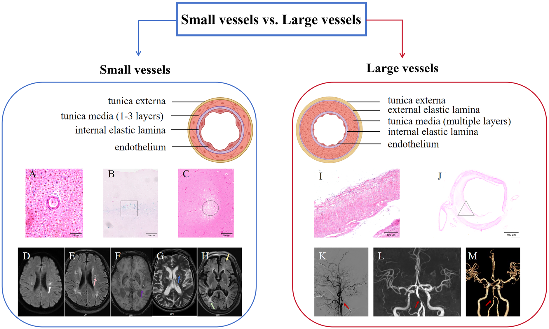
Comparative schematic of small vessels vs large vessels. Left panel: small vessel characteristics. Right panel: large vessel characteristics.
Major Causes and Characteristics of Cerebral Small Vessel Disease.
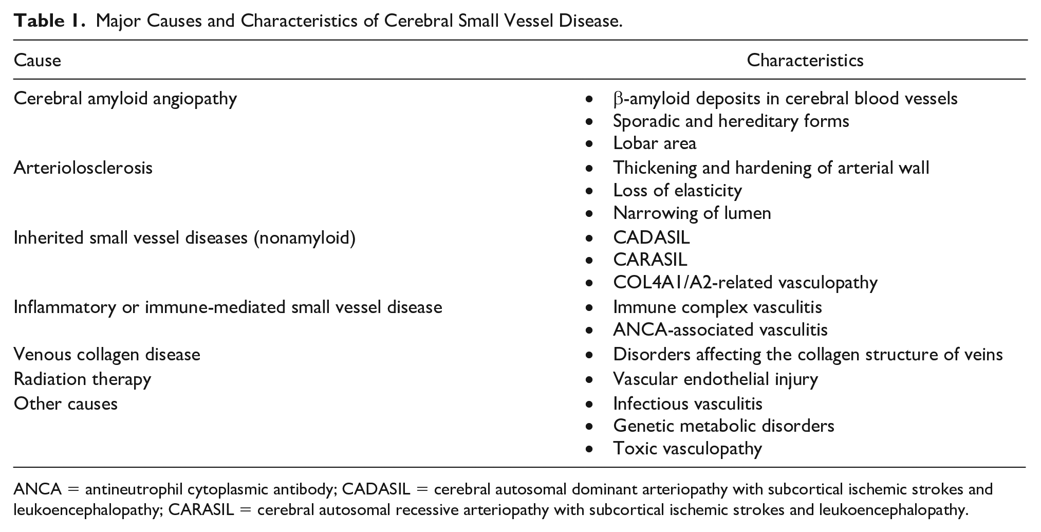
ANCA = antineutrophil cytoplasmic antibody; CADASIL = cerebral autosomal dominant arteriopathy with subcortical ischemic strokes and leukoencephalopathy; CARASIL = cerebral autosomal recessive arteriopathy with subcortical ischemic strokes and leukoencephalopathy.
Arteriolosclerosis is known as small artery sclerosis or hypertensive small vessel disease. From a pathologic perspective, the characteristics of this type of small vessel disease mainly include the loss of smooth muscle in the vascular media, deposition of fibrous material, thickening of the vessel walls, and narrowing of the lumens. Small artery sclerosis type of cSVD has a direct causal relationship with cerebral hemorrhage, as it is a significant risk factor for the latter. The mechanism by which hypertensive small vessel disease causes cerebral hemorrhage can be summarized as follows:
Early stage: Long-term hypertension leads to endothelial dysfunction and increased blood-brain barrier (BBB) permeability.
Intermediate stage: Vascular smooth muscle cells proliferate, leading to thickening and fibrosis of the vascular walls, characterized by the formation of fatty streaks and fibrous plaques.
Late stage: Vascular remodeling occurs, with the lumen gradually narrowing, ultimately culminating in vascular occlusion or rupture (Banerjee and Chimowitz 2017; Hainsworth and others 2024).
This is a long-term chronic accumulation process. cSVD may remain asymptomatic for many years, with only occasional changes detected through imaging examinations (Chojdak-Łukasiewicz and others 2021). A 4-year follow-up study on patients with hypertension found that periventricular white matter hyperintensity (WMH), a typical biomarker of cSVD, progressed over time and was associated with the occurrence of cognitive impairment (Jiménez-Balado and others 2019). In a cohort study with a median follow-up of 6.5 ± 1.4 years, 10% of individuals experienced new microbleeds, 5% developed new cavities, and 21% had progressive expansion of the basal ganglia perivascular space (PVS; Del Brutto and others 2022).
However, recent research challenges the traditional view of deep perforating arteriopathy (often termed hypertension-related vasculopathy), suggesting a more complex pathophysiology. Using mendelian randomization, Koohi and others (2024) concluded that thrombosis is not the primary etiology of lacunar stroke and cSVD (Falcone and Woo 2024). This raises the question: what triggers cSVD development? Kremer and others (2025) proposed endothelial cell dysfunction as the core pathologic mechanism in cSVD, potentially outweighing traditional cardiovascular risk factors. Rajani and others (2018) demonstrated that cSVD can occur independently of hypertension: they observed reduced CLDN5/eNOS preceding hypertension onset in spontaneously hypertensive stroke-prone rats and noted endothelial proliferation with CLDN5 reduction in normotensive Atp11b-knockout (Atp11b-KO) rats. Through genetic analyses, Sun and others (2025) established causal roles for proteins (COL2A1, EPHA2, FLT4) in cSVD, identifying two core pathogenic pathways: endothelial-platelet activation and complement-mediated inflammatory regulation. These findings collectively provide a foundation for more targeted cSVD therapies, such as anti-inflammatory or antiplatelet agents.
Sporadic or hereditary CAA is the second-leading cause of cSVD. Amyloid β (Aβ), particularly Aβ40 and Aβ42, deposits in the small blood vessels of the leptomeninges and cerebral cortex, leading to vascular wall damage through a series of pathophysiologic processes, resulting in imaging manifestations or clinical symptoms (Koemans and others 2023). This process can be categorized into four stages: Aβ deposition on the vascular wall, pathologic changes in the vascular wall, nonhemorrhagic brain injury, and manifestation of cerebral hemorrhage. The exact triggering factors for Aβ deposition on the vascular wall are still unclear, but they may be associated with age, APOE2 or APOE4 genotypes, and other risk factors, leading to decreased vascular reactivity and loss of vascular smooth muscle cells. Studies on transgenic mice have indicated that the reduction of vascular smooth muscle cells is associated with impaired clearance function of brain interstitial fluid around blood vessels (Arbel-Ornath and others 2013), which may manifest as enlarged PVSs (EPVSs) on imaging studies. In the final stage of CAA, cerebral hemorrhage occurs, which can present as acute large hematomas with a diameter exceeding 1 cm, or as cerebral microbleeds (CMBs), cortical superficial siderosis (cSS), and convexal subarachnoid hemorrhage.
cSVD is fundamentally characterized by its chronic and progressive nature, which may extend over a period of decades before clinical manifestations become apparent (Wang and Liu 2023).
Triggering of ICH: From Static to Dynamic
Identify the Origin of Hematoma
Fisher’s (1971) histopathologic paradigm fundamentally redefined ICH as a dynamic manifestation of cSVD. Through systematic analysis of pontine and putaminal hemorrhages, three pillars of cSVD-driven ICH pathogenesis emerged:
Structural vulnerability of hypertensive arteriopathy: Identification of 24 arteriolar rupture points (40-150 μm) colocalized with fibrinoid necrosis and lipohyalinosis—pathognomonic features of hypertensive cSVD.
Multifocal decompensation cascade: Perihematomal fibrin-platelet aggregates provided direct evidence of secondary microvascular recruitment (<100 μm), conceptualizing ICH as a self-propagating cSVD crisis.
Hemodynamic tipping thresholds: The equilibrium between hematoma expansion and tissue counterpressure.
From Fisher’s research, we can see that spontaneous ICH should be positioned as the acute peak of chronic cSVD progression rather than an isolated event.
EPVS: A Potential Pathway of Hematoma Expansion?
Due to the technological limitations of Fisher’s era, the pathways of hematoma expansion could not be explored. A study based on artificial hematoma has provided a new direction for thought. It demonstrated that artificial hematomas spread along the ruptured penetrating arteries and PVSs, proximally and distally. This spread led to the separation of vascular branches from adjacent neural tissue, causing tissue damage and secondary extravasation of blood. Furthermore, some contrast agents extravasated and penetrated into the PVSs surrounding the nonruptured perforating arteries, creating additional sites of extravasation and exacerbating the expansion of the artificial hematoma (Rzepliński and others 2022). PVSs disappear at the level of the anterior capillary artery (Schaeffer and Iadecola 2021). This anatomical feature means that as a hematoma spreads along these spaces toward the distal branches, it may cause separation between the small arteries and surrounding tissue due to mechanical pressure (Figure 1). This interruption of blood supply irreversibly damages the structure of adjacent neural tissue and leads to secondary bleeding.
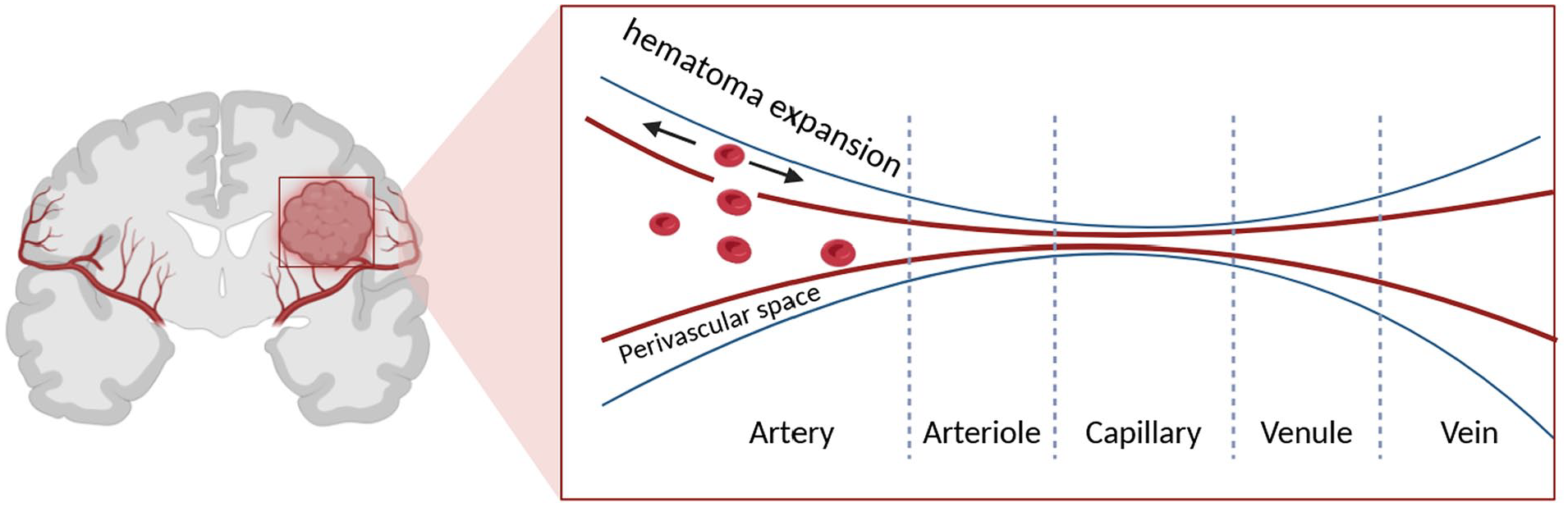
Schematic diagram of enlarged perivascular spaces as a potential pathway for hematoma expansion.
Therefore, PVSs play an important role in the process of hematoma expansion. Excitingly, EPVS is a major imaging marker of cSVD. The existence of EPVS seems to have already provided a pathway for hematoma expansion before the occurrence of cerebral hemorrhage. A study using a rat model of cerebral hemorrhage proposed the “sink phenomenon,” which confirmed the phenomenon of hematoma expansion along the PVSs. The research indicated that blood in a fluid state flowed under pressure differentials into structurally loose tissue spaces and could spread to the subarachnoid space, resulting in secondary subarachnoid hemorrhage, or into the ventricular system, causing secondary intraventricular hemorrhage.
The pathologic studies of cerebral hemorrhage allow us to figure out what happened at the moment of blood vessel rupture. The results of the aforementioned studies indicate that cerebral hemorrhage occurs on the basis of fragile cerebral small blood vessels. In other words, ICH and its subsequent development may be acute manifestations of cSVD.
Diffusion-Weighted Imaging Lesions in ICH
Diffusion-weighted imaging lesions (DWILs) are defined as high signal alterations on diffusion-weighted imaging (DWI) in the region surrounding and distant from the hematoma after ICH (Figure 2; Yang and others 2022).
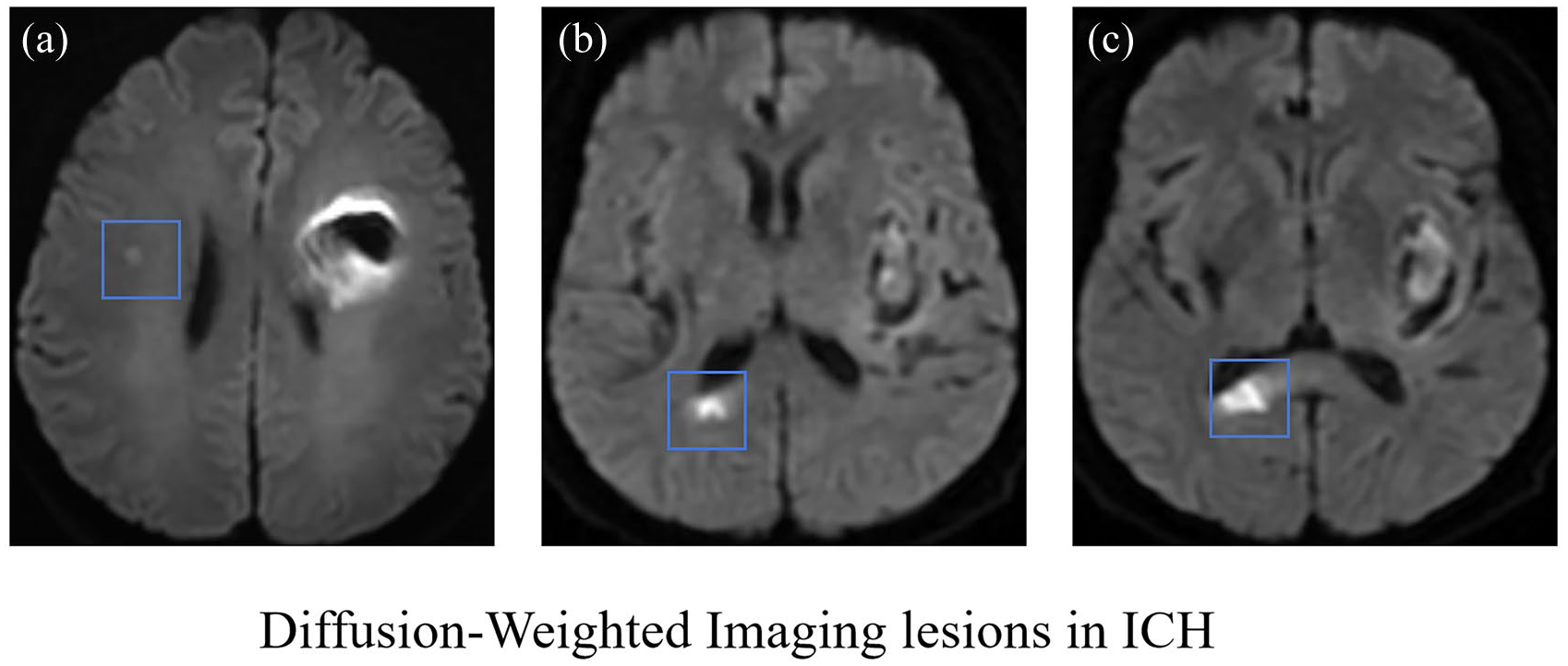
Diffusion-weighted imaging lesions (blue boxes) in intracerebral hemorrhage.
Currently, there are different explanations for the causes of DWILs. Previous studies have suggested that a precipitous drop in blood pressure, microvascular diseases, prethrombotic state, stress hyperglycemia, and EPVS could contribute to DWILs. Some studies proposed that most DWILs were a result of cSVD caused by triggering factors associated with cerebral hemorrhage, such as increased intracranial pressure and impaired cerebral autoregulation (Posener and others 2023). A study about cSVD and DWILs found 50 DWI+ lesions in 39 individuals on 1152 DWI scans, accounting for 3.4% of the cases. The presence of DWI+ lesions was significantly associated with larger WMH volume as well as the annual incidence of lacuna infarction and microbleeds each year.
The DWI+ lesions may evolve into a series of cSVD markers, including WMH, lacuna infarction, or microbleeds (Wiegertjes and others 2019). Furthermore, DWILs are often observed within the initial 48 hours of cerebral hemorrhage. However, new DWILs may still be detected after 7 days and within 1 month, indicating that DWILs may develop on the basis of cSVD (Eitan Auriel 2012). The incidence of DWILs in patients with acute ICH ranges from 15% to 41%, whereas incidentally discovered DWILs are extremely rare in nonhemorrhagic cases. DWILs are also associated with the prognosis of patients with cerebral hemorrhage. A meta-analysis that included 1752 patients with acute cerebral hemorrhage showed that 549 had DWILs, accounting for 31% of the cases, and that DWILs were correlated with higher 3-month modified Rankin scores (4-6; Murthy and others 2020).
The specific mechanisms underlying the occurrence of DWILs in the acute phase of ICH are still not clear. Wu and others (2015) found that EPVSs, particularly those located in the centrum semiovale, and larger hematoma volume were independent predictive factors for the development of DWILs after cerebral hemorrhage. This article proposed an interesting hypothesis that PVSs may act as conduits, transporting toxic blood products along the PVS to distant brain regions away from the bleeding site, resulting in the visible DWILs on MRI. Similarly, a previous study using a rat model of cerebral hemorrhage proposed the concept of “ring hemorrhage,” which referred to hemorrhagic lesions surrounding blood vessels. This study specifically indicated that ring hemorrhage was due to the primary hematoma expanding along the PVSs. Ye and others (2020) found the significant correlation between stress hyperglycemia and the occurrence of DWILs within 14 days after cerebral hemorrhage. The occurrence of stress hyperglycemia often accompanied excessive output of adrenal cortex hormones (e.g., glucagon, growth hormone, catecholamines, and glucocorticoids) and elevated levels of inflammatory cytokines in the circulation, such as tumor necrosis factor α, interleukin 1, and interleukin 6. Can the aforementioned hormones or inflammatory factors act as “toxic blood products” to cause DWILs? The answer is not clear, and further research is needed to figure out this problem.
Imaging Biomarkers of cSVD: Implications for ICH
According to the STRIVE criteria (Standards for Reporting Vascular Changes on Neuroimaging), the neuroimaging markers of cSVD primarily include the following six features: recent small subcortical infarct, lacune of presumed vascular origin, WMH of presumed vascular origin, PVS, CMB, and brain atrophy (Figure 3; Wardlaw and others 2013)
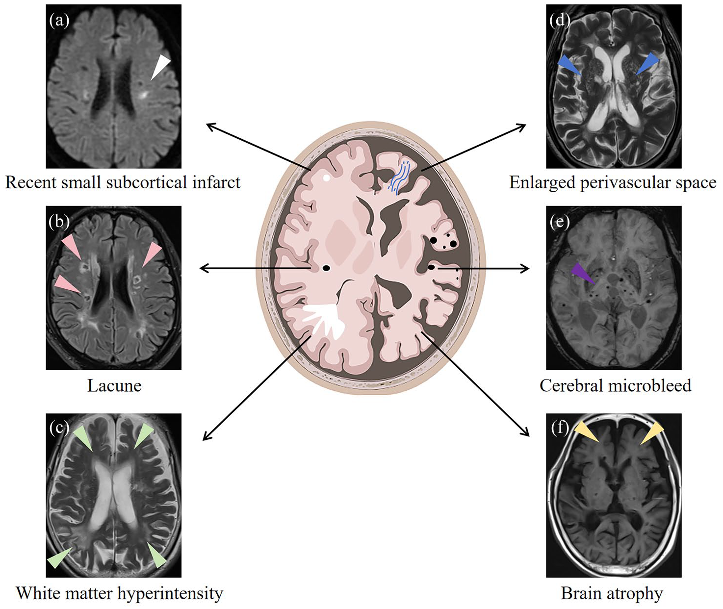
The six imaging markers of cerebral small vessel disease. (a) White arrow = recent small subcortical infarct. (b) Pink arrows = lacunar infarcts (lacunes). (c) Green arrows = white matter hyperintensity. (d) Blue arrows = enlarged perivascular space. (e) Purple arrow = cerebral microbleeds. (f) Yellow arrows = cerebral atrophy.
Additionally, based on the Boston criteria version 2.0, the neuroimaging markers associated with CAA include CMB, cSS, convexal subarachnoid hemorrhage, severe PVSs, and WMHs. These markers help in the diagnosis and characterization of cSVD and CAA-related brain changes through neuroimaging techniques (Charidimou and others 2022). Multiple cSVD imaging markers are frequently detected on the MRI of patients with CAA-related ICH and hypertensive ICH (Figure 4).
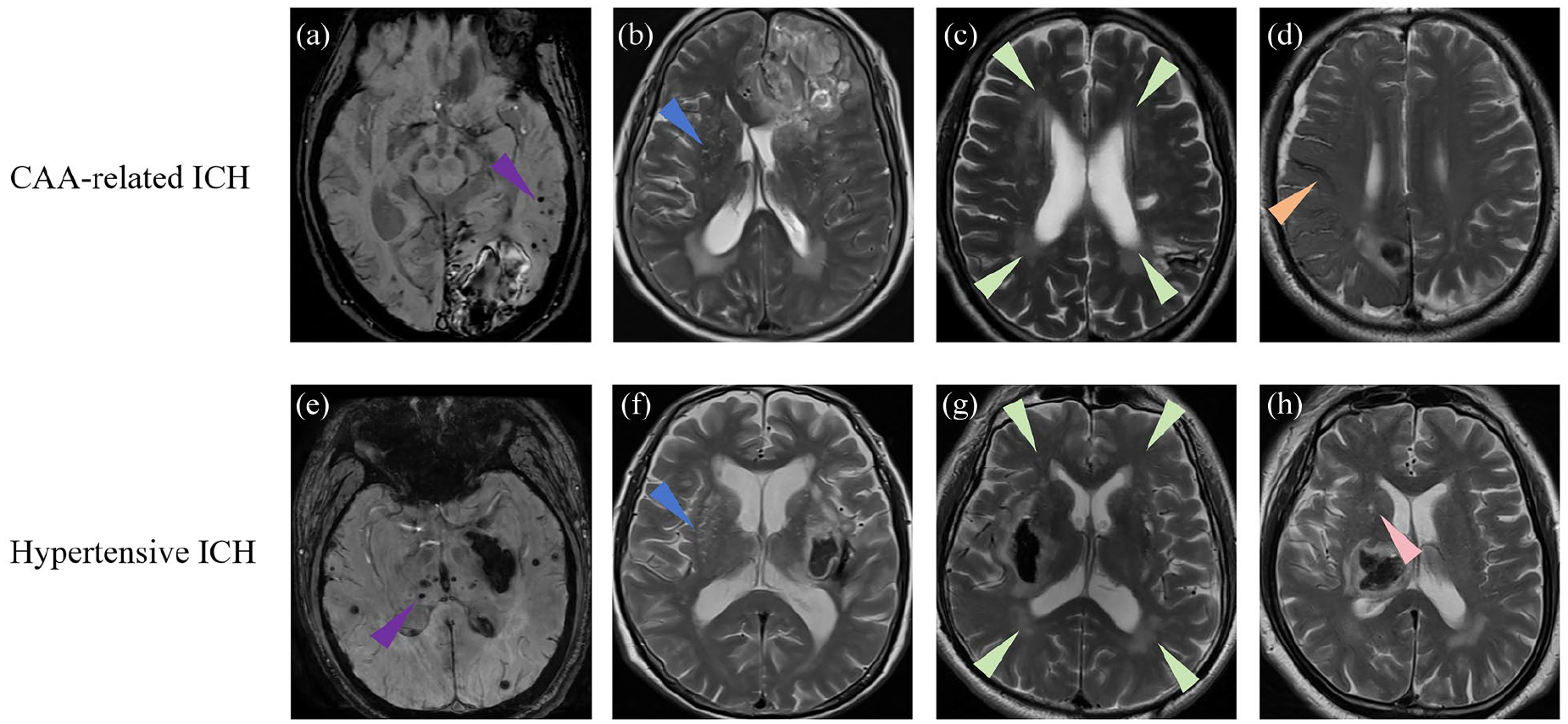
Imaging markers of cerebral small vessel disease in intracerebral hemorrhage (ICH). (a, e) Purple arrow = cerebral microbleeds. (b, f) Blue arrow = enlarged perivascular space. (c, g) Green arrows = white matter hyperintensity. (d) Orange arrow = cortical superficial siderosis. (h) Pink arrow = lacunar infarct (lacune). CAA = cerebral amyloid angiopathy.
cSS refers to the linear deposition of blood degradation product, hemosiderin, localized in the subarachnoid space, leptomeninges, or on the surface of the cerebral cortex. There are three hypotheses regarding the mechanism of cSS formation. First, it is believed that acute convexal subarachnoid hemorrhage occurs due to repeated vascular events in the leptomeninges or superficial cortex affected by CAA. Second, it is proposed that lobar ICH (or microbleeds) leaks or extends to the subarachnoid space, leading to the deposition of hemosiderin. Last, hemorrhagic transformation of small cortical infarctions is considered another possible cause. Therefore, these mechanisms contribute to the formation of cSS in CAA-related conditions.
The presence of cSS serves as an important radiologic marker for the diagnosis of lobar ICH (Charidimou and others 2015). An autopsy of 14 brains diagnosed with CAA indicated that cSS is primarily caused by ruptured blood vessels in the leptomeninges that are severely affected by CAA (Charidimou and others 2020). A study found positive amyloid PET results in 12 patients with cSS (Na and others 2015). Therefore, cSS is considered one of the most specific imaging markers for CAA (Wollenweber and others 2017). Furthermore, a study involving 79 patients with CAA and 69 patients with ICH at baseline indicated that severe progression of cSS is an independent predictor for future hemorrhagic recurrence (Pongpitakmetha and others 2020).
CMBs are small areas of low signal intensity on MRI T2-weighted gradient echo and susceptibility-weighted imaging sequences, typically 2 to 5 mm in diameter, caused by the leakage of red blood cells due to damage to small blood vessels in the brain. These lesions, which appear as small, round, or oval-shaped areas without surrounding edema, are characterized by the deposition of hemosiderin around the blood vessel. Susceptibility-weighted imaging is a special MRI technique that detects these lesions by utilizing the differential sensitivity of ferromagnetic substances, such as hemosiderin, to magnetic fields (Haller and others 2021). Greenberg and others (2009) reported that the volume of hemorrhagic lesions follows a bimodal distribution, with an average volume of 27.5 cm3 (corresponding to a diameter of 3.75 cm) for the higher-volume peak and an average volume of 0.009 cm3 (corresponding to a diameter of 0.26 cm) for the lower-volume peak. Additionally, they observed that patients with CAA with a higher burden of CMBs (>50) demonstrated significantly thicker vessel walls as compared with patients with fewer CMBs (<3). Thickening of the vessel walls is considered a typical characteristic of CAA, which leads to CMBs instead of large hemorrhages. Conversely, in a minority of patients with CMBs, multiple large lobar hemorrhages are observed. This indicates that CMBs and cerebral hemorrhages may have distinct pathologic and physiologic processes. A study examining the correlation between the location of CMBs and the risk of cerebral hemorrhage found that the presence of isolated large lobar microbleeds on MRI typically indicated the presence of severe CAA and a high risk of hemorrhage (van Etten and others 2014). A study on the relationship between CMBs and amyloid protein burden suggested that lobar CMBs and mixed CMBs (involving lobar and deep regions) were associated with a higher amyloid protein burden as compared with isolated deep CMBs. This implies that individuals with lobar CMBs or mixed CMBs are more likely to experience lobar cerebral hemorrhage in the future (Tsai and others 2017).
The PVS, also known as the Virchow-Robin space, refers to the compartment between the extracellular basal lamina of neuroglial cells (outer border, formed by astroglial cell foot processes) and the endothelial basal lamina of blood vessels (inner border). It contains cerebrospinal fluid–like fluid, and the astrocytic sheath forms the outer wall of the PVS, covering a significant portion of the cerebral microvascular system. The PVS plays a crucial role in maintaining homeostasis and regulating neuroinflammation. A survey suggested that the pathogenesis of CAA may involve impaired clearance of Aβ in the perivascular and endothelial pathways, with a reduction in extracellular Aβ degradation. In patients with cerebral hemorrhage, EPVSs are often observed (Held and others 2017). In a study involving 201 patients with primary ICH, 99% showed EPVSs on MRI (Wu and others 2015). The different distribution of EPVSs suggests different underlying mechanisms for cerebral hemorrhage. Charidimou and others (2017) found that severe centrum semiovale EPVS was more common in CAA-related ICH and closely associated with typical imaging markers of CAA, such as lobar CMBs and cSS. Yet, severe basal ganglia EPVS was more commonly observed in hypertensive arteriopathy-related ICH and independently associated with higher WMH volume and deep CMBs, which are indicative of more severe hypertensive arteriopathy–related pathology.
WMH refers to areas of increased signal intensity in the white matter regions of the brain observed on T2-weighted or FLAIR (fluid-attenuated inversion recovery) sequences in MRI. These high-intensity signals are typically associated with pathologic changes in the cerebral white matter, including demyelination, microbleeds, ischemia, inflammation, and edema. WMH is considered one of the characteristic imaging markers of cSVD. A study investigating the relationship between WMH volume and subtypes of cSVD found that larger WMH burden was associated with small vessel strokes as compared with other ischemic stroke subtypes. Furthermore, when compared with other types of stroke, such as ischemic stroke, WMHs had a much stronger association with ICH (Rost and others 2010). A study investigating the relationship between the severity of WMHs and the volume of cerebral hemorrhage concluded that a larger WMH burden was independently associated with a larger volume of cerebral hemorrhage (Chen and others 2018).
Emerging evidence underscores a strong association between neuroimaging markers of cSVD and the occurrence of ICH. Notably, specific markers, such as CMBs and cSS, have been identified as independent predictors of future ICH in population-based studies.
Prevention of ICH: From the Perspective of cSVD
Currently, we have realized that the long-term accumulation of cSVD significantly increases the risk of cerebral hemorrhage. Therefore, can early intervention of cSVD prevent the occurrence of those adverse outcomes? While there are no specific treatment strategies for cSVD at present, a considerable amount of research has provided clues in various areas for the treatment of cSVD, such as antihypertension therapy, antithrombotic treatment, and anti-inflammatory treatment. This research offers potential directions for the treatment and intervention of cSVD, with the aim of preventing the occurrence of devastating events such as cerebral hemorrhage.
Antihypertension Therapy
The etiology of cSVD is complex and involves various factors, including genetics, environment, and lifestyle. Although genetic factors and aging are irreversible, the risk of cSVD can still be effectively reduced through improving lifestyle choices. Among the numerous risk factors, hypertension is considered the most significant one. It is not only associated with the progressive development of cSVD but also plays a crucial role in the acute onset of cerebral hemorrhage. In clinical practice, it is often observed that many patients with cerebral hemorrhage have never undergone regular blood pressure monitoring, and some are even unaware that they have hypertension, let alone maintaining regular medication. The multicenter SPS3 trial (Secondary Prevention of Small Subcortical Strokes), which involved 3020 participants, studied the impact of blood pressure reduction on the secondary prevention of stroke in patients with recent lacunar stroke. The trial aimed to assess the effects of lowering blood pressure on the prevention of recurrent strokes for patients with recent lacunar stroke. The participants were randomly assigned to one of two groups by target systolic blood pressure: <130 mm Hg or 130 to 149 mm Hg. The conclusions indicated that there was no significant reduction in overall recurrent stroke events. However, the group with a target systolic blood pressure <130 mm Hg showed a significant decrease in the occurrence of hemorrhagic stroke.
The 2025 European Stroke Organisation guideline and the 2024 ICH code consensus statement reaffirm the critical importance of blood pressure control (Li and others 2024; Steiner and others 2025). The guideline recommends blood pressure management in adults with prior ICH to reduce the risk of subsequent stroke. Expert consensus recommendations advocate a blood pressure target ≤130/80 mm Hg. The ICH code emphasizes that early intensive blood pressure lowering reduces the risk of hematoma expansion in specific ICH patient subgroups and improves outcomes. Importantly, effective blood pressure control extends beyond its crucial role in the acute phase of ICH to serve as an etiologic treatment for delaying the progression of cSVD (Reddy and others 2025). A number of studies have shown that the intensive blood pressure lowering mitigates the cSVD progression. Yu and others (2025) found that elevated central systolic blood pressure has been directly associated with an increased burden of cSVD on neuroimaging, including larger WMH volumes (β = 0.031, P = 0.003) and a higher prevalence of lobar CMBs (odds ratio, 1.58; P < 0.001) and subcortical CMBs (odds ratio, 1.20; P = 0.023), independent of conventional risk factors. Furthermore, the Trial of Efficacy and Safety of Treatment in Small Vessel Diseases provides direct evidence that specific antihypertensive agents can improve microvascular function, a core pathologic process in cSVD (Kopczak and others 2023). However, excessive blood pressure reduction may precipitate cerebral hypoperfusion, particularly in patients with severe cSVD, potentially increasing risks of cognitive decline. Future research should therefore prioritize precision antihypertensive strategies to achieve individualized blood pressure targets. Controlling blood pressure not only plays a positive role in preventing recurrent cerebral hemorrhage but is also closely associated with a lower risk of cognitive decline or dementia in patients. A meta-analysis examining the relationship between antihypertensive medication and cSVD revealed that antihypertensive treatment has a protective effect on the development of WMHs but did not have an impact on brain atrophy (Rouch and others 2015). Furthermore, a systematic review incorporating 38 studies concluded that antihypertensive medications, especially calcium channel blockers and renin-angiotensin system inhibitors, may be beneficial in preventing cognitive decline and dementia in patients with cSVD. However, these findings still need to be validated through high-quality randomized controlled trials in the future.
Antithrombotic Treatment
Balancing the risks and benefits of antithrombotic therapy represents one of the most challenging aspects of treating patients with cSVD. While these medications are essential for preventing ischemic events, they simultaneously increase hemorrhagic risk, particularly in those with underlying cSVD. The SPS3 study indicated that in the secondary prevention of lacunar stroke, dual antiplatelet therapy (aspirin plus clopidogrel) is not more effective than single antiplatelet therapy (aspirin alone or clopidogrel alone), and it increases the risk of bleeding (SPS3 Investigators 2012). Therefore, for the majority of patients with lacunar stroke, single antiplatelet therapy may be a more appropriate choice. However, for certain patient populations, such as those at high risk of recurrence, further research is needed to determine the optimal treatment approach. The SPS3 study emphasized that the decision regarding antiplatelet therapy in patients with cSVD needs to consider individualized factors, including the patient’s bleeding risk and overall health status. Yet, emerging insights into the pathophysiology of cSVD have challenged conventional antiplatelet therapy (Falcone and Woo 2024). A critical gap exists in the evidence base for individualized treatment decisions: the efficacy and safety of single antiplatelet therapy for secondary prevention in lacunar stroke have never been independently validated. Future studies should address this gap.
Can patients with cSVD with previous cerebral hemorrhage receive antithrombotic treatment? Traditionally, patients with a history of cerebral hemorrhage were not recommended to use antithrombotic medications. However, this conclusion may not apply to all patients with cerebral hemorrhage, such as those with concomitant atrial fibrillation or nonlobar cerebral hemorrhage (Best and others 2023). RESTART (Restart or Stop Antithrombotics Randomised Trial) is a large multicenter prospective randomized trial based on an open-label, blinded end point, parallel-group design that investigated the safety and efficacy of restarting antiplatelet therapy in patients with a history of ischemic stroke who experienced cerebral hemorrhage (Al-Shahi Salman and others 2019). The subgroup analysis of the RESTART study evaluated the impact of brain imaging features, such as CMBs and cSVD, on the risk and effectiveness of antiplatelet therapy. The study results showed that antiplatelet therapy did not confer additional risk for the primary outcomes of cerebral hemorrhage in patients with concomitant CMBs. There was also no interaction observed between antiplatelet therapy and the number or location of CMBs. Therefore, antiplatelet therapy appears to be safe in patients with CMBs and does not increase the risk of recurrent cerebral hemorrhage. While existing research and guidelines provide some guidance on antiplatelet therapy for patients with ischemic stroke with cSVD, further studies are still needed to define the optimal dosage, duration, safety, and efficacy of antiplatelet therapy in different patient subgroups of cSVD.
Anti-inflammatory Treatment
The pathophysiology of cSVD involves complex interactions among vascular risk factors, endothelial dysfunction, BBB disruption, and neuroinflammatory processes. Recent evidence has increasingly positioned inflammation as a consequence and driver of cSVD progression. A recent framework for cSVD progression suggests that early endothelial dysfunction leads to BBB disruption with leakage of fluid and toxic plasma proteins into the vascular media and surrounding tissues, with secondary effects on vascular reactivity, pericyte function, oligodendrocyte proliferation, and perivascular fluid drainage pathways (Smith and Markus 2020).
Inflammatory biomarkers have shown consistent associations with cSVD progression. Systematic reviews have revealed relatively robust associations between cSVD and markers of vascular inflammation, particularly among patients with stroke, suggesting that endothelial and BBB alterations may drive cSVD pathogenesis. Longitudinal investigations have demonstrated that elevated systemic inflammatory markers at baseline predict subsequent cSVD severity and progression. Interestingly, regional analysis has revealed different patterns of association, with markers of vascular inflammation tending to be associated with cSVD in areas typical of hypertensive arteriopathy, while systemic inflammation appears more involved in CAA-related vascular damage (Low and others 2019). The MINERVA study (Minocycline to Reduce Inflammation and Blood-Brain Barrier Leakage in Small Vessel Disease), a phase 2 randomized double-blind trial, demonstrated that minocycline showed no significant improvement in microglial activation or BBB leakage. However, quantitative assessment via dynamic contrast-enhanced MRI revealed a strong correlation between BBB permeability and the cerebrospinal fluid/serum albumin ratio, validating the pathologic relevance of imaging biomarkers in cSVD (Brown and others 2024). This negative result in the MINERVA study contrasts with findings from rodent models, where minocycline effectively restored white matter integrity in spontaneously hypertensive stroke-prone rats (Fu and Yan 2018). In these animal studies, minocycline was associated with reduced white matter lesions, decreased inflammation, and lower BBB permeability. The discrepancy between preclinical promise and clinical results highlights the challenges in translating experimental findings to human cSVD. Given the limitations of broad-spectrum anti-inflammatory drugs such as minocycline, targeting specific immune pathways has become an emerging strategy. Fingolimod, a sphingosine-1-phosphate receptor modulator approved for multiple sclerosis, has shown promise in acute ischemic stroke, though not specifically in cSVD. Fingolimod reduces circulating lymphocytes by preventing their egress from lymph nodes, thereby potentially limiting neuroinflammation (Groves and others 2013; Fu and others 2014). In future research, targeted modulation of S1PR1 signaling may represent a promising strategy to disrupt neuroinflammatory cascades without compromising BBB integrity. Yet, it is noteworthy that, according to the latest European Stroke Organisation guideline, anti-inflammatory interventions are not recommended for adults with acute spontaneous ICH (except within randomized controlled trials; Steiner and others 2025). Clinical trials investigating anti-inflammatory agents such as anakinra, celecoxib, and edaravone are currently ongoing.
Summary
ICH is a significant public health concern, characterized by high incidence and mortality rates. Increasing evidence suggests that cerebral hemorrhage is an acute manifestation of cSVD, precipitated by vascular rupture in a fragile vascular system compromised by cSVD. Recognizing cSVD as the underlying substrate for cerebral hemorrhage can facilitate the identification of high-risk populations and the development of preventive strategies. Future research should focus on elucidating the mechanisms by which cSVD leads to vascular fragility and identifying early biomarkers to enable timely intervention, thereby reducing the incidence of cerebral hemorrhage and improving patient outcomes.
Footnotes
Author Contributions
Li-li Tang and Yu-jia Jin contributed to manuscript writing, literature search, and figure preparation. Xue qun Chen and Peiran Jiang contributed to figure preparation. Feng Gao and Lu-sha Tong were responsible for review and revision of the manuscript.
Declaration of Conflicting Interests
The authors declared no potential conflicts of interest with respect to the research, authorship, and/or publication of this article.
Funding
The authors disclosed receipt of the following financial support for the research, authorship, and/or publication of this article: This work was supported by grants from the National Natural Science Foundation of China (No. 32120103007), the Natural Science Foundation of Zhejiang Province (No. LMS25H090003) and “Leading Goose” R&D Program of Zhejiang (No. 2024C03026).