
Abstract
Studies have linked maternal childhood maltreatment to altered infant brain volumes, but none have examined infant hypothalamic-pituitary-adrenal (HPA) axis function as a mechanism linking the two domains. Further, studies among older children suggest that childhood abuse and neglect may be associated with different developmental outcomes and thus should be studied separately. Study participants were N = 57 mother-infant dyads, stratified for maternal childhood maltreatment. At 4 months, infant cortisol total output (AUCg) and change (AUCi) were assessed across the Still-Face Paradigm. Under natural sleep, infants completed T1-weighted MRI scans (M age = 12.28 months). Whole brain, amygdala, and hippocampal volumes were extracted via automated segmentation. Maternal childhood neglect, but not abuse, was directly associated with higher infant AUCg and AUCi, and was indirectly associated with larger amygdala and hippocampal volumes through infant AUCg. Results suggest that infant cortisol may be particularly influenced by maternal childhood neglect and may be one mechanism further influencing brain development.
Maternal childhood maltreatment (MCM) has been associated with negative offspring outcomes, with children of maltreated mothers exhibiting elevated risk for adverse psychological health (Plant et al., 2018). Alterations in biological stress systems of mother and infant have been proposed as potential mechanisms underlying such intergenerational transmission (Buss et al., 2017). Thus, understanding how maternal childhood neglect and abuse may affect infant brain development is an important scientific priority.
Rodent research has shown that stress response networks, including the hypothalamic-pituitary-adrenal (HPA) axis and limbic brain regions, are highly sensitive to adversity in early infancy. Specifically, low maternal nurturance (Turecki & Meaney, 2016) or maternal unpredictability (Drury et al., 2016) are associated with increased stress responses and altered amygdala volume and activation among rodent pups. The amygdala and hippocampus may be particularly vulnerable to the effects of early stressors due to high glucocorticoid receptor density (Chen et al., 2012) and to a postnatal developmental trajectory characterized by rapid initial growth and gradual pruning (Gilmore et al., 2012). The amygdala, in particular, increases steeply in volume over the first 3 years of life, plateauing at 3.5 years of age, and the hippocampus plateaus at 4.6 years of age (Alex et al., 2023). Such periods of rapid growth are hypothesized to signal experience-expectant sensitivity to environmental inputs, which fosters early adaptation to the affordances in one’s environment (Turecki & Meaney, 2016).
Consistent with the possibility of experience-expectant sensitivity, severity of MCM has been linked to alterations in infant grey matter volume and amygdala volume and connectivity among human infants (Hendrix et al., 2021; Khoury, Ahtam et al., 2021; Lyons-Ruth et al., 2023; Moog et al., 2018). However, potential mechanisms underlying these associations between MCM and altered brain development are only beginning to be evaluated. Randomized rodent studies have identified glucocorticoid levels as one mechanism affecting limbic volumes (Vyas et al., 2004). Thus, it is important to assess whether cortisol regulation may also be one mechanism linking MCM to altered limbic volumes in human infants. Identifying mechanisms opens the way to developing interventions that could interrupt the adverse intergenerational outcomes associated with maternal childhood maltreatment. To evaluate this potential pathway, the current study assessed associations among MCM, infant cortisol levels, and volumes of the infant amygdala and hippocampus.
Direct Effects of Abuse versus Neglect on Neurobiological Development
Evidence indicates that neurobiological outcomes associated with direct experiences of maltreatment may be better understood if abuse and neglect are studied separately (McLaughlin et al., 2019). Direct experiences of childhood abuse have been associated with reduced volume, but increased activity, of the right amygdala later in childhood, whereas neglect has been associated with reduced volume and altered function in frontoparietal regions (McLaughlin et al., 2019). However, few studies have examined whether the mother’s childhood abuse versus neglect history are related to differential effects on offspring limbic volumes. The one study that has assessed such differential effects used the MRI sample from the present study and found that maternal history of childhood abuse was associated with reduced infant right hemisphere amygdala volume, while neglect was associated with lower infant grey matter volume, consistent with the effects of direct experiences of abuse versus neglect (Lyons-Ruth et al., 2023).
A separate literature suggests that direct experiences of neglect or low nurturance in infancy may be associated with enlarged amygdala and/or hippocampal volumes later in childhood (Bernier et al., 2019; Lee et al., 2019; Mehta et al., 2009; Rao et al., 2010; Rifkin-Graboi et al., 2015; Tottenham et al., 2010). An overlapping-cohorts study of institutional-rearing effects on amygdala volume from 4 to 20 years found enlarged amygdala volumes in the institutionally reared group relative to controls prior to 6.5 years of age. However, amygdala volumes among controls continued to increase and became larger than volumes in the institutionally reared group by age 11 (Van Tieghem et al., 2021).
These associations between inadequate care in infancy and larger amygdala and/or hippocampal volumes are also consistent with controlled rodent studies, which find that low maternal nurturance/unpredictability is causally related to elevated stress hormone output among very young rodent pups in genetically randomized designs (e.g., Champagne et al., 2008). These effects are intergenerationally transmitted, with offspring randomly assigned to low-nurturing mothers showing low nurturance to their own offspring and those offspring also showing elevated HPA axis activity (Drury et al., 2016). Additional controlled rodent studies document an effect of elevated HPA activity on larger amygdala volume (Vyas et al., 2004). Thus, human studies of infants in institutional care and intergenerational rodent studies of low-nurturing mothers converge on the hypothesis that experiences of neglect may be related to elevated HPA-axis activity and amygdala volume in infancy. However, it remains unstudied whether the mother’s experiences of childhood neglect are indirectly associated with similar increases in her infant’s limbic volumes through cortisol output.
Maternal Childhood Maltreatment and Infant Cortisol Regulation
As the primary biological stress response system in humans, functioning of the HPA axis and its associated stress hormone, cortisol, are hypothesized to contribute to the intergenerational effects of MCM (Buss et al., 2017). Four studies have examined severity of MCM in relation to infant cortisol response to laboratory stressors. During an arm restraint and noise burst stressor, Brand et al. (2010) found that infants of mothers with more severe MCM exhibited lower baseline cortisol levels but no differences in cortisol trajectories. During a similar stress procedure combined with maternal separation, Duffy et al. (2024) found that male infants of mothers with MCM showed a reduction in cortisol from baseline to post-stress, whereas females showed increases in cortisol post-stressor relative to baseline. Three additional studies used the Still-Face Paradigm (SFP; Tronick et al., 1978), in which the mother disrupts normal interaction with her infant by displaying a “still face” for 2 minutes, then returns to normal interaction (Chasson et al., 2025; Khoury et al., 2021; Martinez-Torteya et al., 2014). None found a classic pattern of cortisol reactivity and recovery to the 2-min Still-Face period, consistent with other studies relating family risk factors to infant cortisol in the SFP (e.g., Grant et al., 2009). Instead, Khoury, Beeney et al. (2021) found that the cortisol levels of infants of mothers with more severe MCM remained elevated over the SFP, whereas cortisol levels of infants of low MCM mothers decreased sharply from baseline. Similarly, Martinez-Torteya et al. (2014) found no association between MCM and infant cortisol reactivity in the SFP, but found that less positive parenting was associated with sustained higher infant cortisol levels over the SFP, while more positive parenting was associated with a decrease in infant cortisol from baseline.
The third study was the only study to examine maternal childhood abuse and neglect separately, as well as to examine the interaction of maternal childhood abuse and neglect with disrupted postnatal caregiving (Chasson et al., 2025). In that study, more severe maternal childhood neglect was associated with sustained higher infant cortisol output to the SFP in the context of maternal disoriented caregiving. In contrast, more severe maternal childhood abuse interacted with higher levels of maternal negative-intrusion to predict lower infant cortisol output. These differential relations to infant cortisol further suggest the importance of analyzing maternal childhood abuse and neglect separately. Notably, the Chasson et al. (2025) study was conducted using the full sample from the MIND study (N = 181), the study from which the current MRI subsample was drawn. However, infant MRI’s were not available for the full sample of 181, leaving open the question of how these alterations in HPA-axis functioning related to maternal childhood neglect versus abuse might further relate to infant limbic volumes. Addressing that question is the focus of the current study.
As noted above, MCM has been associated with reactivity and recovery to a non-maternal stressor (Duffy et al., 2024), to sustained higher levels of cortisol from baseline through recovery to a maternal stressor (Khoury, Beeney et al., 2021), and to both higher and lower cortisol output in interaction with particular aspects of maternal care (Chasson et al., 2025). Therefore, in the present study, both total cortisol output (AUCg, area under the curve with respect to ground) and change in cortisol levels from baseline (AUCi, area under the curve with respect to increase) were assessed.
Maternal Childhood Abuse or Neglect and Infant Limbic Volumes
Among newborns, Demers et al. (2022) found smaller bilateral amygdala volumes associated with maternal childhood adversity at five weeks of age, while Moog et al. (2018) found no association between MCM and infant amygdala and hippocampal volumes among infants at 4 weeks. In previous work with the current sample of 4- to 25-month-old infants, maternal childhood abuse, but not neglect, was associated with smaller right amygdala volume after 18 months of age (Lyons-Ruth et al., 2023). No associations with left amygdala or right or left hippocampal volume were found. Among fetuses (Van Den Heuvel et al., 2023) and among 3-month-olds (Hendrix et al., 2021), MCM and maternal childhood emotional neglect, respectively, were associated with stronger connectivity between amygdala and prefrontal cortex, a potential marker of stress-accelerated development. These studies provide evidence that MCM may be associated with alterations in amygdala volume and connectivity in infancy, but do not assess altered HPA-axis regulation as a potential linking mechanism.
Infant Cortisol Output and Infant Limbic Volumes
Limbic regions have high glucocorticoid receptor density and complex feedback relations to the HPA axis, and thus are particularly vulnerable to early stress exposure (Chen et al., 2012; Vyas et al., 2004). Alterations in amygdala and hippocampal function in relation to HPA activity have been documented in randomized rodent studies (Champagne et al., 2008; Chen et al., 2012; Drury et al., 2016), with elevated stress hormone levels associated with increased amygdala (Vyas et al., 2004) but reduced hippocampal volume (Chen et al., 2012). To our knowledge, only one study has investigated the link between infant cortisol output and infant amygdala and hippocampal volumes among human infants. That study, conducted with the current sample, found that elevated cortisol output in the SFP was associated both with maternal disrupted interaction and with larger amygdala and hippocampal volumes (Khoury et al., 2023). MCM was not included in that study, however, leaving open the question of whether infant cortisol might also mediate relations between MCM and infant amygdala and hippocampal volumes found in previous work (Khoury, Ahtam et al., 2021; Lyons-Ruth et al., 2023).
In summary, previous work with the current MRI sample included three studies. The first linked overall MCM to reduced infant amygdala volume to (Khoury, Ahtam et al., 2021); the second found that maternal childhood abuse, specifically, was responsible for that finding (Lyons-Ruth et al., 2023); the third did not examine MCM, but linked disrupted maternal interaction and elevated infant cortisol output to increased infant amygdala and hippocampal volume (Khoury et al., 2023). Thus, in previous work, MCM was related to reduced amygdala volume, while infant cortisol was related to increased amygdala and hippocampal volumes.
This prior work left unanswered whether cortisol might also be an important mechanism linking the mother’s childhood maltreatment to infant amygdala and hippocampal volumes, as suggested by rodent studies. Those prior studies also pointed to potentially different directions of effect on limbic volumes, with reduction seen in the two papers without cortisol in the model, but larger volumes seen in the one paper with cortisol in the model. Thus, prior work leaves unclear whether to expect abuse and/or neglect to be related to reduced or enlarged limbic volumes when cortisol output is accounted for in the models. The goal of the current study was to assess the role of infant stress hormone levels in intergenerational pathways linking the mother’s childhood neglect and abuse to altered infant limbic volumes.
Current Study
The first aim of the current study was to examine whether maternal childhood abuse or neglect was associated with infant salivary cortisol responses to the SFP, assessed with both AUCg and AUCi. The second aim was to evaluate whether maternal childhood abuse or neglect was associated with infant amygdala or hippocampal volumes. 1 The final aim was to evaluate if infant cortisol levels indirectly linked maternal childhood neglect or abuse to limbic volumes.
Methods and Materials
Participants
Participants were 57 mother-infant dyads from 181 families enrolled in the Mother-Infant Neurobiological Development (MIND) study. Mothers were recruited through community flyers, prenatal classes, and birth records. Using the Adverse Childhood Experiences questionnaire (ACE; Felitti et al., 1998), participants were screened to ensure that at least half experienced one or more forms of childhood maltreatment. Exclusion criteria were (a) English not a primary language spoken at home, (b) maternal age over 44 years at infant birth, (c) infant born before 36 weeks gestation and/or weighing less than 2500 g, and (d) infant congenital disorder or birth defect.
All study participants from the larger cohort (n = 181) were offered participation in the infant MRI. 119 participants agreed to participate but 62 MRIs were unsuccessful, resulting in N = 57 infants with good quality scans. See Supplement for reasons for scan failure. Among mothers in the MRI study, 58% (n = 33) reported one or more forms of childhood maltreatment on the Adverse Childhood Experience (ACE) questionnaire. Infants completing the MRI did not differ from infants not completing the MRI on demographic characteristics (sex, race/ethnicity, family income, maternal education, maternal single parent status, prange = .45–.77) or on maltreatment severity based on the MACE [t(178) = −1.34, p = .18]. Infants were scanned between 4–25 months of age (M = 11.74, SD = 6.12). The study was approved by the Institutional Review Board [Partners Healthcare IRB Protocol #: 2014P002522]. All mothers provided informed consent for participation.
Measures
Still-Face Paradigm (SFP)
Mothers and infants participated in the SFP (Tronick et al., 1978) at 4 months infant age, during which the mother interacted with her infant for 3 min (play period), then displayed a neutral face and did not interact with her infant for 2 min (still-face period), then engaged in a period of interaction for 5 min (reunion period). The SFP was conducted at home to minimize cortisol response to entering the lab. The SFP is a mild infant stressor, eliciting reduced positive and increased negative affect (Mesman et al., 2009).
Infant Cortisol
Saliva was collected from infants three times during the SFP: prior to initiation of the SFP (baseline), 20 minutes after the still-face period ended (+20 min), and 40 minutes after the still-face period ended (+40 min). SFP were conducted between 12:00 and 18:00 h (Mbaseline time 13:58 h [SD = 1 h 38 min]) to minimize effects of the circadian rhythm of cortisol. Infants did not eat or drink 30 min before baseline to avoid saliva contamination. See Supplement for details on assays. AUCg indexed total cortisol output (i.e., difference from zero) and AUCi indexed change in cortisol from baseline (Pruessner et al., 2003).
Maternal Childhood Maltreatment
Mothers completed the 75-item Maltreatment and Abuse Chronology of Exposure scale (MACE; Teicher & Parigger, 2015). The MACE assesses childhood maltreatment severity, including subscales for verbal abuse, non-verbal emotional abuse, physical abuse, sexual abuse, emotional neglect, and physical neglect. The four abuse scales were summed to create an abuse severity index (possible range 0–40); the two neglect subscales were summed to create a neglect severity index (possible range 0–20). The MACE correlates with other CM measures and has high test-retest reliability (Teicher & Parigger, 2015).
Sociodemographic Variables
Variables assessed by maternal interview included infant age, sex, and gestational age at birth, annual family income, and maternal education.
Imaging Data Acquisition and Processing
Image Acquisition
Infant MRIs were completed during natural sleep, without sedation, using a 3.0 T Siemens Skyra scanner with a 64-channel head coil at Boston Children’s Hospital. The T1- weighted acquisition used an advanced version of the Magnetization Prepared Rapid Acquisition Gradient Echo (MPRAGE) sequence, where fast, low-resolution volumetric navigators were played each repetition period for prospective motion correction (Tisdall et al., 2012). Imaging parameters of the MPRAGE sequence included: voxel size = 1 × 1 × 1 mm3, repetition time (TR) = 2500–2540 ms, echo time (TE) = 1.65–2.37 ms, inversion time (TI) = 1450–1470 ms, field of view (FOV) = 192 × 192 mm2 and between 144–173 slices, to cover the entire infant brain. Not all participants had T2-weighted scans, so only T1-weighted images were used for analysis to keep the MRI sequence consistent across participants. T1-weighted sequences with high spatial resolution can be acquired in a relatively short time, unlike T2-weighted sequences with high spatial resolution (Barkovich et al., 2019). Data acquisition time is important when scanning unsedated infants, because T2-weighted sequences are more susceptible to motion artifacts (Barkovich et al., 2019), and motion plays an important role in the quality of the image acquired (Afacan et al., 2016; see Supplement for further detail).
MRI Segmentation
After visual quality control using Freeview software (surfer.nmr.mgh.harvard.edu), T1-weighted volumes were manually aligned along the AC-PC plane, and then underwent N4 bias correction (Tustison et al., 2010), field of view normalization (Ou et al., 2018), and multi-atlas skull stripping (Doshi et al., 2013). Instead of using Infant Freesurfer, the infant T1-weighted MRI was segmented using a publicly-available toolkit known as “Multi-atlas region Segmentation utilizing Ensembles of registration algorithms and parameters, and locally optimal atlas selection” (MUSE; Doshi et al., 2016), that has been extensively validated and adapted to infant brain MRIs (Ou et al., 2014; Ou et al., 2011; Ou et al., 2017; See Supplement for further detail). Quality control of the segmentations was visually performed using the FSLView (https://fsl.fmrib.ox.ac.uk/) software. Whole brain and left and right hemisphere amygdala and hippocampal volumes were extracted.
Data Analytic Strategy
Due to skewness and kurtosis, cortisol levels (baseline, +20 and +40) were log10 transformed and winsorized to reduce the effect of outliers. 2 AUCg/AUCi were then computed to provide summary indices of cortisol. Outliers in infant brain volumes (≥±3SD from mean) were removed and estimated using full information maximum likelihood (FIML), following current practice in imaging studies (Sta. Cruz et al., 2020). One infant had outliers on both amygdala and hippocampal volumes, a second infant had an outlier on hippocampal volume, and another on WBV. These outliers were due to atypical brain anatomy and inhomogeneity in the T1-weighted imaging resulting in inaccurate segmentations. Covariates were included in analytic models if they were associated with dependent variables or if prior research/theory suggested their relevance. Missing data for cortisol, due to insufficient saliva, were estimated using FIML, as follows: baseline n = 4 (7%), +20 min n = 8 (14%), and +40 min n = 3 (5.3%). Regression and mediation models were conducted using the lavaan package in R (Rosseel, 2012). Regression and mediation models were conducted using FIML and robust standard errors to account for missing data, nonnormality, and small sample size (Fox, 2015). Mediation models were rerun with bootstrapped confidence intervals (CIs) (5,000 resamples & 10,000 iterations). CIs that do not contain zero are considered significant at p ≤ .05. For power analyses, see Supplement.
Results
Descriptive Statistics and Covariate Analyses
Sociodemographic Characteristics and Descriptive Statistics
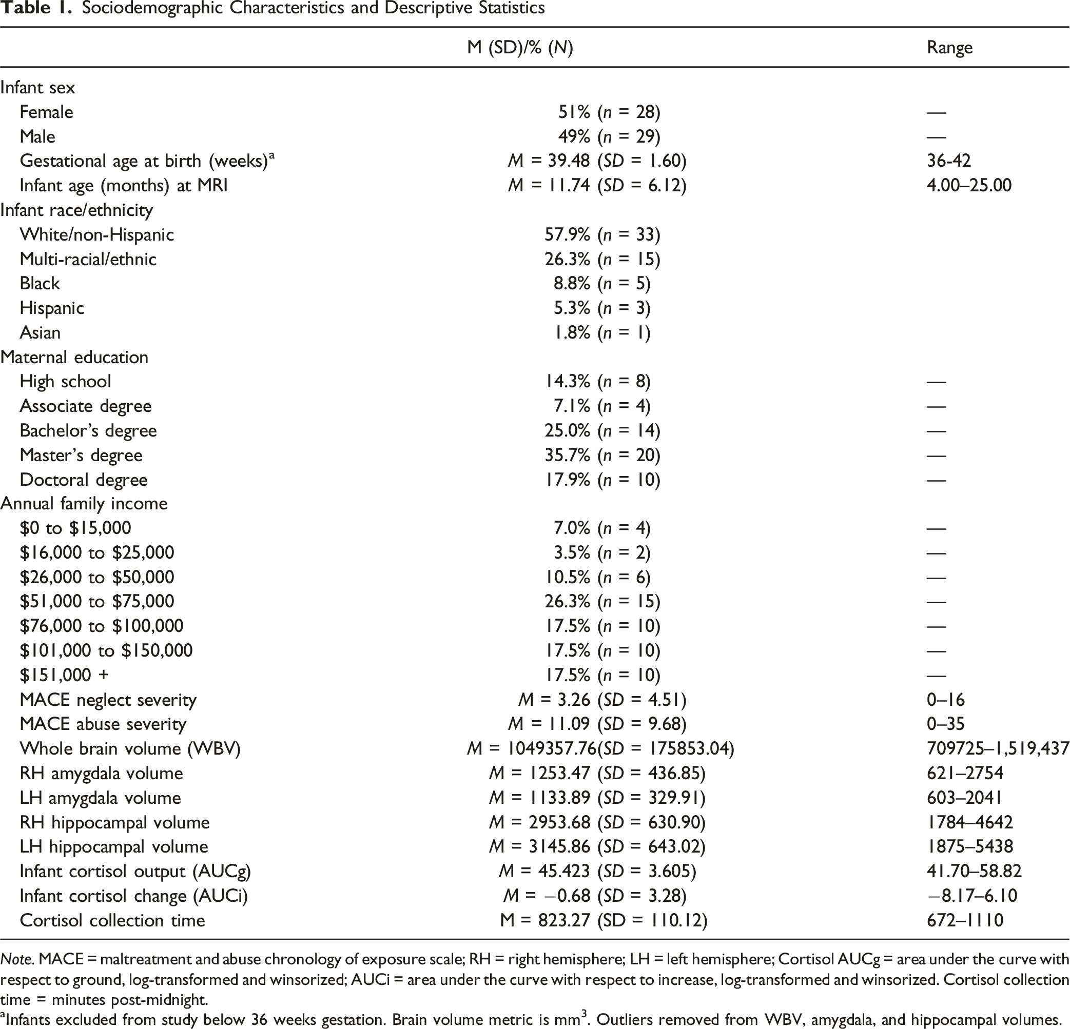
Note. MACE = maltreatment and abuse chronology of exposure scale; RH = right hemisphere; LH = left hemisphere; Cortisol AUCg = area under the curve with respect to ground, log-transformed and winsorized; AUCi = area under the curve with respect to increase, log-transformed and winsorized. Cortisol collection time = minutes post-midnight.
aInfants excluded from study below 36 weeks gestation. Brain volume metric is mm3. Outliers removed from WBV, amygdala, and hippocampal volumes.
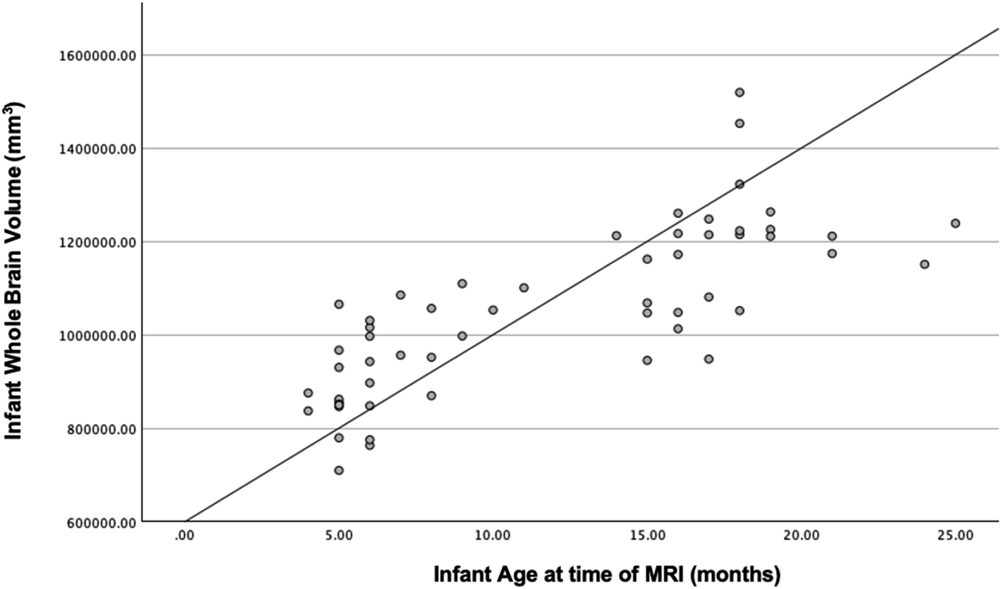
Scatterplot Displaying Infant Age at Scan in Relation to Whole Brain Grey Matter Volume.
Maternal Childhood Abuse, Neglect, and Infant Cortisol Levels
Maternal childhood abuse and neglect were strongly correlated (r = 0.786, p < .001). Given potential multicollinearity, regression models on infant cortisol were first conducted with one type of maternal childhood maltreatment as the independent variable. Regression analyses, covarying saliva collection time, indicated that maternal childhood neglect was associated with higher infant AUCg (β = 0.361, B = 0.359, SE = 0.138, p = .009, CI [0.089, 0.628]), while maternal childhood abuse was not (β = 0.246, B = 0.245, SE = 0.138, p = 0.082, CI [−0.031, 0.520]).
To additionally check if the effects of maternal childhood neglect and childhood abuse on infant cortisol were distinct, a regression analysis was also conducted with maternal childhood neglect and abuse in the same model. Variance Inflation Factor (VIF) and tolerance were calculated, indicating no significant multicollinearity between maternal childhood neglect and abuse in the model (VIF = 2.80, Tolerance = 0.36). Again, maternal childhood abuse was not associated with infant AUCg, with the coefficient now negative (β = −0.078, B = –0.078, SE = 0.216, p = .719, CI [−0.500, 0.345]). Maternal childhood neglect remained positively associated with infant cortisol, with a medium effect size, although the effect did not reach significance (β = 0.423, B = 0.420, SE = 0.219, p = .055, CI [−0.009, 0.848]). Maternal neglect, but not abuse, was also associated with infant AUCi (neglect β = .297, B = 0.295, SE = 0.140, p = .036, CI [0.020, 0.570]; abuse β = .266, B = 0.264, SE = 0.139, p = .058, CI [−0.009, 0.537]). When maternal neglect and abuse were included in the same model, neither were significantly associated with infant AUCi (neglect β = .220, B = 0.219, SE = 0.223, p = .326, CI [−0.217, 0.655]; abuse β = .097, B = 0.096, SE = 0.219, p = .660, CI [−0.333,0.526]).
Because the same values for AUCg or AUCi could be yielded by different patterns of cortisol over the three collection points, average infant cortisol values were plotted over time by higher and lower maternal childhood neglect severity and higher and lower abuse severity (Figure 2). On average, the baseline values of infants of more/less neglected mothers were similar, but the 20- and 40-min post-stress values differed. Thus, on average, the higher AUCg and higher AUCi found among infants of mothers with higher childhood neglect reflected their higher levels of cortisol release during the post-stressor periods. In comparison, mean cortisol levels of infants of mothers with lower levels of neglect declined sharply from baseline to post-stressor, similar to the declines seen in other low-risk groups (Grant, 2009; Martinez-Torteya et al., 2014). In contrast, cortisol trajectories were similar for infants of mothers with higher and lower childhood abuse, with slight declines from baseline to post-stressor periods. Trajectories of Infant Cortisol Levels Over the Still Face Procedure by Severity of Maternal Childhood Neglect and Abuse.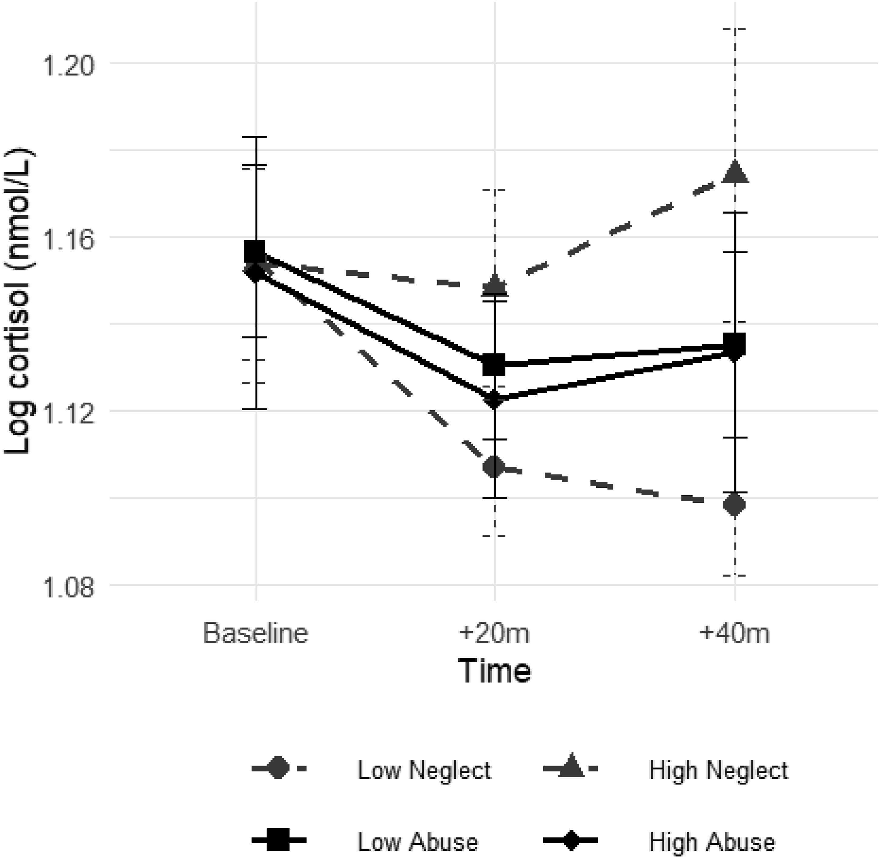
Infant Cortisol Levels and Infant Limbic Volumes
Regression Coefficients From a Single Multivariate Regression Model, With Infant AUCg as the Independent Variable and Infant Right and Left Amygdala and Hippocampal Volumes as the Four Multivariate Dependent Variables
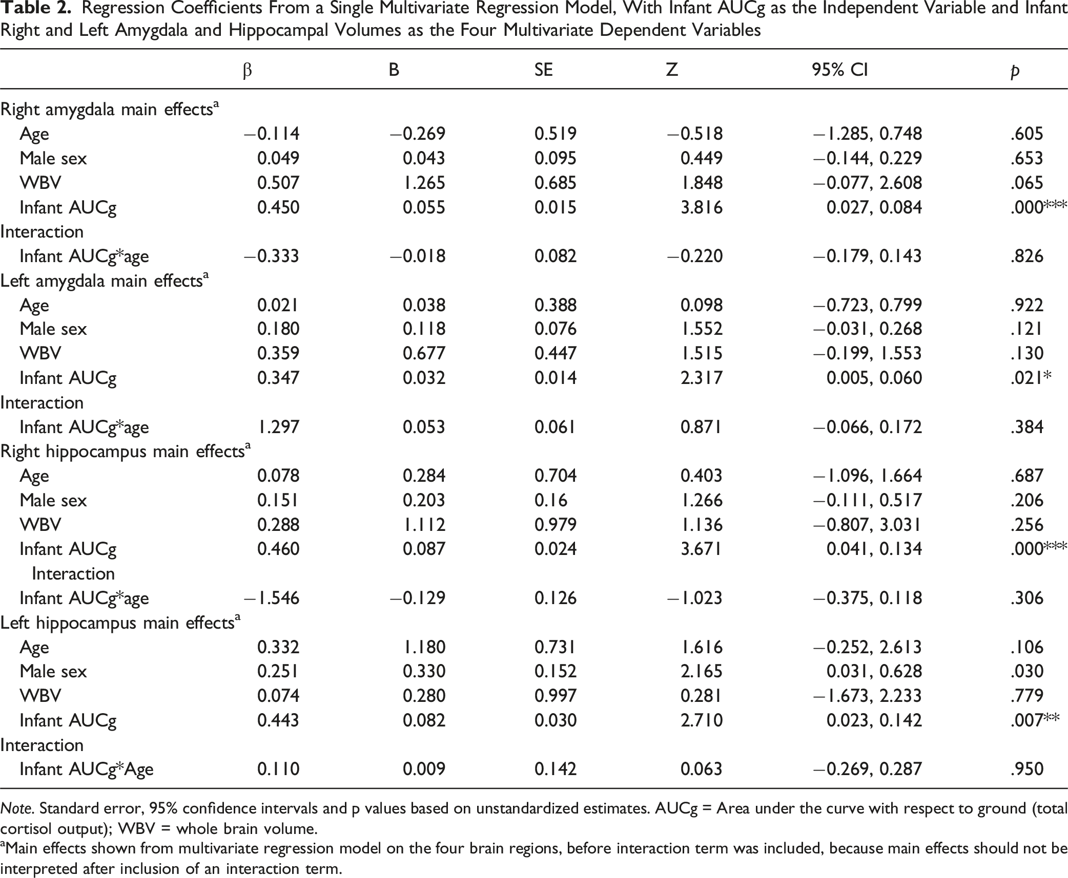
Note. Standard error, 95% confidence intervals and p values based on unstandardized estimates. AUCg = Area under the curve with respect to ground (total cortisol output); WBV = whole brain volume.
aMain effects shown from multivariate regression model on the four brain regions, before interaction term was included, because main effects should not be interpreted after inclusion of an interaction term.
Moderation models were also conducted to assess interactions between infant age at MRI and AUCg on limbic volumes, again using a similar single multivariate model for the four brain regions. Infant age did not moderate associations between AUCg and limbic volumes (Table 2), nor did infant age interact with AUCi to predict limbic volumes (Supplement, Table S1).
Maternal Childhood Abuse, Neglect, and Infant Amygdala and Hippocampal Volumes
Regression Coefficients From a Single Multivariate Regression Model, With Maternal Childhood Abuse and Neglect as the Independent Variables and Infant Right and Left Amygdala and Hippocampal Volumes as the Four Multivariate Dependent Variables
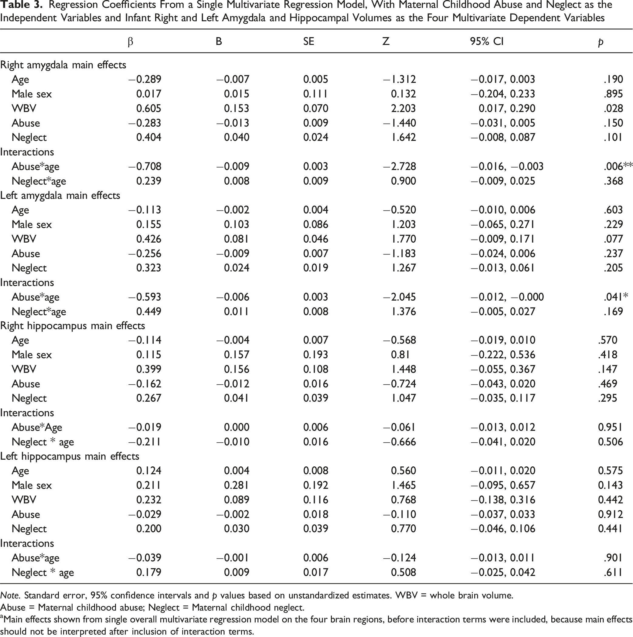
Note. Standard error, 95% confidence intervals and p values based on unstandardized estimates. WBV = whole brain volume.
Abuse = Maternal childhood abuse; Neglect = Maternal childhood neglect.
aMain effects shown from single overall multivariate regression model on the four brain regions, before interaction terms were included, because main effects should not be interpreted after inclusion of interaction terms.
A multivariate moderation model assessed interactions between infant age at MRI and maternal childhood neglect or abuse on infant limbic volumes. There were no significant age by neglect interactions (Table 3). Age moderated the effect of maternal childhood abuse on infant amygdala, but not hippocampus, such that maternal childhood abuse severity was associated with reduced right and left amygdala volumes at older ages (Table 3). Region of significance analyses (Johnson & Neyman, 1936) show maternal childhood abuse was associated with reduced right and left amygdala volume after 425.11 and 421.61 days (14 and 13 months), respectively. 3
Mediation Models
Given that maternal childhood neglect, but not abuse, was associated with higher infant AUCg and AUCi, and that infant AUCg, but not AUCi, was associated with larger limbic volumes, final mediation models were run to evaluate whether infant AUCg mediated indirect associations between maternal childhood neglect and infant amygdala and hippocampal volumes. The indirect paths from maternal childhood neglect to infant right and left amygdala volume (Figure 3(a) and (b)) through infant AUCg were positive and significant. However, these indirect effects were not significant when using bootstrapped CIs (Indirect effect: RH amygdala β = 0.133, B = 0.013, SE = 0.007, p = 0.071, CI [–0.003, 0.026]; LH amygdala β = 0.103, B = 0.008, SE = 0.005, p = 0.123, CI [–0.004, 0.016]). (a, b) Standardized Coefficients and Confidence Intervals, as Well as Unstandardized Coefficients, From Mediation Models With Infant Cortisol Output Mediating the Associations Between Maternal Childhood Neglect Severity and Right Hemisphere (Figure 3a) and Left Hemisphere (Figure 3b) Amygdala Volumes.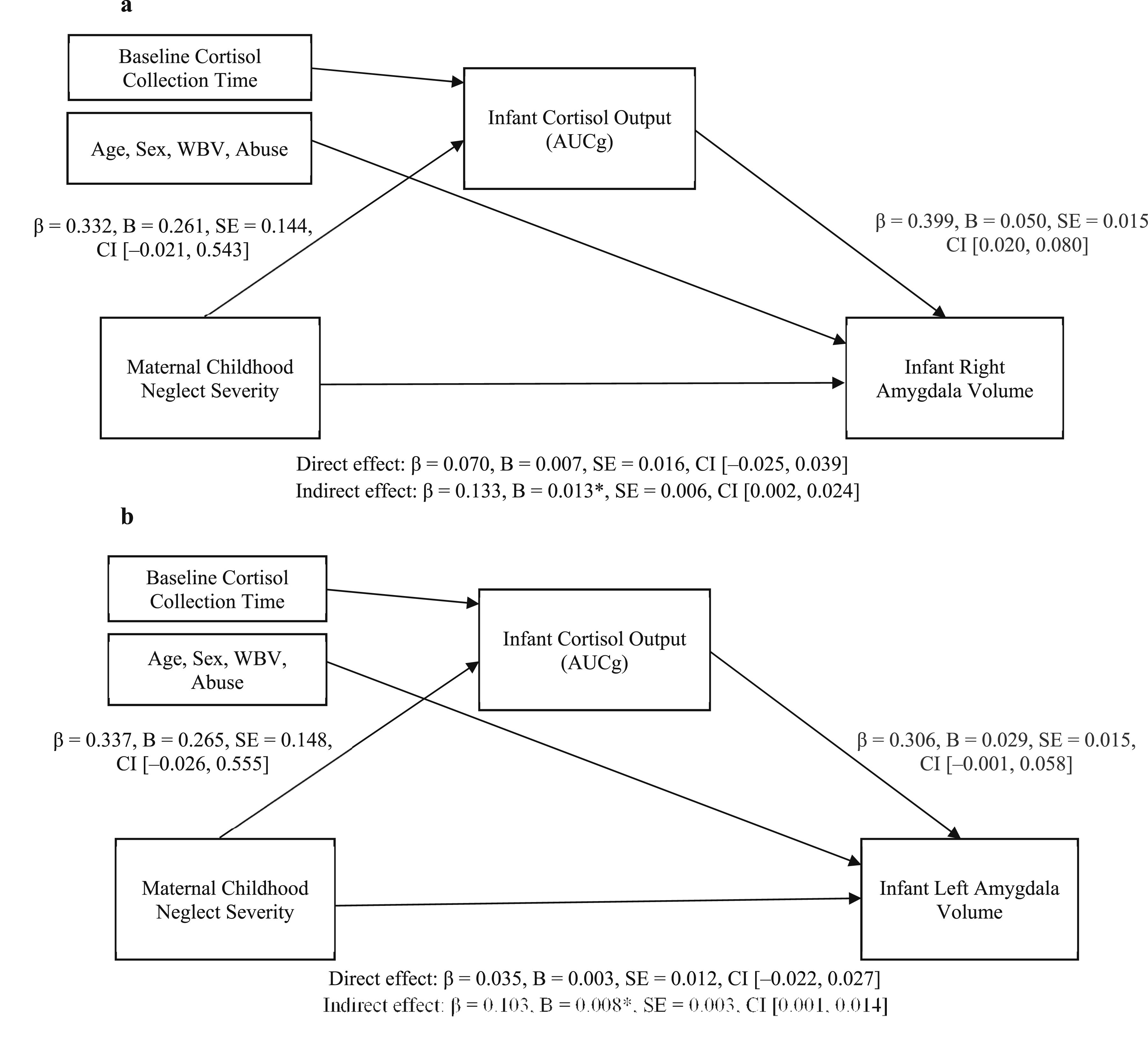
The indirect path to infant right hippocampal volume was also significant (Indirect effect: β = 0.149, B = 0.021*, SE = 0.010, CI [0.002, 0.041], but the path to left hippocampal volume was not significant (Indirect effect: β = 0.137, B = 0.020, SE = 0.011, CI [–0.002, 0.042]). The indirect effect for the right hippocampus was also not significant when using bootstrapped CIs (Indirect effect: β = 0.149, B = 0.021, SE = 0.012, p = 0.082, CI [–0.005, 0.043]). See Supplemental Figure S1a, b for hippocampal mediation results.
Discussion
Understanding how maternal childhood neglect and abuse may affect infant brain development is an important scientific priority (Buss et al., 2017). Results of the present study indicated that maternal childhood abuse and neglect may have opposing effects on infant limbic volumes. Neither maternal childhood neglect nor abuse was directly associated with infant amygdala or hippocampal volumes. However, mediation analyses indicated that maternal childhood neglect was indirectly related to larger infant amygdala and hippocampal volumes through total infant cortisol output. Maternal childhood neglect was also associated with higher cortisol levels. In contrast, maternal childhood abuse was not related to infant cortisol levels but was associated with reduced amygdala volume by the second year of life. Given the modest sample size, the mediated (indirect) relations were not significant with bootstrapped CIs and thus should be considered preliminary until replicated.
The relevance of the mother’s own childhood neglect for her infant’s cortisol levels is consistent with rodent studies, where intergenerational neglectful care (low maternal nurturance, unpredictability) is associated with elevated offspring corticosterone in infancy across generations (Drury et al., 2016). Elevated infant corticosterone levels, in turn, are associated with increased amygdala activity and volume, effects that are also transmitted intergenerationally (Turecki & Meaney, 2016). Direct experiences of early neglect have also been related to larger amygdala volume among human children in the early years of life, with children who had a history of institutional care (severe neglect) also exhibiting early enlarged amygdala volumes (Van Tieghem et al., 2021). Thus, both rodent and human evidence suggests that intergenerational pathways associated with neglect may be associated with larger amygdala volumes among offspring in early life. These enlarged volumes are consistent with accelerated development in frontolimbic regions, which has been further associated with early adversity (Hendrix et al., 2021; Van Tieghem et al., 2021).
It has been proposed that the blunted cortisol patterns seen among neglected children (Gunnar et al., 2020) may represent a physiological downregulation from high cortisol levels starting very early in infancy, consistent with the allostatic load model of downregulation of the HPA axis (McEwen, 2001). Current results converge with those of Chasson et al. (2025) in a larger sample to indicate that infants of neglected mothers may experience higher levels of cortisol early in the first year of life. These findings are also in line with previous research showing that risk factors, such as less positive parenting, are linked to elevated cortisol across the SFP (Grant et al., 2009; Martinez-Torteya et al., 2014). Thus, further work is needed to explore whether the early elevated infant cortisol output seen here precedes later blunting of the HPA-axis response.
Maternal childhood neglect may be linked to infant cortisol and infant limbic volumes through several pathways. Prior work has demonstrated that mothers exposed to childhood neglect exhibit more disrupted parenting, including increased withdrawal, disorientation, and role confusion (Guyon-Harris et al., 2021; Khoury et al., 2022). In a prior report from the current MRI sample, disoriented maternal interaction was directly associated with greater infant cortisol output and indirectly associated with larger amygdala and hippocampal volumes through infant cortisol (Khoury, Ahtam et al., 2023). Thus, the mother’s childhood neglect may be part of a pathway to enlarged limbic volumes in which disrupted postnatal interaction also plays a role.
Maternal childhood neglect may also alter maternal gestational stress hormones that, in turn, influence fetal brain and HPA axis development. For example, maternal prenatal cortisol levels have been associated with larger child amygdala volume and child affect at age 7 years (Buss et al., 2012). However, Buss et al. (2012) did not assess potential postnatal influences, such as maternal caretaking or infant cortisol responses, that might also be associated with maternal prenatal adversity and act as mediating or moderating mechanisms for later developmental effects. Other factors during gestation that are impacted by MCM and that could also act as mechanisms linking MCM and child limbic development include maternal mental health, nutrition, inflammation, and substance exposure (e.g., Buss et al., 2012). Comprehensive models of early neurobiological development that incorporate both prenatal and postnatal mechanisms are needed to fully understand pathways underlying the intergenerational transmission associated with maternal childhood neglect.
In contrast, maternal childhood abuse was associated with smaller rather than larger right and left amygdala volumes, but only at older ages. Notably, studies of older children directly exposed to threat or abuse consistently find smaller right amygdala volumes (Mclaughlin et al., 2019). In addition, the delayed effect of maternal childhood abuse on infant amygdala volume may align with rodent studies, which find that direct experience of maternal abuse impacts amygdala function only after attachment formation and near the time of weaning (Sullivan & Opendak, 2021). This delay in amygdala response is associated with concomitant delay in negative behavioral responses to the mother and thus may support survival by preserving critical attachment behaviors to the mother (Sullivan & Opendak, 2021).
An indirect effect of maternal childhood neglect on larger infant right hippocampal volume also occurred, through elevated infant cortisol output. The higher cortisol output associated with larger hippocampal volume in infancy may lead to the smaller hippocampal volumes at older ages that have been linked to sustained cortisol exposure (Chen et al., 2012; Shang et al., 2025). Additional longitudinal work is needed to understand the interrelations among cortisol output, neglect, and hippocampal development over time.
Limitations and Future Directions
Despite the study’s strengths, limitations must also be considered. First, this study had a modest sample size and assessed infants over a relatively large age range, consistent with difficulties imaging non-sedated infants (Ilyka et al., 2021). In addition, indirect effects were no longer significant with bootstrapped CIs, so that replication in larger samples is needed and mediation models should be considered preliminary. Second, MRIs were conducted at one time point, so conclusions cannot be drawn regarding changes in brain volume over time. In addition, although cortisol samples were collected prior to MRI assessments, causal relations cannot be inferred from correlational data. Further, maternal childhood abuse and neglect experiences were strongly correlated. Cohorts of mothers experiencing only childhood abuse or neglect are needed to better evaluate distinct patterns of infant brain development associated with specific forms of MCM. Finally, diurnal cortisol was not assessed here and would be a valuable addition to future studies. However, as a proof-of-concept study, current findings underscore the importance of separately assessing maternal childhood neglect and abuse and of including infant cortisol in studies of intergenerational pathways affecting infant brain development.
Practical Implications
Understanding the early-life neurobiological stress mechanisms associated with specific types of MCM may help to identify at-risk children, guide intervention, and reduce the intergenerational transmission of child maltreatment. These intergenerational effects may be one evolutionary means to preadapt the infant to the conditions likely to be encountered in the postnatal environment. However, child dysregulated HPA activity and altered limbic volumes have both been linked to later risk of developmental psychopathology (Uy et al., 2025; Wade et al., 2024; Wesarg et al., 2020). In addition, the postnatal environment has been shown to alter prenatal risk (Chasson et al., 2025; Nazzari et al., 2022). Thus, intervening in these intergenerational pathways associated with mothers’ childhood neglect may have the potential to improve outcomes for their children. Randomized trials of parenting supports for currently neglecting mothers, such as the Attachment and Biobehavioral Catch-Up (ABC) intervention, have shown that such supports can reduce disrupted parenting and disorganized attachment (Yarger et al., 2019). Future work should assess whether perinatal supports for mothers maltreated in childhood have the potential to alter the intergenerational transmission of the potentially negative neurobiological effects associated with the mother’s childhood maltreatment.
Conclusion
There is a growing literature supporting the intergenerational effects of maternal childhood maltreatment on infant neurodevelopment. Here, maternal childhood neglect, but not abuse, was associated with higher infant cortisol levels, which were related to larger infant brain volumes in stress-sensitive regions. These associations between maternal childhood adversity and infant neurobiology underline the importance of providing supports for pregnant and postpartum women with a history of adversity, to best support them and their children.
Supplemental Material
Supplemental Material - Maternal Childhood Neglect is Linked to Greater Infant Cortisol Output and Larger Infant Limbic Volumes
Supplemental Material for Maternal Childhood Neglect is Linked to Greater Infant Cortisol Output and Larger Infant Limbic Volumes by Jennifer Khoury, Miriam Chasson, Banu Ahtam, Leland L. Fleming, Yangming Ou, Michelle Bosquet Enlow, P. Ellen Grant, Karlen Lyons-Ruth in Child Maltreatment
Footnotes
Funding
The authors disclosed receipt of the following financial support for the research, authorship, and/or publication of this article: This work was supported by Eunice Kennedy Shriver National Institute of Child Health and Human Development Grant # R01HD079484 to K. Lyons-Ruth, M. H. Teicher, P.E. Grant, M. Bosquet Enlow, a grant from the Mental Wellness Foundation to K. Lyons-Ruth, post-doctoral research fellowships from the Rothschild Foundation and the Haruv Institute to M. Chasson, and a Tier II Canadian Research Chair (CRC) in Interdisciplinary Studies in Neurosciences awarded to J. Khoury.
Declaration of Conflicting Interests
The authors declared no potential conflicts of interest with respect to the research, authorship, and/or publication of this article.
Data Availability Statement
The data that support the findings of this study are available from the corresponding author upon reasonable request.
Supplemental Material
Supplemental material for this article is available online.
Notes
References
Supplementary Material
Please find the following supplemental material available below.
For Open Access articles published under a Creative Commons License, all supplemental material carries the same license as the article it is associated with.
For non-Open Access articles published, all supplemental material carries a non-exclusive license, and permission requests for re-use of supplemental material or any part of supplemental material shall be sent directly to the copyright owner as specified in the copyright notice associated with the article.