
Abstract
Purpose
The aim of this study was to determine the compatibility of epirubicin-loaded DC bead™ with different non-ionic contrast media over a period of seven days when stored light protected under refrigerated conditions.
Methods
DC bead™ (2 ml) (Biocompatibles UK Ltd) of the bead size 70–150 µm ( = DC bead M1) or bead size 100–300 µm were loaded with 75 mg epirubicin powder formulation (Farmorubicin® dissolved in 3 ml water for injection to a concentration of 25 mg/ml) or 76 mg epirubicin injection solution (Epimedac® 2 mg/ml) within 2 h or 6 h, respectively. After removal of the excess solution, the epirubicin-loaded beads were mixed in polypropylene syringes with an equal volume (∼1.5 ml) of contrast media, i.e. Accupaque™ 300 (Nycomed Inc.), Imeron® 300 (Bracco S.p.A), Ultravist® 300 (Bayer Pharma AG), Visipaque™ 320 (GE Healthcare) and agitated in a controlled manner to get a homogenous suspension. Syringes with loaded beads in contrast media were stored protected from light under refrigeration (2–8℃). Compatibility was determined by measuring epirubicin concentrations in the suspensions in triplicate on day 0, 1, and 7. A reversed phase high-performance liquid chromatography assay with ultraviolet detection was utilized to analyze the concentration and purity of epirubicin.
Results
Mixing of epirubicin-loaded beads with different non-ionic contrast media released 0.1–0.5% of epirubicin over a period of 24 h, irrespectively, of the DC bead™ size or type of contrast media. No further elution or degradation was observed after seven days when the admixtures were stored protected from light under refrigeration.
Conclusion
Compatibility of epirubicin-loaded DC bead™ with an equal volume of different contrast media in polypropylene syringes is given over a period of seven days. Due to a maximum elution of 0.1–0.5% of epirubicin from loaded DC bead™, admixtures with contrast media can be prepared in advance in centralized cytotoxic preparation units. Microbiological aspects have to be considered when determining the expiration date of the product.
Keywords
Introduction
Hepatocellular carcinoma (HCC) is a widespread primary liver tumor that ranks worldwide second in cancer-related death.1,2 Depending on the stage of HCC, different treatment options are used which comprise surgical resection, transarterial chemo embolization (TACE), systemic chemotherapy, radio frequency ablation (RFA), selective internal radiation therapy (SIRT) and others. 3 TACE is also employed in liver metastases of colorectal carcinoma. The combination of the cytotoxic drug substance and the embolic agent is locally administered to the tumor site via a microcatheter. The embolic agent blocks the artery which feeds the tumor, leading to oxygen and nutrient deficiency in the tumor bed. Simultaneous administration is feasible with drug-loaded microspheres, such as DC bead™. DC bead™ consist of non-degradable hydrogel microspheres, which are copolymerized from polyvinyl alcohol (PVA) and unsaturated monomer 2-acrylamido-2-methylpropane sulfonate (AMPS). 4 The negatively charged sulfonate moieties of cross-linked PVA-AMPS permit to load and release positively charged drugs, for instance anthracyclines like doxorubicin and epirubicin (acting by DNA intercalation and topoisomerase II inhibition) or camptothecin derivatives like irinotecan or topotecan (acting by topoisomerase I inhibition). 5 The ion exchange capacity of DC bead™ loaded with cationic cytotoxic drugs represents the mechanism of controlled release in the tumor. The loading level and release of cationic drugs are limited by the number of sulfonate moieties in the microspheres. Stability and compatibility of drug-loaded beads are essential for safe administration. Epirubicin represents the epimer of doxorubicin differing in the configuration of the hydroxyl group at C4 in the sugar moiety. It exhibits the same antitumor activity like doxorubicin but less cardiotoxicity.6,7 The physicochemical properties of epirubicin and doxorubin are highly similar, and stability of epirubicin in aqueous solutions depends mainly on the pH. Acid induces hydrolysis of the glycosidic bond and in alkaline solutions degradation of the aglycone structure takes place.8–10 Prior to injection the drug-loaded DC bead™ suspension is mixed with non-ionic contrast medium to guide the injection to the targeted tumor. The aim of the study was to determine the compatibility of epirubicin-loaded DC bead™ with different non-ionic contrast media over a period of seven days when stored light protected under refrigeration (2–8℃). The contrast media selected are those commonly used during the TACE procedures by interventional radiologists.
Material and methods
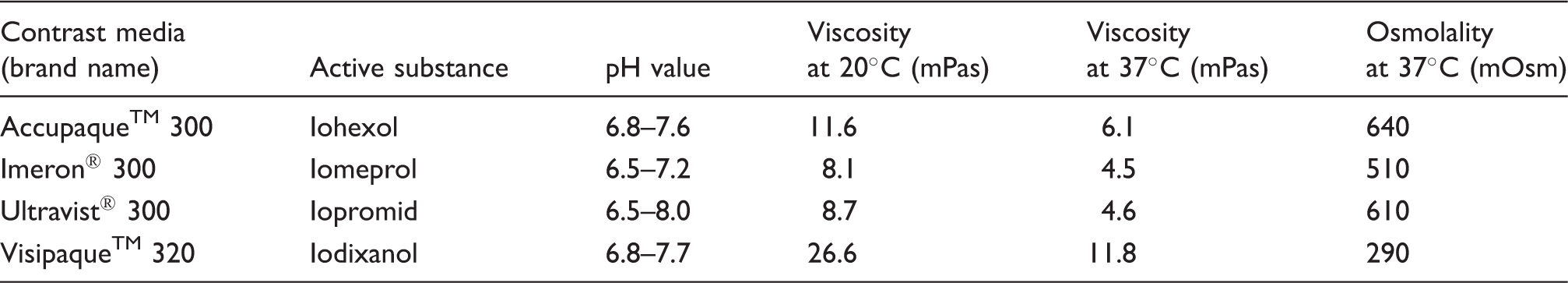
Physiochemical characteristics of the non-ionic contrast media.
High-performance liquid chromatography method
Each sample was assayed three times by a validated stability indicating high-performance liquid chromatography (HPLC) assay with photodiode array detection (PDA) to analyze the concentration and purity of epirubicin. The HPLC system consisted of a Waters 717 plus Autosampler, a Waters 510 HPLC-pump, and a Waters 996 photodiode array detector. Waters Empower pro, Empower 2 software, version 6.10.01.00 was used to acquire and analyze the data. The concentrations of epirubicin were determined by using a Symmetry® column C18 (250 × 4.6 mm) with a particle size of 5 µm (lot 022338079, MZ-Analysentechnik, Mainz, Germany). The mobile phase consisted of 27.5% acetonitrile (ACN) HPLC Gradient Grade (lot 134 622, 2.5 L, Promochem, Wesel, Germany) and 72.5% 0.05 M potassium dihydrogen phosphate buffer solution (PBS) (pH = 4.6). The PBS pH = 4.6 was prepared by solving 6.8 g potassium dihydrogen phosphate (Merck, Darmstadt, Germany, lot A0185177031) in 1000 ml water HPLC Gradient Grade (Applichem, Darmstadt, Germany, 2.5 L, lot 4Q009403). The pH was adjusted by using 85% ortho phosphoric acid (AppliChem GmbH, Darmstadt, Germany, lot 7A005123). The washing solution consisted of 95% water HPLC grade and 5% ACN HPLC grade. The flow rate was set at 1.5 ml/min, and the injection volume was 10 µL. PDA wavelength was 190–600 nm with the detection wavelength of 479 nm. Under these conditions, the retention time of the epirubicin peak was about 5–7 min (see Figures 1 to 3). The identity of the epirubicin peak was confirmed by concentration-dependent changes of the peak area and PDA chromatograms. Run time was set at 10 min.
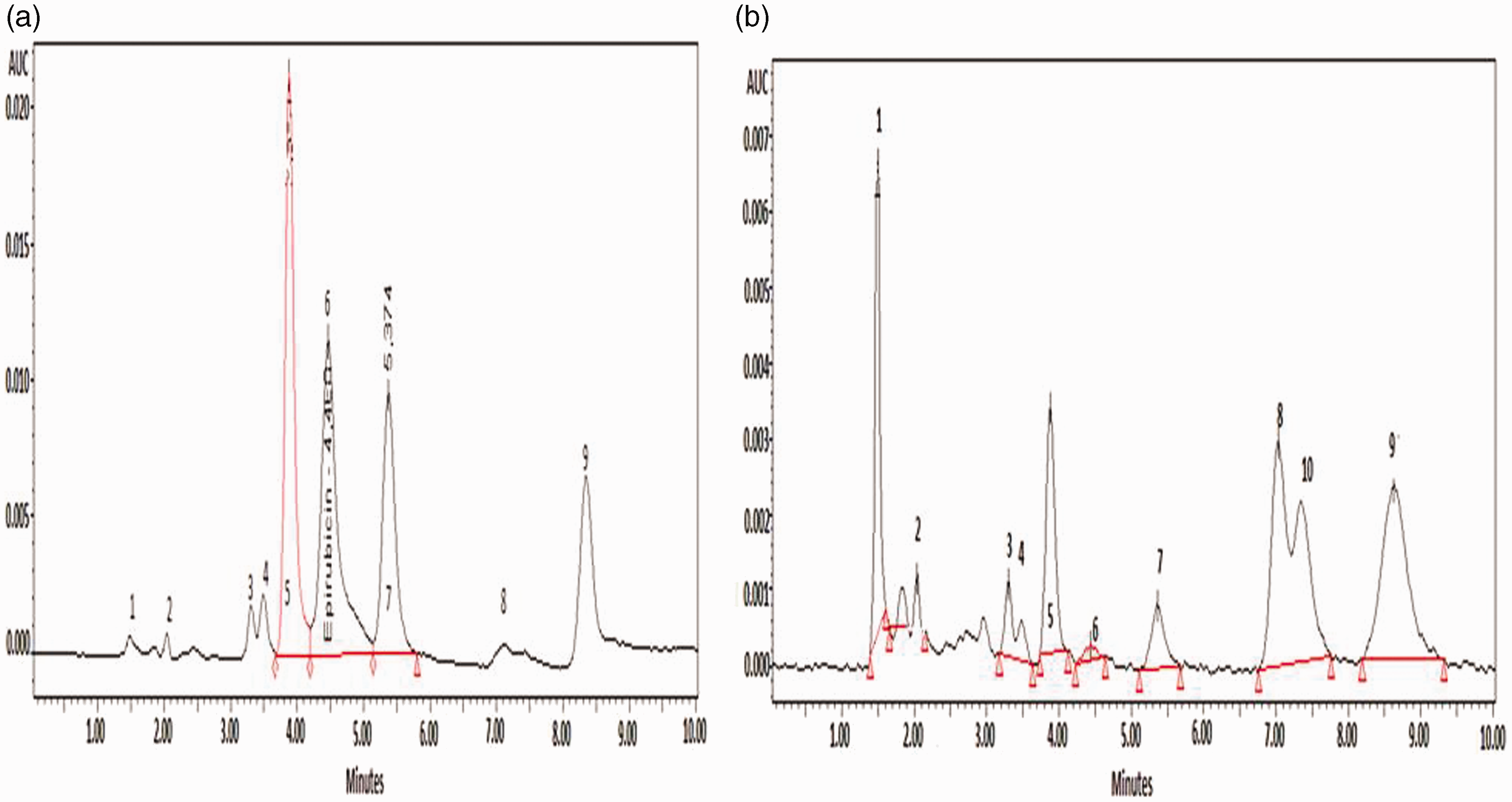
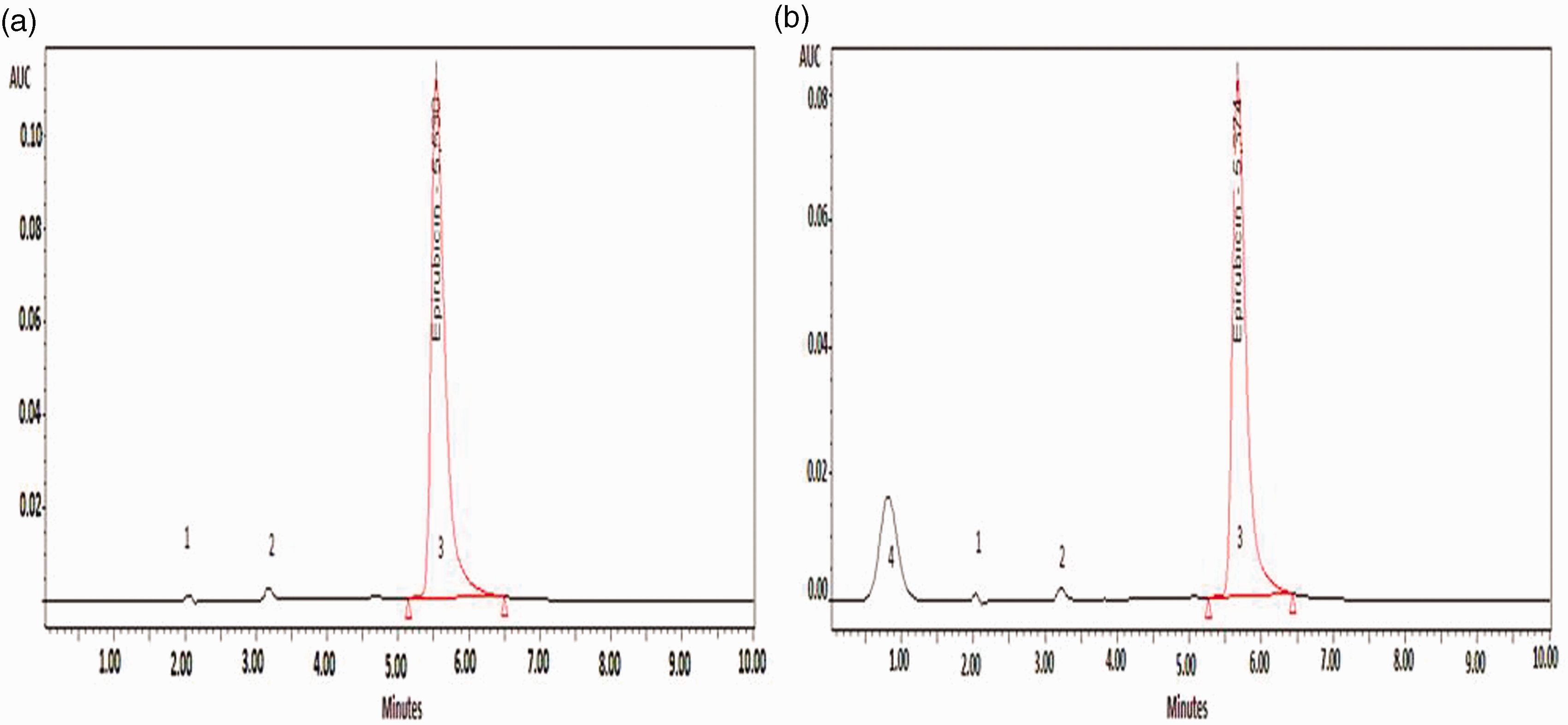
HPLC PDA chromatogram of epirubicin. Chromatograms of alkaline (1 mol NaOH, pH 13) degradaded epirubicin solution (2 mg/ml); (a) without heating, (b) after heating. Chromatograms of acid (HCl, pH 0.2) degradaded epirubicin solution (2 mg/ml); (a) without heating, (b) after heating.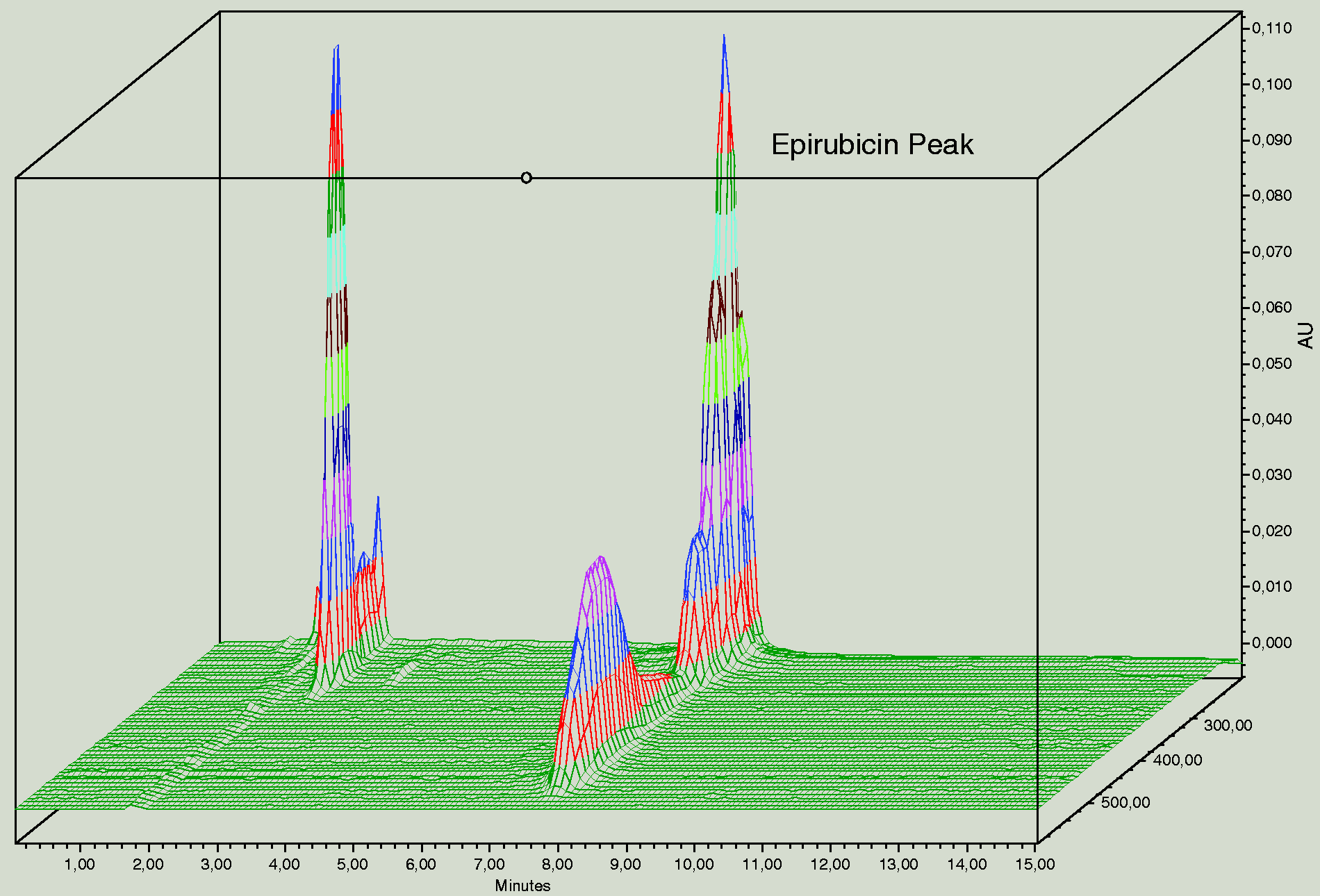
Suitability of the HPLC assay
Suitability of the HPLC method was proven by analyzing forced degraded samples of epirubicin injection concentrate. The injection solution (2 mg/ml epirubicin) was acidified with hydrochloride acid in order to achieve pH values of 0.2, 1.0, 2.0, and 3.6 and was heated in a water bath at 60℃ for 2 h. The injection solution (2 mg/ml epirubicin) was alkalinized with 1 mol sodium hydroxide solution to pH = 13 and heated in the same manner. Solutions were diluted with phosphate buffer 1:5 and assayed. Under alkaline pH conditions, the color of the epirubicin solution changed from red to deep purple. The peaks of the degradation products did not interfere with epirubicin parent peak. After heating, intact epirubicin was not detectable any longer (see Figure 2). Acidic degradation resulted in the hydrolysis of the glycosidic bond and formation of the deep red-colored aglycone doxorubicinon. The peak of the aglycone degradation product did not interfere with the epirubicin parent peak (see Figure 3). These results confirm the suitability of the HPLC method implemented.
Validation of the HPLC assay
The method was validated following the ICH Harmonised Tripartite Guideline for Validation of analytical procedures: text and methodology Q2 (R1).
Linearity and calibration curve
In order to study the linearity of the calibration curve, epirubicin solutions of different concentrations (n = 15) were prepared by diluting a 1:10 dilution Epimedac® injection concentrate with calculated amounts of PBS in order to achieve the nominal epirubicin concentrations 3, 5, 10, 20, 30, 50, 80, 90, 100, 110, 120, 150, 170, 180, and 200 µg/ml. Aliquots of the calibration standards were injected in triplicate. The calibration curve was constructed by analyzing plots of the peak area versus epirubicin concentrations. The correlation coefficient of R2 = 0.998375 proved linearity over the concentration range. The equation of calibration curve was y = 6.9464 x + 40.283.
Accuracy
Accuracy was evaluated with four different quality control solutions (20 µg/ml, 50 µg/ml, 100 µg/ml, 150 µg/ml) and five-fold injection. The mean recovery was 100.05% ± 0.27% (n = 20). The accuracy was 100.57 ± 0.24% for 20 µg/ml, 99.84 ± 0.71% for 50 µg/ml, 99.64 ± 1.72% for 100 µg/ml, and 100.19 ± 0.57% for 150 µg/ml.
Intra-day precision
Intra-day precision was determined by five-fold injection of four quality control solutions nominally containing 20 µg/ml, 50 µg/ml, 100 µg/ml, and 150 µg/ml epirubicin. Intra-day precision expressed as relative standard deviation was 0.99% for 20 µg/ml, 1.54% for 50 µg/ml, 1.53% for 100 µg/ml, and 1.94% for 150 µg/ml epirubicin.
Inter-day precision
Inter-day precision was determined by four-fold injection of four quality control solutions (20 µg/ml, 50 µg/ml, 100 µg/ml, 150 µg/ml epirubicin) on five different days. Inter-day precision expressed as relative standard deviation was 3.25% for 20 µg/ml, 0.95% for 50 µg/ml, 0.75% for 100 µg/ml, and 0.89% for 150 µg/ml.
Limit of detection (LOD)
The limit of detection was calculated by the equation LOD = 3.3 SD/s, where s represents the slope of the calibration curve and SD the standard deviation of the peak area. The LOD amounted to 0.025 µg/ml of epirubicin.
Limit of quantification (LOQ)
The limit of quantification was calculated by the equation LOQ = 10 SD/s, where s represents the slope of the calibration curve and SD the standard deviation of the peak area. The LOQ amounted to 0.075 µg/ml of epirubicin.
Experiments
Bead-slurry (8 ml) either DC bead™ 100–300 µm or DC beadM1™ was transferred via an 18-gauge needle into an empty 50 ml or 10 ml syringe made of polypropylene (PP). After sedimentation of the beads, the excess solution was expelled via a 5 µm filter needle; 2 ml beads remained in the syringe.
Loading of DC bead™ with 2 mg/ml epirubicin hydrochloride solution
38 ml epirubicin injection solution 2 mg/ml was transferred into a 50 ml syringe. The syringe was connected by a female/female connector to the 50 ml syringe containing the bead slurry and the epirubicin solution was pushed into the syringe. These experiments had to be performed with 76 mg epirubicin referring to clinical practice and volumetric measurement of the drug solution with a 50 ml syringe calibrated in milliliters. Loading was performed under static conditions over a period of 6 h. Each test suspension was prepared in triplicate.
Loading of DC bead™ with 25 mg/ml epirubicin hydrochloride solution
Each vial of epirubicin powder for reconstitution 50 mg was reconstituted with 2 ml water for injection. The resulting epirubicin concentration amounted to 25 mg/ml; 3 ml of reconstituted epirubicin injection solution 25 mg/ml was drawn up into the bead containing 10 ml syringe to load the beads with 75 mg epirubicin under static conditions over a period of 2 h. Each test suspension was prepared in triplicate.
At the end of the loading period, the color of the beads had changed from blue to red and the color of excess solution had become lighter. The excess solution was removed from syringes by pushing it into empty bags. Samples were withdrawn from the excess solutions and the concentration of epirubicin measured by HPLC. Prior to the HPLC assay, samples were diluted with PBS (pH = 4.6) 1:3 when Epimedac® was used for loading or 1:40 when Farmorubicin® was used for loading in order to obtain concentrations in the range of calibration curve. The loading rate was calculated by using equation (1).
Compatibility of epirubicin-loaded beads with different non-ionic contrast media
After removal of the excess solutions, the epirubicin-loaded beads were mixed with different contrast media (Accupaque™ 300, Imeron® 300, Ultravist® 300, Visipaque™ 320) in a 1:1 ratio and agitated in a controlled manner to obtain a homogeneous suspension. Samples (0.3 ml) were withdrawn via a 5 µm filter needle and diluted 1:5 with PBS pH = 4.6 on day 0, 1, and 7. Aliquots were injected three times and assayed by HPLC in order to determine the remaining loading level of epirubicin in the beads.
Calculation of the percentage rate eluted was performed by the following equation:
Results
Epirubicin loading levels of epirubicin-loaded DC Bead™ (bead size 70–150 µm, 100–300 µm) mixed with different contrast media and stored protected from light under refrigeration.
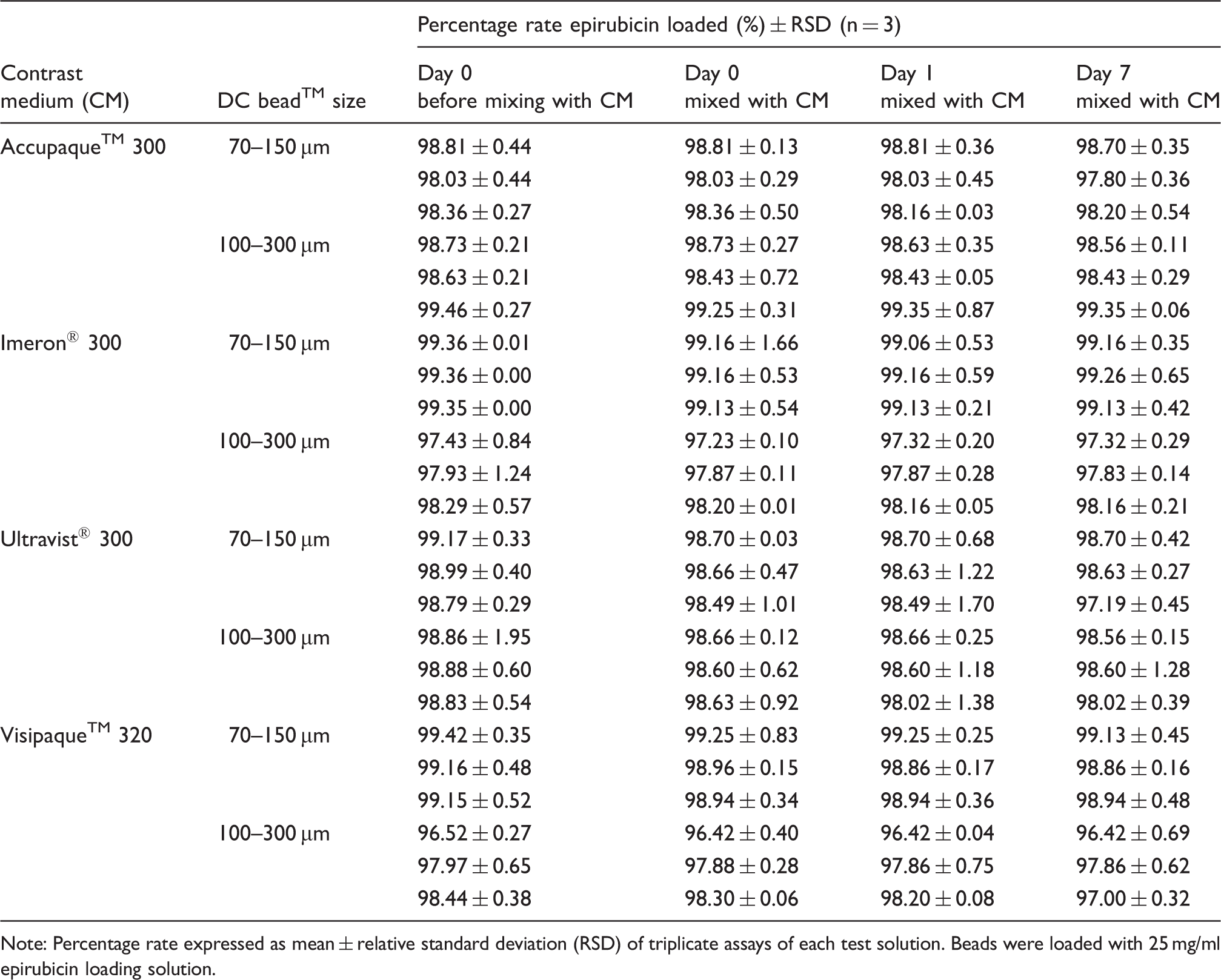
Note: Percentage rate expressed as mean ± relative standard deviation (RSD) of triplicate assays of each test solution. Beads were loaded with 25 mg/ml epirubicin loading solution.
Epirubicin loading level of DC Bead™ (bead size 70–150 µm, 100–300 µm) mixed with different contrast media and stored protected from light under refrigeration.
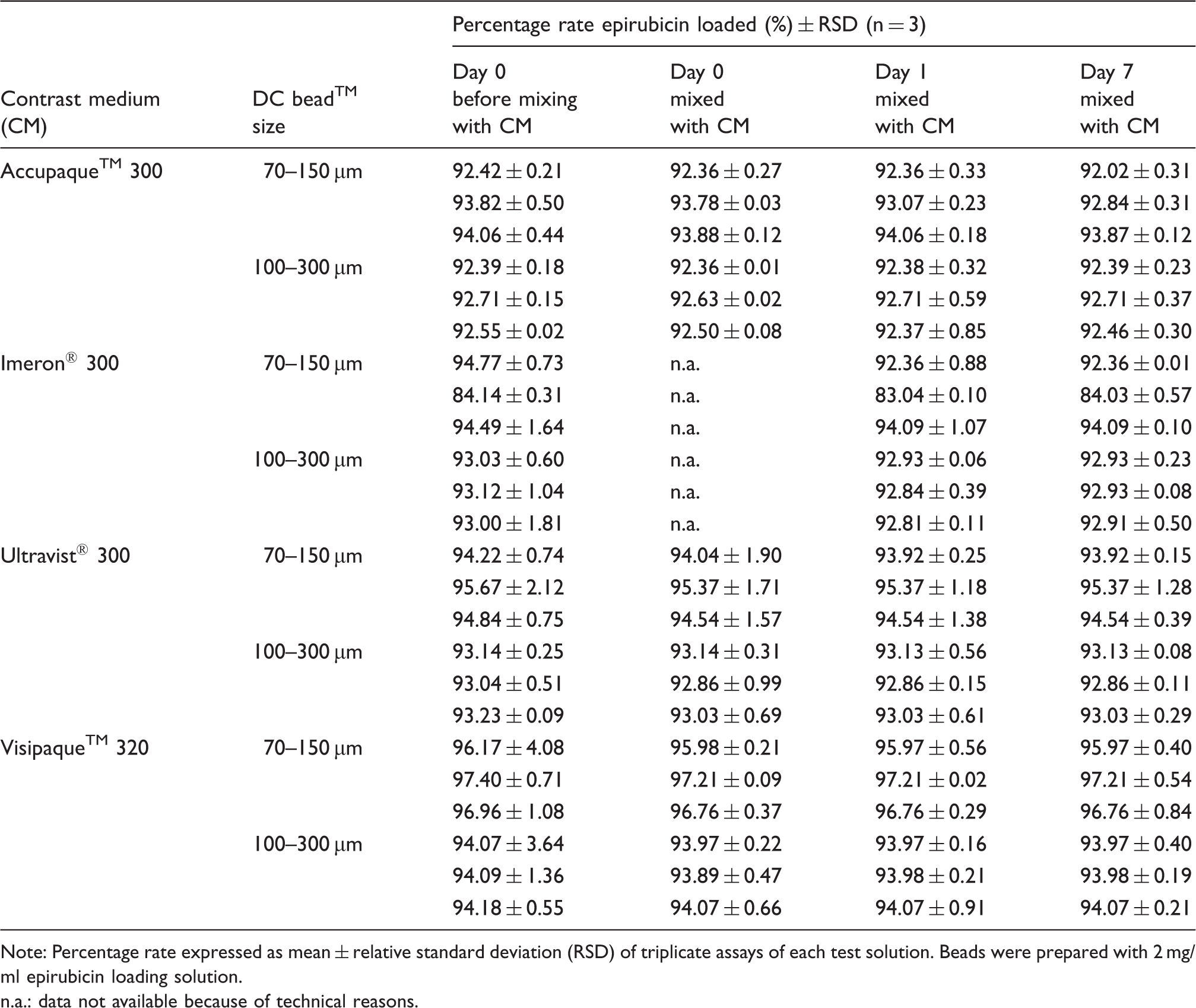
Note: Percentage rate expressed as mean ± relative standard deviation (RSD) of triplicate assays of each test solution. Beads were prepared with 2 mg/ml epirubicin loading solution.
n.a.: data not available because of technical reasons.
Discussion
In order to determine the concentration and purity of epirubicin, at first the HPLC method previously reported by Sobczak et al. was implemented. 14 But the resulting epirubicin peak was not symmetric and feasible for accurate peak area calculation. Therefore, an alternative HPLC assay was developed by testing different types of columns and mobile phases (ACN: Water and ACN: PBS in different ratios and different pH values) as well as different flow rates in the range from 0.7 ml/min to 1.5 ml/min. Finally, a Symmetry® column C18, a mobile phase consisting of 72.5% PBS (pH 4.6) and 27.5% ACN, and a flow rate of 1.5 ml/min revealed to be suitable for the separation and quantification of epirubicin. This HPLC assay was shown to be stability indicating and valid. Of note, minimal variations of the liquid phase caused shifts in the retention time (see Figures 1 to 3). The small differences between the epirubicin loading levels of the different test solutions when the same bead sizes and the same concentration of the loading solution were used are to be explained by the loss of a few beads during the loading procedure and preparation of samples. Therefore, the results are given for each test combination on its own and not for the average of three test combinations. However, it is obvious that the loading level of epirubicin increases about 1% when DC bead™ M1 is loaded and otherwise unchanged parameters. This phenomenon results from the larger surface area of beads with a lower diameter and more efficient loading during the same period. Using a 2 mg/ml concentrated epirubicin solution for loading the same loading efficiency is only reached after a longer loading period. After 6 h, loading was not completed, but sufficient in order to test the admixture compatibility with contrast media.
Like other cytotoxic preparations, the loading of DC bead™ is in general performed in a centralized pharmacy-based cytotoxic preparation unit. As the drug-loaded beads are administered admixed to contrast media, the question arises whether compatibility of the admixtures is given and whether admixtures with contrast media can be prepared in advance by the pharmacy staff. To give a valid answer to these questions, the compatibility and stability of epirubicin-loaded beads with four different non-ionic contrast media which are conventionally used by the interventional radiologists, were studied over a maximum period of seven days.
The results confirm compatibility and stability of the admixtures with different non-ionic contrast media. The minimal percentage rates (0.1–0.5%) of epirubicin eluted did not allow the identification of differences caused by the bead sizes or the loading levels. Of note, any influence of the viscosity or osmolality of the various contrast media on the elution rate was not verifiable for the same reason. Potential degradation products of epirubicin are not to be expected and most probably existing only below the LOD. Adsorption of epirubicin to the PP syringes or HPLC glass vials can also be excluded. Hecq et al. also reported stability of doxorubicin after admixture of doxorubicin-loaded DC bead™ with Omnipaque 350 and storage over seven days under refrigeration. 12 In their experiments, higher concentrations of doxorubicin and impurities were measured in the samples utilized after seven days. However, the experiments clearly show that there is no significant elution of epirubicin or doxorubicin from loaded DC bead™ after admixture and storage with non-ionic contrast media. The ionic binding of the positively charged anthracycline drugs to the sulfonate groups of DC bead™ is much stronger than this of irinotecan, which is eluted by contrast media in a percentage rate up to 10% after mixing. 13
Conclusion
Compatibility of epirubicin-loaded DC bead™ with an equal volume of different contrast media in PP syringes is given over a period of seven days. Due to a maximum elution of 0.1–0.5% of epirubicin from loaded DC bead™, admixtures with contrast media can be prepared in advance in centralized cytotoxic preparation units. Microbiological aspects have to be considered when determining the expiration date of the product.
Footnotes
Acknowledgements
We are grateful to Rachel Holden, Biocompatibles UK Ltd, a BTG International Group Company for reviewing the manuscript.
Declaration of Conflicting Interests
The author(s) declared no potential conflicts of interest with respect to the research, authorship, and/or publication of this article.
Funding
The author(s) disclosed receipt of the following financial support for the research, authorship, and/or publication of this article: This study was supported in part by a grant of Biocompatibles UK Ltd, a BTG International Group Company.