






Abstract
Abstract
Introduction
We aimed to assess the effect of VEGF-A, PDGF-BB, and c-Met expression levels on survival in patients with metastatic colorectal cancer receiving bevacizumab therapies.
Patients and methods
A total of 105 patients diagnosed with metastatic colorectal cancer between the years 2006 and 2016 were included in the research retrospectively.
Results
The progression-free survival (PFS) durations of patients with high expression levels of VEGF-A and with low expression levels of VEGF-A were 11 months and 10 months (p = 0.44), respectively. The PFS durations of patients with high PDGF-BB expression and low PDGF-BB expression were 12 months and 10 months (p = 0.16), respectively, while the PFS durations of patients with high and low c-Met expression were 8 months and 13 months (p = 0.005), respectively. Metastatic overall survival was 27 months and 18 months (p = 0.05) in patients with high and low VEGF-A expression levels, respectively, 31 months and 21 months (p = 0.16) in patients with high and low PDGF-BB expression levels, respectively, and 21 months and 26 months (p = 0.11) in patients with high and low c-Met expression levels, respectively.
Conclusion
The results of this research revealed a high c-Met expression relationship with worse PFS and low VEGF-A expression associated with poor metastatic overall survival in patients with metastatic colorectal cancer receiving bevacizumab therapies.
Introduction
Colorectal cancer (CRC) is the third most prevalent malignant neoplasm in both women and men. The prognosis is better recently compared to years ago, and the median survival time has reached 25 months in metastatic patients with targeted therapies. 1 The combination of chemotherapy and bevacizumab is one of the standard therapies for advanced CRC. Angiogenesis plays a significant role in carcinogenesis, metastasis and neoplasm growth in patients with CRC. 2
Among the molecular signals that initiate new blood vessel formation, vascular endothelial growth factor-A (VEGF-A) is one of the most efficacious molecules, which induces many downstream effects in endothelial cells. 3 VEGF-A is increased in many types of malignant tumors and is related to poor disease prognosis and angiogenesis. VEGF-A is an anti-apoptotic survival factor for vascular endothelial cells as well as permeability and the proliferating factor. 4 Inhibiting VEGFR signaling could have a therapeutic effect not only by preventing angiogenesis but also by leading to regression of vascular endothelial cells in the tumor microenvironment. Bevacizumab is a recombinant monoclonal antibody against VEGF-A that inhibits its binding to VEGFR and the activation of downstream signaling. 5
The platelet-derived growth factor receptor (PDGFR) modulates tumor progression by affecting the tumor endothelial cells, the stromal cells, and the tumor cells. Also, PDGFR signaling has been identified as enhancing the proliferation of malignant cells in an autocrine effect, inducing angiogenesis, recruiting pericytes (which regulate the tumor vascularity), and controlling the interstitial fluid pressure to effect trans vascular transfer of chemotherapeutics in a paracrine effect. 6
A tyrosine kinase receptor, c-Met, for which HGF is the only ligand, is activated, overexpressed, amplified, or mutated in different types of cancers. 7 HGF is produced by mesenchymal cells—including macrophages, endothelial cells, and fibroblasts. 8 In various cancers, c-Met plays an extensive role in malignant tumor cell proliferation, invasion, and antiapoptotic effects. 7 The c-Met overexpression was associated with poor prognosis in patients with metastatic CRC. 9 In the subgroup analysis of this study, who was treated with bevacizumab-containing systemic chemotherapy, c-Met overexpression was associated with worse PFS. 9
Despite the enhanced number of proteins and signaling pathways that have been discovered to be closely linked with tumor angiogenesis, 10 the role of chemokines in the tumor microenvironment that bolster CRC angiogenesis remains unknown. We do not have a biomarker to predict patients who will benefit from anti-angiogenic treatment in metastatic CRC. The effects of cytokines and biomarkers, which are effective in the process of angiogenesis in different cancers, including CRCs, have been evaluated previously. However, there is no study evaluating VEGF-A, PDGF-BB, and c-Met together. The goal of the present study is to detect the predictive importance of VEGF-A, PDGF-BB, and c-Met in patients with advanced CRC treated with first-line bevacizumab combined chemotherapy.
Materials and methods
A total of 105 metastatic CRC patients who developed metastasis at the time of diagnosis or during follow-up between the years 2006 and 2017 were included in this research. In the first line, the patients who received chemotherapy + bevacizumab combined therapy were included in the study retrospectively. The research was endorsed by the Ethics Committee (ethics committee no: 2015/567). The VEGF-A, PDGF-BB, and c-Met expression levels analyzed in tumor paraffin blocks of patients by immunohistochemical (IHC) analysis were compared with survival times.
Immunohistochemical analysis
For IHC staining, a paraffin-embedded block fixed in 10% formalin that belongs to the tissue best representing the tumor was selected, and the 5 µm thick sections were taken onto poly-L-lysine-coated slides. After the sections were kept in the oven for 1 h at 60 °C, they were passed through xylol and decreasing concentrations of alcohol and washed with distilled water. They were boiled with sodium citrate (pH = 6.0) for 20 min in the microwave. Preparations that were placed on the immunohistochemical staining device (Ventana-Benchmark XT) were stained by IHC.
Primary antibodies and control tissues
In immunohistochemical staining, cytoplasmic, cell membrane and extracellular matrix staining for VEGF-A, cytoplasmic staining for PDGF-BB, and membranous staining for c-Met were evaluated as positive. The antibodies used in IHC staining, control tissues, and cut-off values are shown in Table 1.11–13 The prepared sections were stained, and light microscopic examination was performed (Nikon Eclipse Ni). The evaluation was performed by a pathologist who was unaware of the groups.
Antibodies for IHC.

Clinical response evaluation and survival times
Patients with a basally measurable lesion were included in the study. The evaluation was made according to the RECIST 14 criteria and clinically, such as FIRE-3 study. 15 The time from diagnosis to death or the last check-up date was accepted as overall survival (OS); the time from metastasis to death or the last check-up date was considered as overall metastatic survival (mOS). The period from the onset of palliative chemotherapy to progression was evaluated as progression-free survival (PFS).
Statistical analysis
SPSS 22 (SPSS, Chicago, IL, USA) was used for statistical analysis. Qualitative variables were compared using the chi-square/Fisher’s exact tests. Quantitative variables were compared using the Mann–Whitney U test. The association with survival was analyzed by Kaplan–Meier plot and log-rank test. p Values < 0.05 were considered statistically significant. Univariate analyses for OS and PFS were performed using the Cox proportional hazards model.
Results
A total of 105 patients with histopathologically confirmed CRC were included in the study. The median age was 60 (24–83). Sixty-eight (64.8%) of the patients had stage-4 metastasis at diagnosis, and 37 of them developed metastasis during follow-up. The number of patients undergoing primary surgery was 88 (83.8%). The general demographic features of patients are shown in Table 2.
Patient characteristics.

VEGF-A, PDGF-BB, c-Met expression levels
High VEGF-A expression was determined in 78 (74.3%), high PDGF-BB expression was determined in 24 (22.9%), and high c-Met expression was determined in 52 (49.5%) of the patients. However, due to technical reasons, the results could not be determined in one patient for VEGF-A staining, in two patients for PDGF-BB staining, and in five patients for c-Met staining (Table 3). No significant correlation was found between VEGF-A, PDGF-BB, c-Met expression and clinicopathological parameters (Table 4).
Molecular stainings of patients’ tumor tissue.

The relationship between VEGF-A, PDGF-BB, and c-Met expression levels and other parameters.

The relationship between VEGF-A, PDGF-BB, c-Met expression levels
In 21 (27.6%) of 76 patients with high VEGF-A expression, high PDGF-BB expression was also detected (p = 0.09). Of the 50 patients with high c-Met expression, 15 (30%) had a high expression of PDGF-BB (p = 0.11). Of the 47 patients with low c-Met expression, 27 (57.4%) showed high VEGF-A expression (p < 0.001).
Survival analysis (PFS, mOS)
In a group with low expression of VEGF-A, PFS was 10 months (95% CI 8–12), whereas in a group with high expression of VEGF-A, PFS was 11 months (95% CI 7–13) (p = 0.44).
PFS in a group with low expression of PDGF-BB was 10 months (95% CI 8–12), whereas PFS in a group with high PDGF-BB expression was 12 months (95% CI 6–18) (p = 0.16).
In a group with low expression of c-Met, PFS was 13 months (95% CI 10–16), and in patients with high c-Met expression, PFS was eight months (95% CI 7–9) (p = 0.005) (Figure 1).
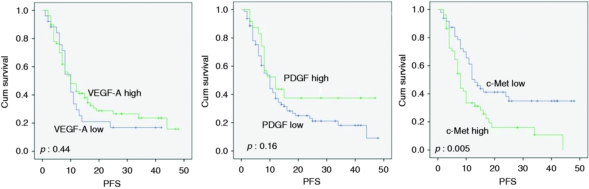
Progression-free survival (PFS) according to VEGF-A, PDGF-BB and c-Met expressions.
Metastatic overall survival (mOS) in a group with low expression of VEGF-A was 18 months (95% CI 14–22), whereas it was 27 months (95% CI 19–35) (p = 0.05) in a group with high expression of VEGF-A.
In a group with low expression of PDGF-BB, mOS was 21 months (95% CI 16–26), and in a group with high expression of PDGF-BB, mOS was 31 months (95% CI 5–57) (p = 0.16).mOS was 26 months (95% CI 16–36) in a group with low expression of c-Met, and it was 21 months (95% CI 15–27) in a group with high c-Met expression (p = 0.11) (Figure 2).
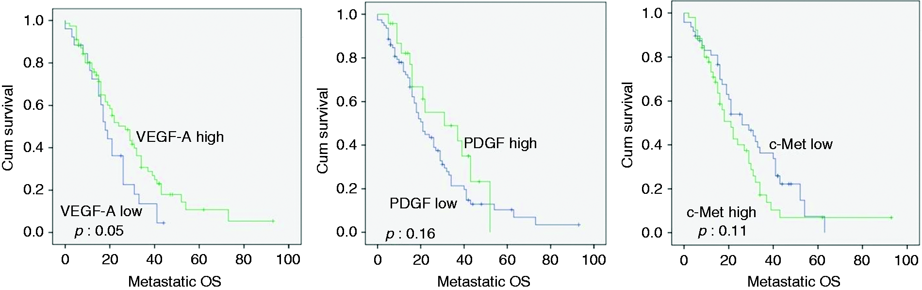
Metastatic overall survival curves according to VEGF-A,PDGF-BB and c-Met expressions.
Univariate analysis of parameters affecting mOS and PFS
In univariate analysis, the patients were evaluated according to the status of being over/under 65 years, gender, having primary surgery, single/multiple site metastasis, right/left colon localization, liver/lung metastasis, VEGF-A, PDGF-BB, and c-Met expression status.
There was a 1.6-fold increase in the risk of death in those with low expression of VEGF-A compared to those who showed high expression (p = 0,05; 95% CI 0,97–2,62); while patients with high c-Met expression showed a 1.9-fold increase in the risk of progression compared to those with low expression (p = 0,008; 95% CI 1,18–3,08). In univariate analyses, there was found only one significant parameter. Hence, multivariate analysis was not performed.
Discussion
In the literature, there are no data in mCRCs on the effect of VEGF-A subgroup expression on survival in patients receiving bevacizumab treatment. However, our study was the first research to evaluate the impact of VEGF-A expression on survival in tumor tissues.
Recently, newly developed targeted therapies have led to significant improvements in the survival of metastatic CRC patients. The most important of these therapies is the bevacizumab monoclonal antibody developed against VEGF-A, which is effective in the inhibition of angiogenesis. This agent showed a significant improvement in survival when combined with chemotherapeutic treatments.16,17 However, all patients receiving bevacizumab treatment do not show the same effect. This effect may be associated with different molecular features of the tumor. Pre-determination of the groups that will benefit from bevacizumab treatment will help in patient selection.
Determining the prognostic factors associated with chemotherapy response in CRC patients may allow us to predict the course of the disease. Previously, it was reported in CRC patients that plasma concentrations of angiogenesis-related molecules were significantly increased and associated with prognosis.18,19
VEGF-A is the most widely studied and pharmacologically targeted VEGF family member in the process of angiogenesis. VEGF is produced in tumors by hypoxic tumor cells, endothelial cells, and tumor-associated macrophages. Blocking the VEGF pathway with antibodies, recombinant fusion proteins, or tyrosine kinase inhibitors has been shown to prolong survival in stage-4 CRC patients. In a study, a significant correlation was found between tumor size and stage and VEGF expression. 20 But, there was no relationship with VEGF-A expression and age, gender, colon localization, K-RAS status, multiple site metastasis, liver, lung, and brain metastasis in our study. It was investigated whether VEGF had predictive value in mCRC patients receiving bevacizumab; no significant difference was found. 21
CRC is the first malignancy where clear evidence was shown for the efficacy of anti-VEGF strategy in randomized trials. Recently, newly developed targeted therapies have led to significant improvements in the survival of metastatic CRC patients. VEGF serum levels were used in the evaluation of chemotherapy responses and significantly decreased VEGF levels were observed in patients who showed partial response. 19
In our study, progression-free survival of patients with high VEGF-A expression tended to be better than those with low expression, while there was a meaningful increase in mOS. This may be explained by the fact that 32 (53.3%) of the patients who received second-line palliative chemotherapy received a treatment protocol with bevacizumab.
PDGF is a potential growth factor for mesenchymal cells. PDGF-R expression in the stroma has been reported to be related to worse prognosis in various malignant tumors, including breast, prostate, and pancreatic cancers. 22 Of patients with early-stage CRC, those with high PDGF-BB levels had a higher incidence of vascular invasion, and those with high serum PDGF-BB levels had lower life expectancy compared to those with low levels. Therefore, in early-stage CRC patients with high serum levels of PDGF-BB, the cancer is spreading faster, and they show worse prognosis. 23 Similarly, in a study of triple-negative breast cancer, a high PDGF expression level was found to be correlated with lymph node metastasis, recurrence, and central nervous system metastasis. 24 On the other hand, PDGF-BB levels did not correlate with PFS and OS in patients with sarcomatoid non-small cell lung cancer. 25 Similarly, in our study, in patients with high PDGF-BB expression, PFS and mOS tended to increase compared to those with low PDGF-BB expression but did not reach statistical significance. Also, PDGF-BB expression did not show correlation with age, gender, colon localization, K-RAS status, multiple site metastasis, liver, lung, and brain metastasis.
c-Met, a hepatocyte growth factor receptor, is a tyrosine kinase receptor that can activate different signaling pathways leading to proliferation, motility, invasion, and metastasis. This is done especially by triggering angiogenesis. It increases the growth and movement of vascular endothelial cells. It initiates angiogenesis by playing a role in neovascularization in tumor tissue and the release of VEGF. It has been found that c-Met is associated with a metastatic character in many types of cancers. 26 In particular, the association of c-Met ligand HGF with disease progression has been reported in the evaluation of chemotherapy response in patients with mCRC. 19 It has also been reported that c-Met or HGF overexpression in cervical cancer lesions is associated with worse prognosis and has a value as a prognostic biomarker. 27 High c-Met expression levels have been shown to increase the risk of recurrence and death in patients with triple-negative breast cancer. 13 In another study, patients with c-Met overexpression had a worse prognosis than those with low c-Met expression. 28 Similarly, we found that progression-free survival was shorter in patients with high expression of c-Met compared to those with low expression. However, mOS was also shorter, but it did not reach significance. In another study, c-Met expression was not correlated with histological grade, perineural invasion, tumor stage, or metastasis in diagnosis. 29 Similarly, we could not find a correlation between c-Met expression and age, gender, colon localization, K-RAS status, multiple site metastasis, liver, lung, and brain metastasis.
In the study of Chen B et al., the correlated expression level of PDGF-BB and VEGFR-3 was shown to have a strong value in the prognosis of hepatocellular cancer patients. 30 In the correlation analysis between VEGF-A, PDGF-BB, and c-Met, there was only relationship between high VEGF-A and low c-Met expression. Similarly, no correlation was found between PDGF-BB and c-Met expression. However, a positive correlation was found between high VEGF-A expression and low c-Met expression.
In conclusion, while a few biomarkers that predict bevacizumab treatment response in mCRC patients have been studied in the literature, our study is the first in which VEGF-A, PDGF-BB, and c-Met levels were evaluated together. High VEGF-A and PDGF-BB expression levels in tumor tissue can be evidence of active angiogenesis. Long-term survival with bevacizumab treatment can be predicted in patients with high VEGF-A expression. However, the high c-Met expression may be responsible for resistance to antiangiogenic therapy. The use of molecular genetic tests including VEGF-A, PDGF-BB and c-Met in colon cancer will allow individualization of treatments in the near future.
Footnotes
Declaration of Conflicting Interests
The author(s) declared no potential conflicts of interest with respect to the research, authorship, and/or publication of this article.
Funding
The author(s) received no financial support for the research, authorship, and/or publication of this article.
