
Abstract
Background
Leukocytoclastic vasculitis (LCV) is a vasculitic inflammation against blood vessels. Various anticancer therapies can cause vasculitis, but capecitabine-induced LCV is an unusual entity. Here, we describe an LCV case associated with neoadjuvant capecitabine use for locally advanced rectal cancer (LARC).
Case report
A 70-year-old man presented with rectal bleeding. A colonoscopic biopsy revealed rectal adenocarcinoma and he was diagnosed with LARC after imaging studies. Capecitabine plus radiation therapy was started as a neoadjuvant treatment.
Management and outcome
Seven days after the first capecitabine dose, the patient was admitted with a rash. The LCV diagnosis was histopathologically proven. Capecitabine was withheld. After the patient's rash began to regress under corticosteroid pressure, capecitabine was started at a lower dose. His treatment was completed successfully with oral corticosteroids plus low-dose capecitabine.
Discussion
We aimed to point out a rare and unusual adverse effect of a frequently used drug in oncologic practice.
Keywords
Introduction
Locally advanced rectal cancer (LARC) patients are treated with a combination of chemotherapy and radiotherapy followed by surgical resection. 1 Infusional 5-FU or oral capecitabine are equally effective alternatives if the chemoradiotherapy (CRT) modality is chosen. 2 Because of its convenience for oral use, capecitabine is the clinically preferred option. Leukocytoclastic vasculitis (LCV) is a small vessel vasculitis that can develop due to various factors including infections, drugs, malignancies, and systemic diseases. 3 Capecitabine is an antineoplastic agent with prominent side effects such as emesis, diarrhea, mucositis, and hand-foot syndrome. 4 LCV is a rare and unexpected side effect of capecitabine treatment. Here, we report a patient with LARC who develops histologically confirmed cutaneous LCV under neoadjuvant capecitabine treatment.
Case report
A 70-year-old man presented with mild abdominal pain and rectal bleeding. A colonoscopic examination showed a fragile, solid tumoral mass in the distal rectum (1 to 5 cm), and a biopsy revealed an adenocarcinoma. The patient did not have distant metastases in the imaging studies except for the locoregional lymph nodes, so he was evaluated as LARC. Concurrent neoadjuvant CRT (capecitabine 825 mg/m2 twice daily, five days per week—3000 mg/day—plus 50.4 Gy radiation to the pelvic site) was started.
Seven days after the first capecitabine dose, the patient was admitted with a rash. He suffered from fatigue and dysuria but denied other systemic symptoms such as cough, fever, arthralgia, myalgia, abdominal pain, dyspnea, hemoptysis, and hematuria. There was no newly initiated medical treatment except for capecitabine. Physical examination revealed a palpable maculopapular rash on the lower extremities (figure 1). The liver and renal functions, as well as the thrombocyte count, were all within normal limits. C-reactive protein (CRP) was 113 mg/L (normal, 0-5). Urine analysis showed glycosuria due to the patient's diabetes history, but there was no evidence of infective parameters or proteinuria, and the urine culture was negative. Capecitabine was withheld, and 40 mg daily of methylprednisolone was started. Anti-neutrophil cytoplasmic antibody (ANCA) was detected as positive, while antinuclear antibodies and anti-glomerular basal membrane antibodies were negative.
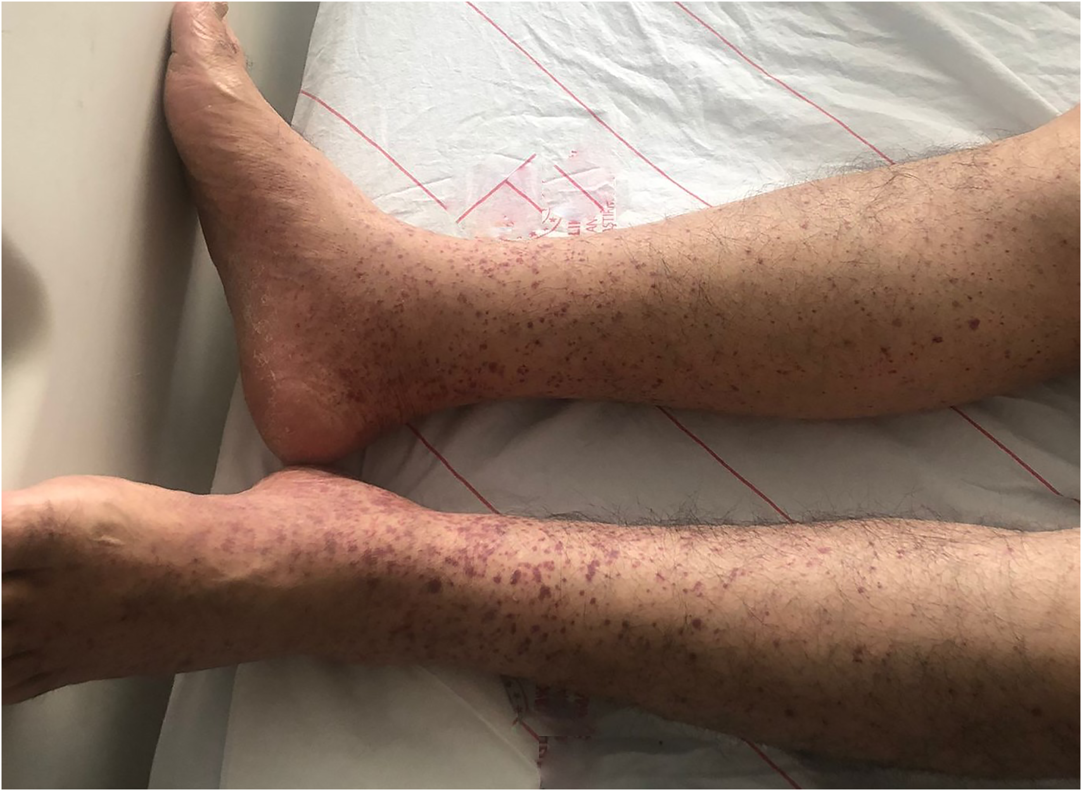
Maculopapular rash in lower extremities.
Based on the clinical and laboratory findings and the temporal relationship between the signs and the administration of capecitabine, drug-induced vasculitis (DIV) was suspected, and a tissue biopsy was performed. Pathology confirmed the diagnosis of LCV due to vasculopathic findings in the biopsy (figure 2). Five days later, the patient's rash regressed under methylprednisolone treatment (figure 3). CRP level decreased to 7.8 mg/L on day 12, and the steroid dose tapered to 20 mg/day. Capecitabine was started at a lower dose (500 mg twice a day) then he was discharged on the 18th day of hospitalization with 16 mg/day methylprednisolone. Radiation treatment was continued for three weeks with low-dose capecitabine, and methylprednisolone treatment was tapered gradually and stopped.

Skin biopsy. Leukocyte infiltration, fibrinoid necrosis and leukocytoclasis with erythrocyte extravasation in small vessel walls of superficial dermis (arrows). (H&E, 20×).
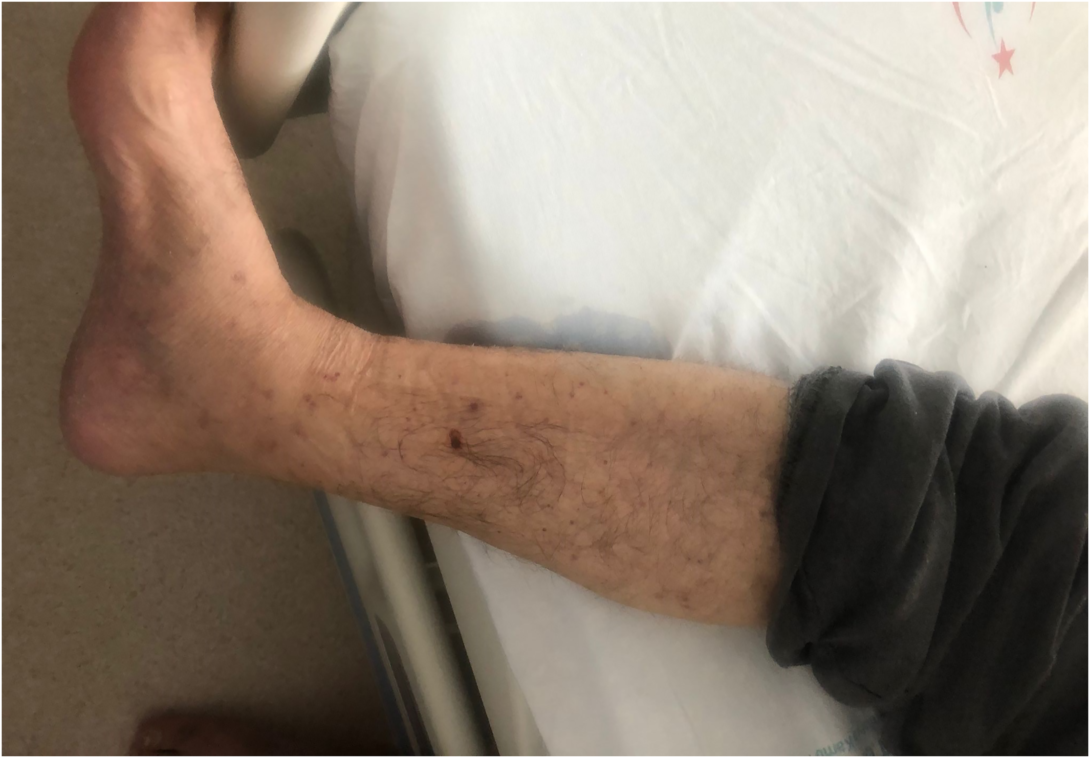
Resolution of rash after steroid therapy.
Approximately three weeks after CRT, the patient was admitted with chest pain while he was waiting for surgery. Cardiac vegetation was detected on his echocardiography. He went into septic shock during his follow-up with infective endocarditis. The patient died in the intensive care unit despite all treatments.
Discussion
LCV is a vasculitic inflammation against blood vessels that is histopathologically characterized by neutrophil infiltration with leukocytoclasia. 3 The main clinical presentation is palpable purpura and mostly lower limbs are affected. While up to 50% of cases are idiopathic; drugs, infections, systemic inflammatory diseases, or malignancies are major secondary causes. 5
Infections are one of the most common triggers for LCV. 3 Our patient did not have any systemic infectious signs and his urine culture was negative for infection despite the complaint of dysuria.
LCVs can be seen secondary to underlying systemic autoimmune diseases and chronic infections such as hepatitis B and C. 3 Our patient had no suspected systemic autoimmune or vasculitic symptoms and no history or evidence of viral or bacterial infection.
LCVs may occur as a paraneoplastic syndrome in the course of malignancies. Paraneoplastic vasculitides account for less than 5% of all vasculitis cases and are mainly associated with hematological malignancies.6,7 In addition, failure of conventional treatments for vasculitis and the disappearance of symptoms with effective treatment of malignancy are the factors indicating paraneoplastic vasculitis.6,8 In our case, the temporal relationship of vasculitis and capecitabine intake, and rapid response to steroid therapy with discontinuation of the suspected drug mostly supports the capecitabin-induced vasculitis. As well as he had not yet received enough treatment to cure his malignancy.
Systemic vasculitis syndromes were another possible diagnosis since ANCA was positive in our patient. However, the presence of ANCA is not diagnostic of systemic vasculitis syndromes, as up to 60% of patients with cutaneous LCV can have a positive ANCA. 5 Besides this, most drugs stimulate ANCA formation as the pathogenesis of DIV. 9 Moreover, drug-induced LCVs tend to occur 7–10 days after exposure to a drug, and withdrawal of the suspected drug helps to improve the symptoms. 3 There were neither systemic symptoms nor laboratory findings indicating ANCA-positive vasculitis in our case.
Medications are another important factor in LCV development.3,9 Various antineoplastic-induced vasculitis cases have been reported including oxaliplatin, 5-FU, immune checkpoint inhibitors, and oral tyrosine kinase inhibitors such as erlotinib and sorafenib.10–14 While the most common capecitabine-induced dermatologic reaction is hand-foot syndrome, 15 LCV is an unusual and rare side effect of capecitabine treatment. To our knowledge, only one capecitabine treatment-associated LCV case has been reported to date. 16 As in that case, during the first week of treatment with capecitabine, histologically proven LCV was detected in our patient. Capecitabine was interrupted for a short time and was continued with a lower dose under corticosteroid treatment. CRT was completed without any problems.
We analyzed the probability of adverse drug reaction using Naranjo's algorithm, 17 and found a score of 3 which means “possible” causality. Our patient had not taken any newly initiated drug other than capecitabine in the last two weeks, and withdrawal of capecitabine relieved the symptoms with the help of steroids. When we evaluated all these findings and data together, we concluded that LCV was related to capecitabine in our patient.
Conclusion
Early recognition, rapid diagnosis, and appropriate treatment with corticosteroids are key points in vasculitis. In all visits, every sign and symptom has to be handled with care. Like in our case, clinicians should be aware of rare and unusual adverse effects of frequently used chemotherapeutics.
Footnotes
Authors’ contributions
DKS designed the case; DKS, MOA and EA collected the data about the case; OK reviewed the pathologic diagnosis; DKS and MOA drafted the manuscript. All authors reviewed and approved the final version of the manuscript.
Declaration of conflicting interests
The author(s) declared no potential conflicts of interest with respect to the research, authorship, and/or publication of this article.
Funding
The author(s) received no financial support for the research, authorship, and/or publication of this article.
Statement of ethics
Written informed consent was obtained from the patient for publication of this case report and accompanying images.