
Abstract
Introduction
Immune checkpoint inhibitors can cause immune-related toxicity in various systems, with myocarditis being the most severe and life-threatening manifestation. This report presents a case in which myocarditis developed following administration of programmed cell death protein-1 (PD-1) inhibitors therapy. We describe the diagnosis and treatment of this patient in detail.
Case report
We present the case of a 59-year-old female diagnosed with post-operative esophageal cancer and hepatic metastases. The patient underwent second-line treatment with domestically-made PD-1 inhibitor, camrelizumab, in combination with paclitaxel (albumin-bound) and carboplatin for two cycles. During the course of treatment, an electrocardiogram (ECG) revealed ST segment elevation in leads II, III, aVF, V2, V3, and V4, along with T wave changes in leads I and aVL. Laboratory examinations showed abnormal levels of N-terminal pro-B-type natriuretic peptide (NT-proBNP) and cardiac troponin T (cTnT). Despite the absence of clinical symptoms, the patient was routinely hospitalized three weeks later. Based on the findings from the ECG, cardiac biomarkers, echocardiography, echocardiogram, cardiac magnetic resonance, and angiography, she was diagnosed with immune-checkpoint-inhibitors-related myocarditis.
Management and outcome
The patient received immunoglobulin (0.5 g/kg/day) and was initially given methylprednisolone (1000 mg/day). Methylprednisolone was gradually reduced to 40 mg/day in 2 weeks. During this time, the levels of biomarkers indicative of myocardial injury also exhibited a simultaneous decline.
Discussion
This case highlights the importance of early detection and prompt intervention, including initiating appropriate steroid therapy and discontinuing of immune checkpoint inhibitors. Such measures can effectively prevent morbidity and mortality, ultimately leading to an improved prognosis.
Keywords
Introduction
Immunotherapy has emerged as a prominent field in oncology treatment in recent years. With the increasing clinical utilization of of various immune checkpoint inhibitors (ICI), the incidence of ICI-related cardiovascular toxicity has been reported frequently in recent studies.1–3 Although the incidence of ICI-related myocarditis is low (about 1%) in clinical research, the consequences may be serious, with a mortality rate of about 50%. 4 However, immune myocarditis is often encountered in the real world. Camrelizumab received approval from China in 2019. Its mechanism of action involves the binding of programmed cell death protein-1 (PD-1) to PD-ligand 1 (PD-L1), and PD-ligand 2 (PD-L2), which are expressed by tumor cells. This binding inhibits the activation and proliferation of T-cells, as well as the production of pro-inflammatory cytokines, leading to a decrease in immune activation. 5 In China, camrelizumab has been approved for various indications, including Hodgkin's lymphoma, advanced hepatocellular carcinoma, non-small cell lung cancer, and esophageal cancer. 6 There have been reports of new-onset arrhythmia, which could potentially be induced by camrelizumab.7,8 This article discusses the clinical features of myocarditis caused by ICI, changes in laboratory examination and immunosuppressive treatment strategies through the analysis of a patient received cancer treatment in our hospital.
Case
We reviewed the patients who received tumor treatment in our hospital from January 2021 to present. A 59-year-old female patient, weighing 51 kg, was diagnosed with esophageal cancer accompanied by extensive hepatic metastases. She presented with mild fatigue. The patients received fluorouracil combined with cisplatin as first-line treatment. The primary lesion of esophagus has been removed. The enhanced MRI of abdomen showed a huge mass (53.29 mm×59.81 mm) in the right lobe of the liver with metastatic lesion (March 2021, Figure 1), who received PD-1 monoclonal anti-body camrelizumab (200 mg intravenous injection, every 3 weeks) combined with paclitaxel albumin-bound (260 mg/m2, every 3 weeks) plus carboplatin (400 mg/m2, every 3 weeks) as second-line treatment. 9 After two cycles of treatment, the patient was routine hospitalized. The patient has no palpitations, chest pain, shortness of breath, and other discomforts except fatigue. Hypertension, diabetes, hyperlipidemia, coronary heart disease, and cerebrovascular diseases were excluded. Tumor control was described as a stable disease. Yet the patient has a unique manifestation of reactive cutaneous capillary endothelial proliferation (RCCEPs) with camrelizumab (Figure 2). Over time, we dynamically monitored her heart index and electrocardiogram (ECG). Laboratory examinations showed carcinoembryonic antigen (CEA) 7.08 ng/ml (non-smoker ≤ 5.00, smoker ≤ 6.50), carbohydrate antigen 72-4 (CA72-4) 8.170 U/ml (0.000–6.900 U/ml), N-terminal pro-B-type natriuretic peptide (NT-pro BNP) 1545.00 pg/ml (reference range < 300 pg/ml) (Figure 3), creatine kinase-MB (CK-MB) 50.8 U/L (reference range < 25 U/L), cardiac troponin I (cTnI) 225.9 pg/ml (reference range < 17.5 pg/ml), and cTnT 665.6 pg/ml (reference range < 14 pg/ml). Compared with the baseline (Figure 4(A)), the ECG showed ST-segment gradually elevation in chest leads V1 to V3 (Suspected front wall and inferior wall myocardial infarction), ST segment change in limb leads I, AVL, II, III, AVF, and chest leads V1–V6 (Figure 4(B)–(D)). Echocardiography showed that the left ventricular ejection fraction (LVEF) was 62%, E/A 1.34, no enlargement in each compartment (Figure 5). No abnormal clinical manifestations were found.
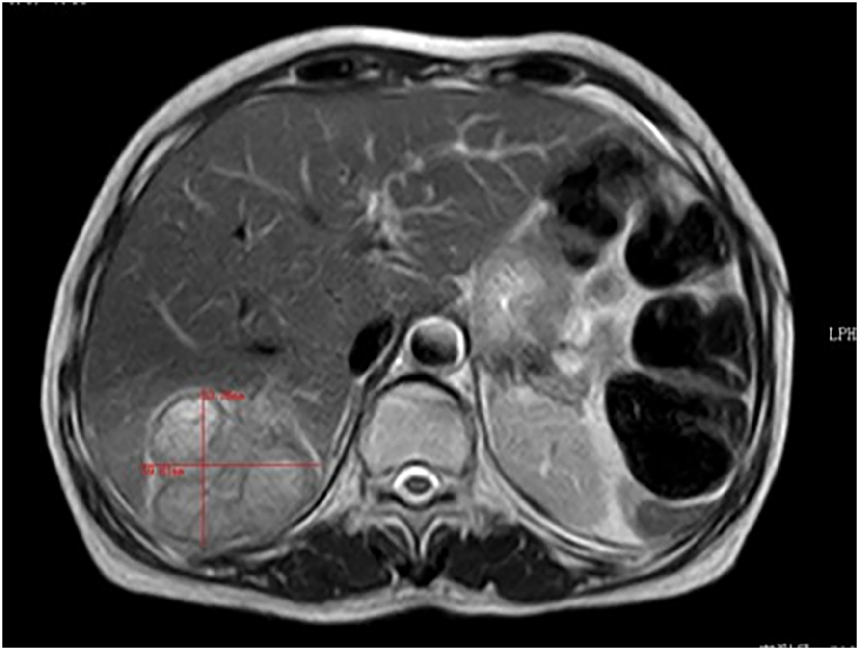
Enhanced MRI of abdomen reveals a huge mass in the liver.
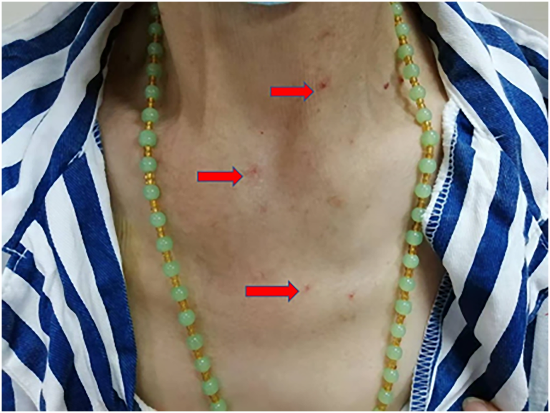
Angiogenesis with camrelizumab.
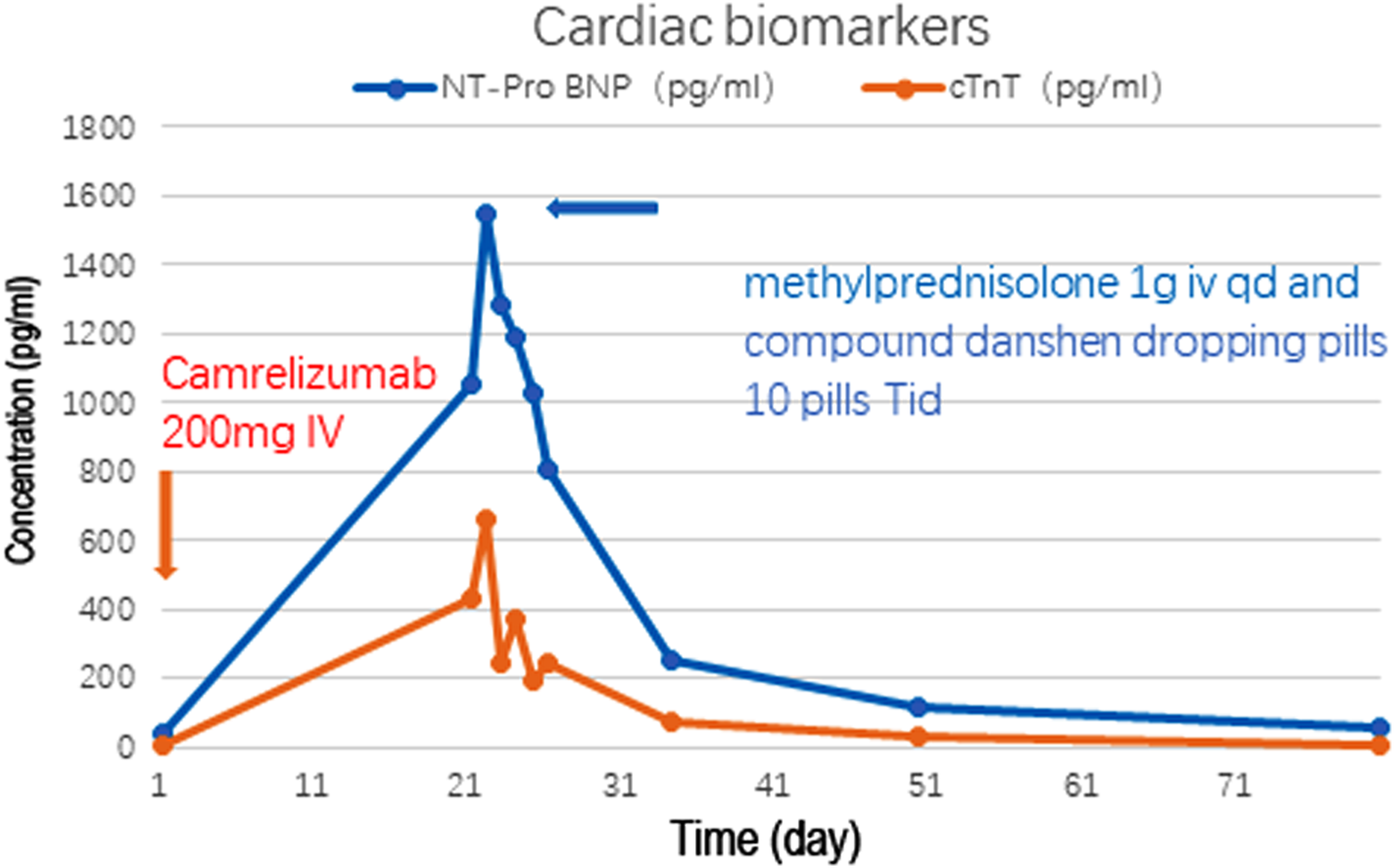
Changes of cardiac biomarkers in the case.

Changes after ICI treatment and follow-up: (A) before camrelizumab treatment, (B and C) after 2 cycles of camrelizumab treatment, ST-segment gradually elevation in chest leads V1–V3 (suspected front wall and inferior wall myocardial infarction), ST segment change in I, AVL, II, III, AVF, V1 to V6. (D) After immunoglobulin, methylprednisolone, and compound danshen dropping pills treatment in day 5. (E) Normal ECG in follow-up 2 weeks. (F) Normal ECG in follow-up 1 year).
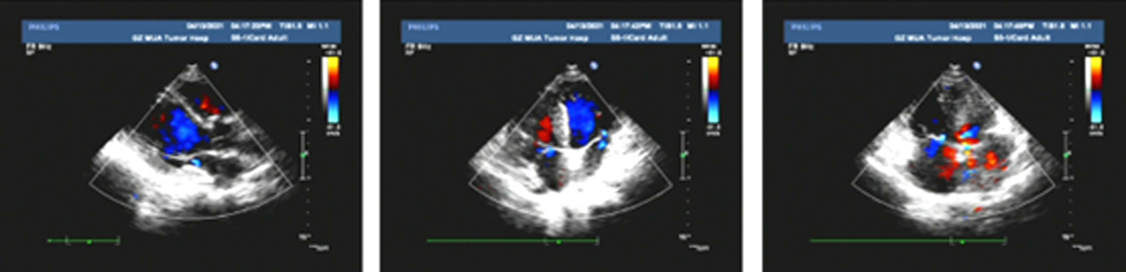
Echocardiography changes after ICI treatment in echocardiography. The %LVEF is 62%.
According to the “Naranjo Algorithm Assessment” 10 to evaluate the possibility of adverse drug reactions, the Naranjo score was 5, which indicated that myocarditis may be caused by camrelizumab (Table 1).
ADR probability scale.
Holter monitor showed that the total number of ventricular premature beats throughout the day was 14, no supraventricular tachycardia, and mean heart rate 76 bpm.
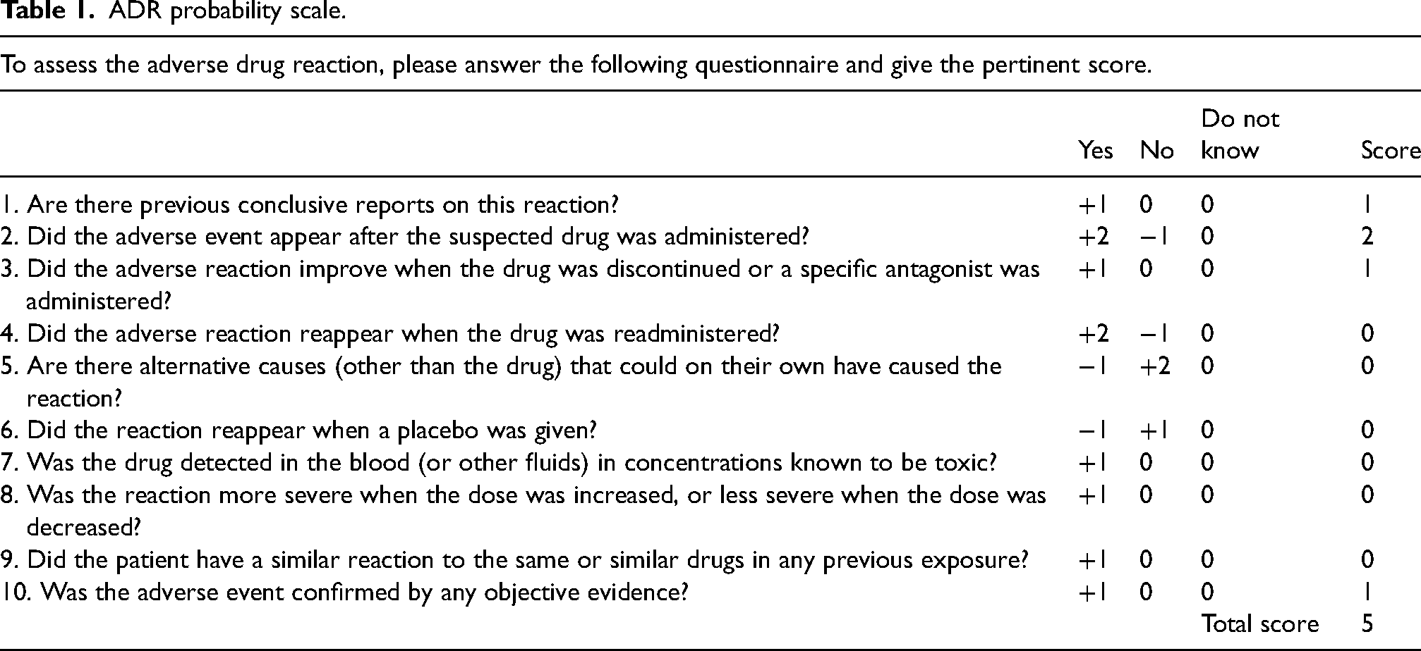
In order to rule out myocardial infarction, the patient was transferred to the department of cardiology for coronary angiography (CAG) (Figure 6). The results of CAG showed that the left coronary artery, including left main coronary trunks (LMT), left anterior descending branch (LAD), left circumflex branch (LCX) and right coronary artery (RCA) were smooth, and no occlusion or obvious abnormality was found. Blood flow TIMI was grade 3. Left ventriculography: left ventricular wall movement was normal and ejection was sufficient. Left ventricular catheterization: left ventricular pressure 109/41 mmHg, aortic pressure 118/72 mmHg.
CAG reveals the left coronary artery and the RCA were smooth.
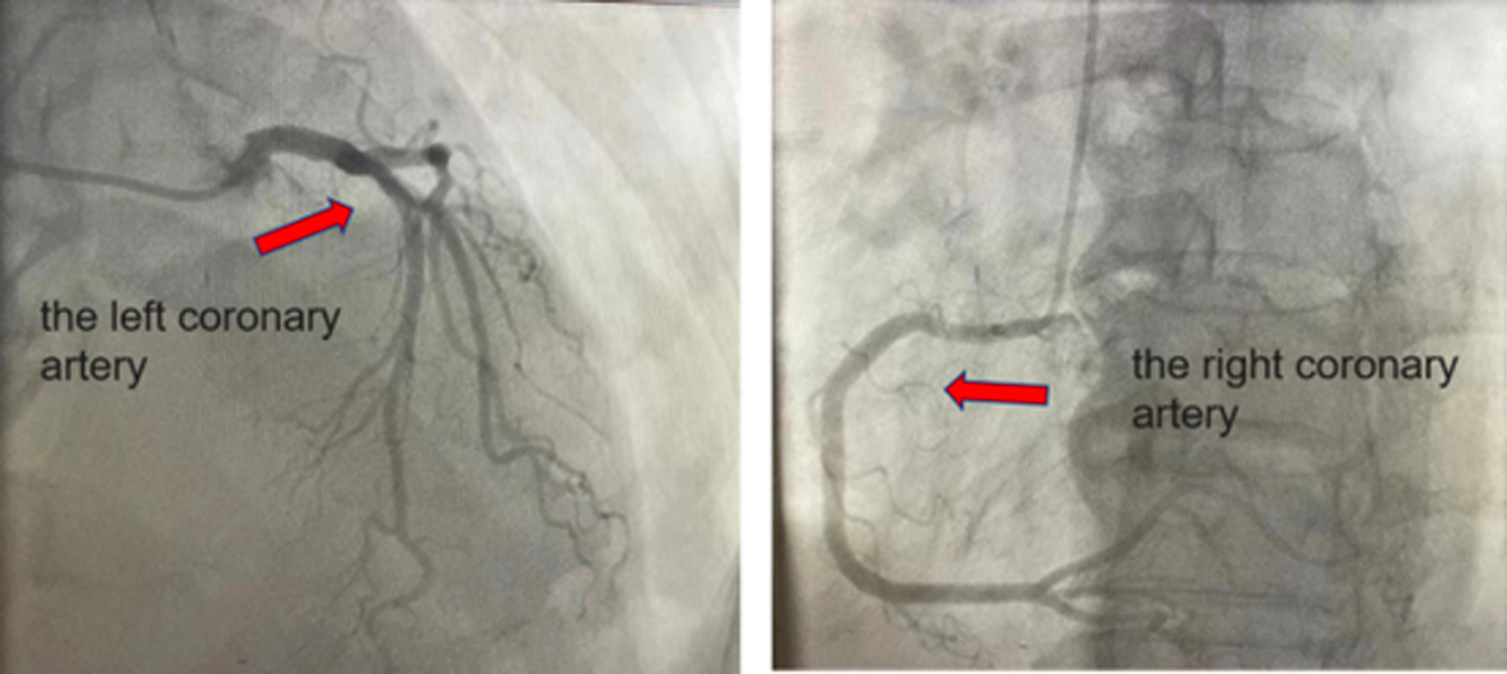
Enhanced cardiac MRI reveals the delayed perfusion signal of left ventricle was not uniform, so it was possible to consider artifacts. No abnormality of left ventricular systolic function was found (Figure 7).
Enhanced cardiac MRI reveals no abnormality of left ventricular systolic function.
After consultations in the hospital's multidisciplinary treatment (MDT) team, the diagnosis of acute myocarditis with normal ejection fraction was considered to be induced by an anti-PD-1 immunotherapy camrelizumab, and the ICI-related myocarditis was classified as grade 3 according to the Common Terminology Criteria for Adverse Events, version 5.0 (CTCAE v 5.0). 11 The patient received immunoglobulin (0.5 g/kg/day) 12 and 1000 mg/day of methylprednisolone was injected intravenously, and traditional Chinese medicine compound danshen dropping pills (10 pills Tid) to suppress immunotherapy, and camrelizumab was withheld during the management of myocarditis. After the treatment, the patient's NT-ProBNP and cTnT gradually decreased, but for rule out myocardial infarction happening, further inspection such as coronary angiography was performed.
After three days of high-dose intravenous hormone shock therapy and other symptomatic treatment, day 4, methylprednisolone was reduced to 500 mg/day, day 7, methylprednisolone was reduced to 250 mg/day, and day 10, methylprednisolone was reduced to 80 mg twice daily, after 3 days, it was further reduced to 40 mg twice daily, and gradually intravenous drip reduced to 40 mg once a day. Immunoglobulin therapy gradually intravenous drip reduced to 2.5 g once a day. Compound danshen dropping pills was continuous oral administration. In the process, the patient's cardiac marker of NT-ProBNP and cTnT gradually decreased (Figure 3). After 13 days of continued treatment, the methylprednisolone was discontinued and oral dexamethasone (DXM) tablets (20 mg qd×5 day) were given. Following discharge, the patient continued to take an adjusted dose of DXM orally 10 mg days 19–25; and 5 mg days 25–30, and then the medication was withdrawn. Laboratory examinations showed that cTnT 32 pg/mL, and NT-Pro BNP 115.40 pg/mL, both of which were close to normal values (Figure 3). During and after the prolong corticosteroid treatment, the level of procalcitonin was < 0.05 ng/ml (reference range < 0.05 ng/ml), indicting no opportunistic infection prophylaxis. The patient made a good recovery. Camrelizumab was not rechallenged after successful treatment of myocarditis. At the 1-year follow-up, the patient was asymptomatic, with a normal cardiac function.
Discussion
Recent studies have highlighted the significant advantages and potential of ICIs in the clinical treatment of various malignancies. PD-1is one of the most important immune checkpoints, 13 and it negatively regulates the activation of effector T lymphocytes through its interaction with PD-L1. 14 PD-1 inhibitors function by specifically binding to the PD-1 molecule, thereby blocking the PD-1/PD-L1 pathway and reinvigorating anti-tumor activity. This pathway primarily regulates the local cytotoxic lymphocyte (CTL) response and can give rise to immune-related adverse events (IRAEs) as a result of ICI administration. 15
Camrelizumab is an ICI against PD-1 and PD-L1. Camrelizumab has demonstrated efficacy in treating advanced or metastatic esophageal squamous cell carcinoma. Studies have shown that when combined as a first-line treatment with paclitaxel and cisplatin, camrelizumab significantly prolongs the median overall survival of patients with advanced esophageal cancer (15.3 months vs. 12.0 months) and reduces the risk of death by 30%. 16 In the second-line setting, camrelizumab has also improved overall survival in patients with advanced or metastatic esophageal squamous cell carcinoma, offering a potential new treatment option. 9 Clinical trials have revealed a manageable safety profile for camrelizumab. Common adverse reactions include fatigue, nausea, vomiting, anemia, hypertension, and respiratory tract infections, among others. Serious adverse reactions such as cardiac toxicity, pulmonary toxicity, and hepatic toxicity have been reported but occur at a relatively low incidence rate. 17 A study investigating camrelizumab-induced myocarditis retrospectively evaluated cardiac toxicity events in multiple myeloma patients undergoing camrelizumab treatment, revealing that myocarditis occurred in 0.27% of patients treated with ICI, and is serious adverse event associated with camrelizumab treatment. 18 ICI-induced myopathy (ICIM) is a complex disorder encompassing various manifestations such as polymyositis, 19 myocarditis, 20 and dermatomyositis, 21 among others. Among these, myocarditis poses a particularly grave risk and can be a fatal complication. The patient included in this study was a 59-year-old female with esophageal squamous cell carcinoma and hepatic metastases. She underwent second-line treatment with domestically-made PD-1 inhibitor, camrelizumab, in combination with paclitaxel (albumin-bound) and carboplatin. The patient received a diagnosis of immune-checkpoint-inhibitors-related myocarditis based on multiple diagnostic methods. Nevertheless, we need to be cautious that carboplatin and paclitaxel for cancer treatment can lead to cardiovascular toxicity in patients, 22 although the specific association between these treatments and myocarditis in cancer patients remains unknown or unreported.
Over the past few decades, significant advances have occurred in cancer treatment. The therapeutic approach to cancer has shifted from standard chemotherapy in the early days to targeted therapies now as well as immunotherapy. As the number of patients treated with immunotherapy continues to increase, so does the number of adverse events associated with it. Biomarkers of high risk of ICI myocarditis are not well defined, but the PD-1/PD-L1 pathway is known to be associated. 23 In 2016, a study reported the first case of acute lymphocytic myocarditis after using nivolumab. 1 Two retrospective studies have shown that patients treated with ICIs have a higher rate of cardiac events: 1.0% and 1.14%, respectively,24,25 in which cases of myocarditis are also constantly increasing. Signs and symptoms of ICI-related myocarditis are nonspecific. Myocarditis generally presents with physical deterioration, chest pain, dyspnea, and headache. Except for fatigue, the patient in this study did not have any discomfort such as shortness of breath and chest pain, and the effects of myocardial infarction, hypertension, diabetes mellitus, and other diseases can be excluded. Most patients with myocarditis present with an abnormal electrocardiogram (ECG) and cardiovascular symptoms, usually accompanied by elevated N-terminal prohormone B-type natriuretic peptide (NT-Pro BNP /BNP) levels of troponin, creatinine kinase MB.26,27
The patient has markedly increased cardiac troponin, which gradually decreased after receiving glucocorticoid. NT-proBNP/BNP reflects the heart function status of tumor patients, and when it is significantly elevated, it indicates the deterioration of heart function. In this case, NT-pro BNP increased after using ICI, but the increase was not large, the symptoms of dyspnea and chest pain were not found, and the recovery after treatment was better. This case underwent echocardiography before and after the occurrence of myocarditis, and the LVEF was reduced from 66% to 62%, but it was still in the normal range, indicating that normal LVEF does not exclude the diagnosis of myocarditis.
Myocarditis may present early with transient wall thickening with edema, impaired left and/or right ventricular function, preserved EF, or ventricular dilation. Cardiac MRI can quantify tissue damage, including edema, congestion, and fibrosis, and can support the diagnosis of myocarditis (Lake Louis criteria).28,29 Endomyocardiac biopsy is the gold standard for judging myocarditis according to the ESC guidelines, especially in patients with life-threatening arrhythmias, but it has limited its clinical application because of some operational risks. 30 In addition, cardiac PET/CT imaging is helpful in confirming the diagnosis of myocarditis. 31 The ECG of ICI-related myocarditis may show atrioventricular block, bundle branch block, and atrial and ventricular arrhythmias. 32 The patient in this study had significant arrhythmia characterized by myocardial infarction, mainly manifested as ST-segment elevation, and had no clinical manifestations of dyspnea and chest pain after using ICI. This indicates that we need to pay attention to the possibility of myocardial inflammation when there is potential arrhythmias.
There are currently very limited therapeutic agents for ICI-related myocarditis, and glucocorticoids are currently the most commonly used immunotherapy drugs that effectively suppress overactivated autoimmunity. CTLA-4 agonist abatacept, alemtuzumab, anti-thymocyte globulin, intravenous immunoglobulins, tacrolimus, azathioprine, cyclophosphamide, and infliximab or plasmapheresis have been used in steroid-refractory cases of myocarditis.33,34 There is currently no consensus on the optimal sequence, combination, and duration of immunosuppressive therapy for hormone-refractory cases. In this study, the patient was treated with an infusion of 1000 mg/day of methylprednisolone and immunoglobulin therapy (0.5 g/kg per day), and was oral administration traditional Chinese medicine compound danshen dropping pills (10 pills Tid). During this process, the patient's NT-pro BNP and cTnT cardiac markers gradually decreased, the ECG ST-T abnormalities recovered, gradually improved after treatment, and clinical cure was discharged. At 1-year follow-up, the patient was asymptomatic, with a normal cardiac function.
Taken together, ICI therapy can trigger severe cardiotoxicity and should be given clinical attention. Medical staff should assess the patient's vascular status before using ICIs, monitor closely during use, and treat promptly if the patient has cardiovascular complications. This report can remind medical personnel to be alert to the possibility of rare adverse reactions when using camrelizumab.
Footnotes
Availability of data and materials
All the data are available upon reasonable request from the corresponding author.
Declaration of conflicting interests
The author(s) declared no potential conflicts of interest with respect to the research, authorship, and/or publication of this article.
Ethics approval and consent to participate
The study was conducted in accordance with the principles of the Declaration of Helsinki, and the study protocol was approved by the ethics committee of the Affiliated Cancer Hospital and Institute of Guangzhou Medical University (number 2020–50).
Funding
The authors disclosed receipt of the following financial support for the research, authorship, and/or publication of this article: This work was supported by the Guangzhou Health Science and Technology Project, the Traditional Chinese Medicine Bureau Research Project of Guangdong Province in China (grant number 20202A010014, 20211A011091 and 20212A011024, 20202111).
Informed consent
Informed consent was obtained from the patient to participate.
Patient consent for publication
Informed consent was obtained from the patient to publish the images in an online open-access publication.