
Abstract
Introduction
Bevacizumab is a humanized monoclonal anti-VEGF antibody widely used in oncology and ophthalmology for the treatment of age-related macular degeneration and diabetic macular edema. Its intravitreal administration requires galenic repackaging, raising concerns regarding physicochemical stability and sterility assurance during storage. This study evaluated the structural and thermal stability of repackaged bevacizumab stored at 4 °C for up to four weeks.
Methods
Stability was assessed using intrinsic fluorescence spectroscopy and differential scanning fluorimetry (DSF) performed on a real-time PCR platform. Structural integrity was monitored through fluorescence emission spectra, while thermal stability was determined by melting temperature (Tm) analysis over time.
Results
Intrinsic fluorescence showed a constant emission maximum at 338 nm throughout the storage period, indicating preservation of the tertiary structure. DSF analysis demonstrated stable thermal unfolding profiles, with no significant variation in Tm values between baseline (67.4 ± 0.3 °C) and four weeks (67.2 ± 0.4 °C; p > 0.05).
Conclusions
Repackaged bevacizumab maintained structural integrity and thermal stability for up to four weeks under refrigerated storage conditions. These findings support refrigerated batch preparation under the tested conditions and highlight RT-PCR-based DSF as a practical quality-control tool for hospital pharmacy settings.
Keywords
Background
Bevacizumab (Mvasi®) is a humanized monoclonal antibody that inhibits vascular endothelial growth factor (VEGF), a key molecule involved in the formation of new blood vessels (angiogenesis). This mechanism of action makes it essential in several clinical applications, both in oncology, such as in the treatment of solid tumors, and in ophthalmology, where it is mainly used for diseases such as age-related macular degeneration (AMD) and diabetic macular edema (DME).1,2 In ophthalmology, bevacizumab is characterized by its anti-angiogenic, anti-edematous, and anti-exudative efficacy. However, unlike other anti-VEGF drugs approved for intravitreal use, such as ranibizumab and aflibercept, bevacizumab is not specifically formulated for this purpose, necessitating a galenic preparation that involves repackaging of the drug into sterile unit doses.3,4 Multiple randomized clinical trials and meta-analyses have demonstrated that intravitreal bevacizumab provides clinical efficacy comparable to approved anti-VEGF agents such as ranibizumab and aflibercept in terms of visual acuity improvement and anatomical outcomes.5–7 Despite similar therapeutic performance, bevacizumab offers a substantial economic advantage, as the cost per intravitreal dose is markedly lower than that of licensed alternatives. This cost-effectiveness has resulted in the adoption of bevacizumab in several healthcare systems, including the United States, the United Kingdom, and many European countries, where it represents a significant fraction of intravitreal anti-VEGF treatments.8,9 Consequently, hospital pharmacies play a central role in ensuring the continuous supply of repackaged bevacizumab, while maintaining high standards of quality and safety. However, bevacizumab is not commercially formulated for intravitreal administration and therefore needs to be repackaged in a sterile environment into single-dose syringes by hospital pharmacies. This process poses critical pharmaceutical challenges related to physicochemical stability and microbiological safety. Monoclonal antibodies are structurally complex biomolecules that are sensitive to temperature changes, mechanical stress, light exposure, and surface interactions, all of which may induce partial unfolding, aggregation, and loss of biological activity.10,11 These stability concerns directly impact drug management strategies, including shelf-life assignment, centralized compounding models and batch preparation planning. The repackaging of bevacizumab represents a challenge in terms of physicochemical stability and maintenance of aseptic handling conditions. The repackaging process and subsequent storage conditions could compromise drug integrity, reduce therapeutic efficacy, and increase the risk of adverse events. In this context, standardized repackaging procedures are essential to ensure controlled handling conditions and preservation of structural stability during refrigerated storage.10,12
Several studies have investigated the stability of repackaged bevacizumab using chromatographic and spectroscopic approaches.10,12 However, most analytical methods require specialized instrumentation that is not routinely available in hospital pharmacy laboratories. At the same time, differential scanning fluorimetry (DSF) has emerged as a sensitive and rapid technique for monitoring protein conformational stability, but its application has been mainly restricted to pharmaceutical development and drug discovery settings. 13 To date, the potential of DSF as a routine quality control method for the hospital preparation of monoclonal antibodies remains largely unexplored.
From a regulatory point of view, AIFA Note 98 regulates the intravitreal use of bevacizumab, defining it as a therapeutic option deliverable by the National Health Service (NHS) for AMD and DME. However, due to the absence of ready-to-use formulations, the preparation of bevacizumab for intravitreal use is delegated to highly specialized galenic laboratories. This requires the adoption of strict operating procedures in accordance with Good Preparation Practices (GPP) and international Good Manufacturing Practices (GMP) standards. In addition, the centralization of repackaging in hospital laboratories offers significant economic advantages, reducing the cost of treatment compared to commercial alternatives, while maintaining a comparable efficacy and safety profile. 12
In this context, real-time PCR platforms represent a widely available analytical infrastructure in clinical laboratories and offer the opportunity to perform DSF-based thermal stability measurements using fluorescence detection. The integration of RT-PCR-based DSF into hospital quality control workflows could provide a scalable and cost-effective solution for routine monitoring of repackaged biologics, supporting pharmaceutical quality and sustainable drug management.
Objective
Therefore, the aim of the present study was to evaluate the structural and thermal stability of repackaged bevacizumab (Mvasi®) stored at 4 °C for up to four weeks using intrinsic fluorescence spectroscopy and RT-PCR-based differential scanning fluorimetry. The study was conceived as a preliminary translational investigation aimed at evaluating whether rapid and widely accessible analytical techniques could support routine monitoring of the structural stability of repackaged monoclonal antibodies in hospital pharmacy settings.
In parallel, aseptic process validation by media fill test simulation was performed to verify the reliability of the aseptic compounding workflow. The study also explored the potential applicability of these analytical approaches to routine quality-control activities in hospital pharmacy settings. Furthermore, this work analyses the organizational and management model of bevacizumab repackaging, highlighting how a standardized and centralized approach may improve patient safety and economic efficiency.
Methods
Sample preparation
Bevacizumab biosimilar (Mvasi®, Amgen Europe B.V., Netherlands; concentration 25 mg/mL; AIC 045925017/E) was aseptically repackaged under GMP-compliant conditions in a certified hospital compounding facility. All operations were performed inside a negative-pressure isolator (ISOMATE CYTO®, DIN 12980:2017 compliant), specifically designed for high-risk sterile compounding and cytotoxic drug manipulation. The isolator provided an ISO Class 5 (Grade A) controlled environment according to EN ISO 14644–1, with vertical unidirectional airflow and H14 HEPA filtration, ensuring continuous protection of the critical working area from particulate and microbiological contamination.
The commercial bevacizumab formulation (25 mg/mL) was aseptically fractionated into sterile 0.15 mL polypropylene syringes, corresponding to a final dose of 3.75 mg per unit, in accordance with standard intravitreal administration practices.
Three independent batches were prepared during separate production sessions to ensure reproducibility of the aseptic process. After preparation, the samples were stored under controlled refrigeration conditions (4 ± 2 °C) and protected from light until analysis. The storage temperature was continuously monitored using calibrated digital data loggers to ensure cold chain compliance throughout the study period.
Aseptic process validation by media fill simulation test
The aseptic compounding and repackaging process of intravitreal bevacizumab was validated through a media fill simulation test in accordance with European Pharmacopoeia14,15 requirements and Good Preparation Practices (GPP). The test was designed to simulate routine aseptic operations, including drug preparation, repackaging and filling, under real working conditions and maximum routine workload. 16 Tryptic soy broth (TSB) was used as the culture medium. The experiment was conducted in a clean room by setting the recommended operating parameters of the ASCCA guidelines (2017). After filling, all units were incubated using a two-step protocol (7 days at 20–25 °C followed by 7 days at 30–35 °C) to allow detection of bacterial and fungal contamination, in accordance with pharmacopeial guidelines.14,15 At the end of the incubation period, samples were visually inspected for microbial growth. Process acceptance criteria were defined as the absence of contaminated units.
Drug utilization analysis
The analysis of drug utilization trends was performed by extracting data relating to purchases of intravitreal anti-VEGF agents (aflibercept, ranibizumab and brolucizumab) for the first quarter of 2022, 2023 and 2024 from the hospital management software C4H Dedalus. Data extraction parameters included product type, time interval, reference warehouse and cost center. Due to the dual oncological and ophthalmological indication of bevacizumab, utilization data for intravitreal Mvasi® were retrieved using the Log80 clinical management system through the “patient report” function, selecting the time interval, clinical unit (Retina Surgery Unit) and intravitreal bevacizumab (Law 648/96). This analysis was limited to 2023 and 2024, as intravitreal bevacizumab compounding was suspended in 2022. Economic evaluation was performed exclusively on drug acquisition costs
Drug stability analysis
The analysed samples, stored at 4 ± 2 °C and protected from light, were prepared in standard solutions at 1.66 µM in phosphate-buffered saline (PBS) and stored at 4°C. Storage temperature was continuously monitored using calibrated digital data loggers. Stability was assessed through steady-state fluorescence and thermal unfolding analyses using a fluorescent probe that binds to exposed hydrophobic regions of the protein, thereby providing information on its conformational state during four weeks of storage
Spectrofluorimetric analysis
The stability of bevacizumab was assessed by analysing the fluorescence of a 1.66 µM bevacizumab solution in PBS at physiological pH (7.4) using a SPEX-FluoroMax spectrometer (Horiba Scientific, Piscataway, NJ, USA). Fluorescence emission spectra were recorded from 300 to 500 nm with excitation at 278 nm using a 1 cm path length quartz cuvette at a temperature of 25°C. The excitation and emission slits were both set to 2 nm, and scan speed was 120 nm·min−1. Analyses were conducted at baseline (T0) and weekly for up to one month. 17 To quantitatively evaluate potential spectral shifts and broadening effects, the spectral center of mass (SCM) of the fluorescence emission spectra was calculated, according to Hawe et al. (2008). 18 The SCM was defined as the intensity-weighted average wavelength according to the equation “SCM = ∑(λ i ⋅I i )/∑I i ”, where λ i represents the emission wavelength and the I i represents the corresponding fluorescence intensity at each spectral point. The calculations were performed in the emission range between 300 and 500 nm, which includes the main fluorescence emission region of tryptophan.
Thermal unfolding (differential scanning fluorimetry-DSF)
Protein stability was tested by means of the fluorescence-based thermal stability assay or differential scanning fluorimetry (DSF), developed by Pantoliano et al., 2001. 13 DSF records the fluorescence emission signal from the binding of a dye (SYPRO orange) to the exposed hydrophobic patches upon protein unfolding. Fluorescence measurements were performed using a CFX96 Connect RT-PCR-instrument (BioRad). 18 μl aliquots of protein solution (2 μM), freshly diluted in Tris-buffered saline (50 mM Tris-HCl, pH 8.0, 150 mM NaCl) containing 0.1 mM dithiothreitol (DTT), 0.2 mM EDTA, and 1: 1000 dilution of SYPRO Orange (50 mM stock solution in DMSO), were distributed into a 96-well plate. Melting point analysis was performed by increasing the temperature from 25 to 80°C with an increment of 0.2°C every 5 s and reading, at each temperature, the fluorescence in fluorescence resonance energy transfer mode (FRET). To calculate Tm values, DSF data from the melting curve were exported and fitted to the Boltzmann equation using GraphPad software. Each measurement represented the average of three replicate wells.
Statistical analysis
All experiments were conducted in triplicate using three independent batches. Data are presented as mean ± standard deviation (SD). Statistical comparisons between different storage times were performed using one-way analysis of variance (ANOVA). Differences were considered statistically significant at p < 0.05. Statistical analyses were performed using XLSTAT software (Paris, France) integrated with Microsoft Excel (Office).
Results
Drug utilization trends and pharmaceutical impact of centralized bevacizumab repackaging
In 2022, during the temporary suspension of hospital bevacizumab repackaging activities, intravitreal therapy relied exclusively on licensed anti-VEGF agents, resulting in an annual expenditure exceeding €1.03 million (Table 1). Following reactivation of centralized bevacizumab repackaging in 2023, repackaged Mvasi® was progressively introduced into clinical practice, accounting for approximately 14.7% of intravitreal administrations in 2023 and increasing to over 29% in early 2024.
Intravitreal drug utilization and pharmaceutical expenditure (2022–2024).
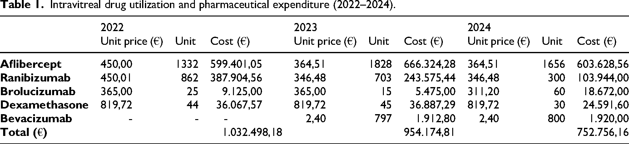
Validated stability data may support refrigerated batch preparation of repackaged monoclonal antibodies, improving centralized workflow efficiency and reducing product wastage. Centralized bevacizumab repackaging was associated with a marked reduction in per-dose treatment costs, with an estimated acquisition cost of approximately €2.40 per syringe compared with €311–450 for licensed anti-VEGF alternatives.
As shown in Figure 1, this progressive adoption was associated with a redistribution of intravitreal treatment patterns, characterized by a relative reduction in the use of higher-cost licensed anti-VEGF agents, particularly ranibizumab, which showed a marked decline in prescription share. These observations are consistent with previous pharmacoeconomic studies reporting superior cost-effectiveness of intravitreal bevacizumab compared with approved anti-VEGF therapies, while maintaining comparable clinical efficacy.7,8
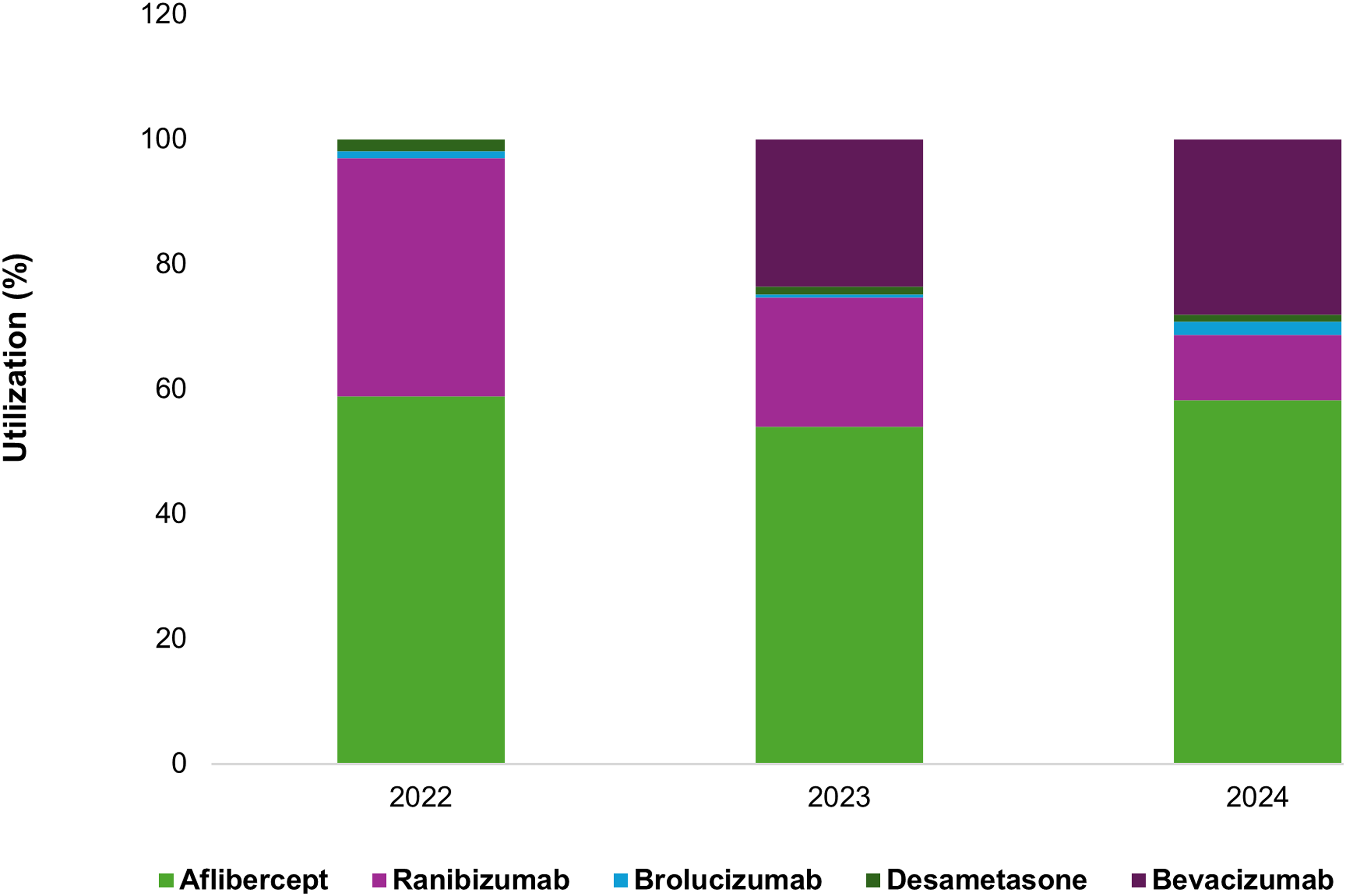
Temporal distribution of intravitreal anti-VEGF therapies and impact of centralized bevacizumab repackaging (2022–2024).
In several healthcare systems, including the United States, United Kingdom and parts of Europe, bevacizumab accounts for more than 60% of intravitreal anti-VEGF injections, primarily driven by its favorable cost–benefit profile.9,19
Overall, these findings suggest that centralized bevacizumab repackaging, supported by structural stability assessment and aseptic process validation, may represent a practical strategy for supporting refrigerated batch preparation in hospital pharmacy settings.
Steady-State fluorescence emission
Aseptic process validation through media fill simulations provided an essential complementary quality assurance measure alongside the structural and thermal stability data obtained in this study. All simulation runs resulted in the absence of microbial growth, confirming the robustness of the aseptic workflow and environmental control strategy. This aspect is particularly relevant for high-risk aseptic manipulations involving intravitreal biologics, where microbiological contamination represents a major clinical safety concern. 20 However, it should be noted that media fill testing validates the aseptic preparation process and environmental control conditions but does not constitute direct evidence of microbiological stability during prolonged refrigerated storage.
The study focused on rapid and operationally accessible methods suitable for routine hospital pharmacy quality control. Although intrinsic fluorescence spectroscopy and DSF provide sensitive information regarding tertiary structure integrity and thermal unfolding behavior, the present study does not include complementary analyses such as aggregation studies, subvisible particle assessment, concentration stability measurements, or bioactivity evaluation. Therefore, the findings should be interpreted as evidence of preserved structural and thermal stability under the tested storage conditions rather than a complete physicochemical stability characterization. Furthermore, stress conditions including freeze–thaw cycles, transport-related mechanical stress, syringe-material interactions, and light exposure were not investigated. The selected storage interval was based on the physicochemical stability data already reported in the Summary of Product Characteristics (SmPC/RCP) for diluted bevacizumab preparations under refrigerated conditions.
Intrinsic fluorescence spectroscopy was employed to investigate potential tertiary structure alterations of repackaged bevacizumab during refrigerated storage. The emission spectra consistently exhibited a fluorescence maximum (λmax) centered at 338 nm throughout the entire observation period of four weeks (Figure 2). This spectral position is characteristic of bevacizumab under native conditions and is consistent with previously reported measurements performed at physiological pH and moderate temperatures, where λmax values between 338 and 340 nm have been observed.17,21
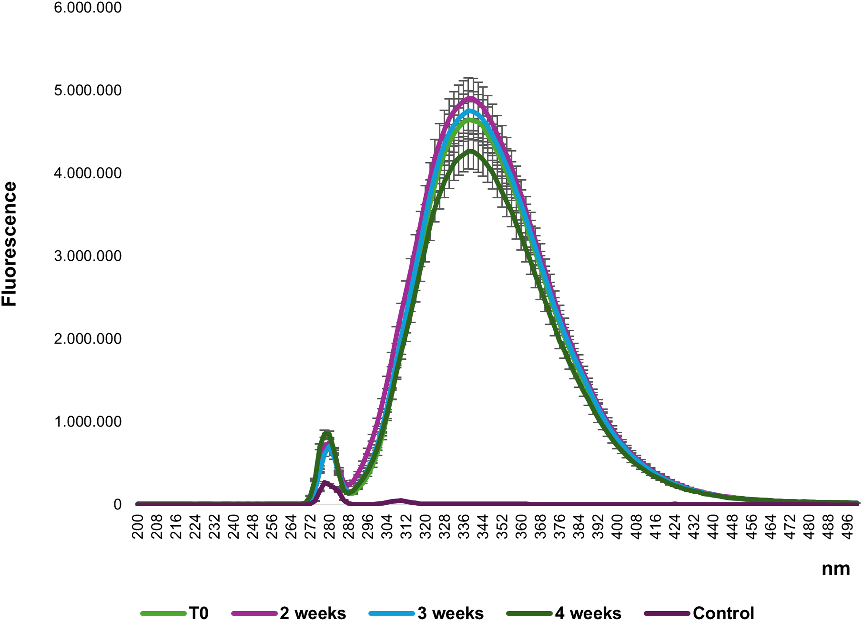
Intrinsic fluorescence emission spectra of repackaged bevacizumab during refrigerated storage. Steady-state fluorescence emission spectra of bevacizumab recorded at pH 7.4 and 25 °C following excitation at 278 nm. Spectra were acquired at baseline and during refrigerated storage (4 °C) for up to four weeks. All samples showed a stable fluorescence emission maximum centered at approximately 338 nm, with no detectable red-shift or spectral broadening over time. Preservation of the emission profile indicates maintenance of the native tertiary structure and absence of major conformational rearrangements or aggregation phenomena during storage.
From a biophysical perspective, the intrinsic fluorescence of proteins is a sensitive indicator of the local microenvironment surrounding aromatic residues, particularly tryptophan, which is highly sensitive to changes in protein folding, exposure to solvents, and rearrangements of the tertiary structure.22,23 The stability of the maximum emission observed in this study indicates that the microenvironment of tryptophan residues remained essentially unchanged during storage, suggesting preservation of the native IgG folding architecture and maintenance of a compact globular conformation.
Previous investigations have demonstrated that conformational destabilization and aggregation phenomena in monoclonal antibodies are often accompanied by pronounced spectral shifts toward longer wavelengths, typically in the range of 350–352 nm, reflecting increased burial of aromatic residues within hydrophobic aggregates or altered tertiary packing.18,21 The absence of such red-shifts in the present study provides strong evidence that neither early-stage aggregation nor significant unfolding events occurred during refrigerated storage of repackaged bevacizumab.
Although minor variations in fluorescence intensity were detected over time, these changes remained within experimental variability and were not associated with detectable spectral displacement. This observation is consistent with previous reports indicating that absolute fluorescence intensity is influenced by instrumental factors, photophysical effects and minor concentration variations, and therefore should not be interpreted as a direct indicator of protein degradation in the absence of concomitant spectral shifts.17,18
Intrinsic fluorescence analysis enables identification of early structural alterations that may precede visible protein aggregation. In this study, the stability of the emission spectra suggests that repackaged bevacizumab retained not only its native conformation but also favorable colloidal stability during refrigerated storage. To further quantify spectral stability, the temporal evolution of the fluorescence emission maximum was analysed. As shown in Figure 3 (Panel A), the λmax values remained essentially constant throughout the entire storage period, with no statistically significant deviations between independent batches. This quantitative analysis reinforces the qualitative interpretation derived from spectral overlaps and confirms the absence of progressive rearrangements of the tertiary structure. In addition to λmax monitoring, the spectral center of mass (SCM) was calculated as an integrated parameter describing the overall emission profile. SCM analysis provides a more robust metric than peak position alone, particularly in the presence of subtle spectral broadening phenomena. 22 As reported in Figure 3 Panel B, SCM values remained stable during refrigerated storage, further supporting preservation of the native folding state and excluding early-stage aggregation processes that may not yet produce visible macroscopic changes. Minor SCM variations (<1 nm) were observed; however, no systematic temporal drift was detected, indicating preservation of spectral stability and absence of progressive conformational rearrangements.
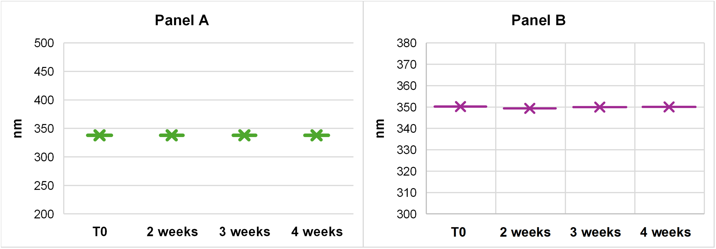
Quantitative analysis of fluorescence spectral stability of repackaged bevacizumab during refrigerated storage. (A) Time-dependent evolution of the fluorescence emission maximum (λmax) of repackaged bevacizumab stored at 4 °C for up to four weeks. λmax values remained stable throughout the observation period, indicating preservation of the tryptophan microenvironment and absence of detectable tertiary structure alterations. (B) Spectral center of mass (SCM) calculated from fluorescence emission spectra acquired between 300 and 500 nm. SCM values showed no significant temporal drift, supporting maintenance of the global spectral profile and excluding early-stage unfolding or aggregation processes. Data are presented as mean ± SD from three independent batches.
Overall, steady-state intrinsic fluorescence spectroscopy proved to be a robust, rapid, and low-sample-consumption analytical tool for monitoring the structural stability of repackaged monoclonal antibody formulations. Its integration into hospital quality control frameworks, in combination with complementary thermal stability assays, represents a practical approach for routine stability monitoring in real-world compounding environments.
Thermal unfolding and melting temperature (Tm)
To further evaluate formulation stability, bevacizumab samples were subjected to thermal unfolding analysis by differential scanning fluorimetry (DSF). Quantitative analysis of melting temperatures revealed highly reproducible values throughout the storage period. The mean Tm remained essentially unchanged between freshly prepared samples and those stored for up to four weeks at 4 °C (Tm = 67.4 ± 0.3°C at time 0 and 67.2 ± 0.4°C at week 4; ΔTm < 0.5 °C, p > 0.05) (Figure 4). The observed melting curves exhibited a single cooperative unfolding transition, characteristic of IgG1 monoclonal antibodies and reflective of a highly ordered tertiary and quaternary structural organization. 24 Previous studies have shown that bevacizumab displays thermal unfolding transitions in the range of 65–70 °C depending on buffer composition and formulation conditions, consistent with the Tm values obtained in the present study.10,17 Variations below 1 °C are generally considered within experimental variability for DSF measurements and are not indicative of significant structural destabilization in monoclonal antibody formulations.25,26 From a formulation science perspective, thermal stability represents an important surrogate marker of overall protein robustness. Changes in Tm have correlated with increased aggregation propensity, reduced colloidal stability and enhanced sensitivity to mechanical or thermal stress. 27 Therefore, the stable Tm profile observed in this study suggests that the repackaging process and subsequent refrigerated storage did not promote aggregation-prone conformations or destabilizing intermolecular interactions. The concordance between intrinsic fluorescence spectroscopy and DSF thermal unfolding analysis provides complementary insight into both local tertiary structure integrity and global unfolding resistance. While fluorescence measurements probe the microenvironment of aromatic residues, DSF captures large-scale conformational transitions associated with protein unfolding.17,22 The agreement between these independent techniques further strengthens confidence in the preserved structural and thermal stability of repackaged bevacizumab.
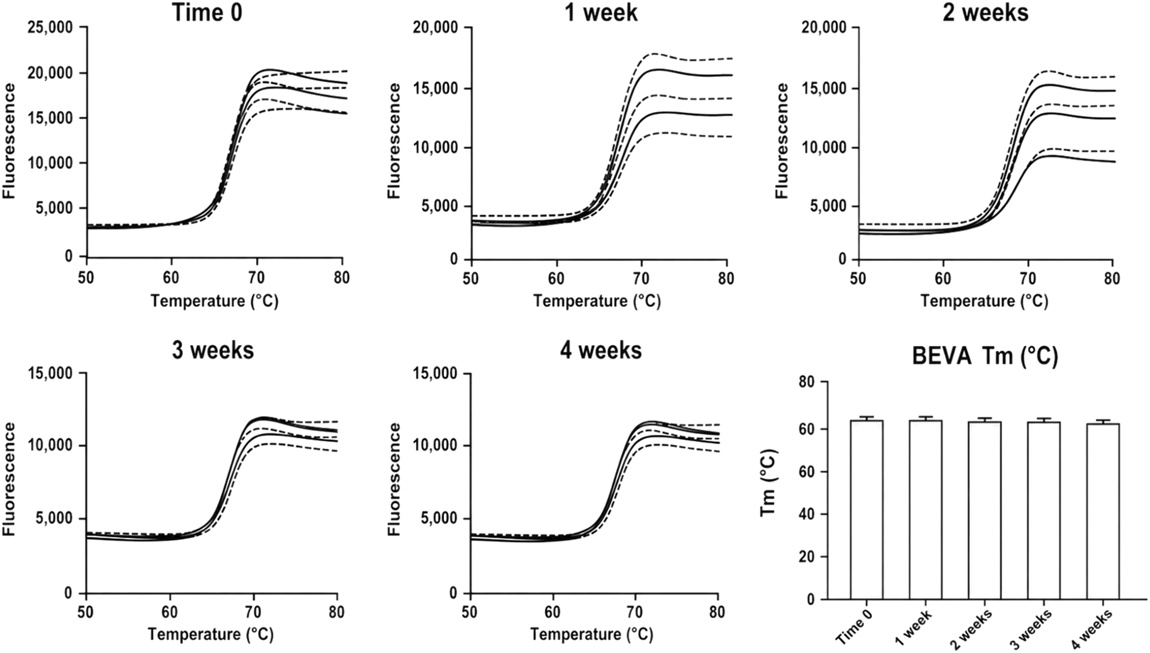
DSF data of bevacizumab after 1–4 weeks of storage at 4°C. Each DSF panel shows the thermal profile of three aliquots of the same sample (solid lines). To calculate Tm values, DSF data were fitted to the Boltzmann equation using GraphPad software (dotted curves).
Another strength of this study is the implementation of DSF using a real-time PCR platform. RT-PCR-based DSF has increasingly been recognized as a versatile and high-throughput tool for protein stability screening, offering advantages in terms of accessibility, speed, and minimal sample consumption. 28
From an operational perspective, the availability of stability data under the tested refrigerated conditions may support batch preparation strategies in hospital pharmacy settings. 10 By providing quantitative evidence of thermal stability, the present study may help reduce this barrier and support the development of stability-based organizational models.
Study limitations
The present study was primarily designed to assess the structural and thermal stability of repackaged bevacizumab under routine hospital pharmacy storage conditions using rapid and operationally accessible analytical techniques. Although intrinsic fluorescence spectroscopy and RT-PCR-based differential scanning fluorimetry provided complementary evidence of preserved tertiary structure and thermal stability, the study does not represent a complete physicochemical characterization of the formulation. Additional analyses including subvisible particle assessment, aggregation profiling, concentration stability evaluation and functional bioactivity assays were beyond the scope of the present investigation. Furthermore, stress conditions potentially relevant to real-world handling, such as repeated freeze–thaw cycles, transportation-related mechanical stress, prolonged light exposure and syringe-material interaction studies, were not investigated. Despite these limitations, the study provides preliminary translational evidence supporting the feasibility of RT-PCR-based DSF and intrinsic fluorescence spectroscopy as practical quality-control tools for monitoring the structural stability of repackaged monoclonal antibody formulations in hospital pharmacy practice.
Conclusions
The present study was intentionally designed to reflect routine clinical practice conditions in a hospital pharmacy setting, focusing on refrigerated storage under controlled aseptic handling rather than forced degradation or stress-testing conditions. This study demonstrates that repackaged bevacizumab (Mvasi®) maintains structural integrity and thermal stability during refrigerated storage for up to four weeks when prepared under validated aseptic conditions. Further studies including aggregation analysis, particle characterization, and bioactivity assays are warranted to provide a more comprehensive physicochemical stability assessment. Overall, the combined use of intrinsic fluorescence spectroscopy and RT-PCR-based differential scanning fluorimetry provided complementary and robust evidence of preserved tertiary structure and thermal stability, with no detectable conformational destabilization within the sensitivity limits of the applied spectroscopic methods. A key strength of this work lies in the successful implementation of DSF on a standard real-time PCR platform, highlighting the feasibility of adopting rapid, low-cost and low-sample-consumption stability assessment tools within hospital pharmacy environments. This approach represents a practical alternative to more complex biophysical techniques, enabling routine quality control of repackaged monoclonal antibody formulations without requiring specialized instrumentation.
From an operational perspective, the availability of stability data may support refrigerated batch preparation strategies in hospital pharmacy settings. The combined use of intrinsic fluorescence spectroscopy and RT-PCR-based DSF may therefore represent a practical approach for routine structural stability monitoring of repackaged monoclonal antibody formulations.
Footnotes
Acknowledgments
The authors gratefully acknowledge Sapienza University of Rome for institutional support and ASL-Roma 1 for their collaboration and assistance in this study.
Ethical approval and informed consent
This study did not involve human participants or animals. Therefore, ethical approval and informed consent were not required. All experimental procedures were conducted in vitro in accordance with relevant institutional and laboratory guidelines.
Credit author contribution statement
LP: Conceptualization, Investigation, Formal analysis. LPag: Conceptualization, Writing – Review & Editing. GM: Formal analysis, Writing – Review & Editing. FA: Formal analysis, Writing – Review & Editing. AI: Writing – Original Draft, Writing – Review & Editing. SC: Writing – Original Draft, Writing – Review & Editing; FC: Conceptualization, Methodology, Investigation, Formal analysis, Data curation, Writing – Original Draft, Supervision All authors reviewed and approved the final version of the manuscript.
Funding
The authors received no financial support for the research, authorship, and/or publication of this article.
Declaration of conflicting interests
The authors declared no potential conflicts of interest with respect to the research, authorship, and/or publication of this article.
Data availability statement
The data supporting the findings of this study are available from the corresponding author upon reasonable request.
Anonymity statement
All identifying information related to the authors, institutions, funders, and approval committees has been removed or anonymized to ensure a blinded peer review process.