
Abstract
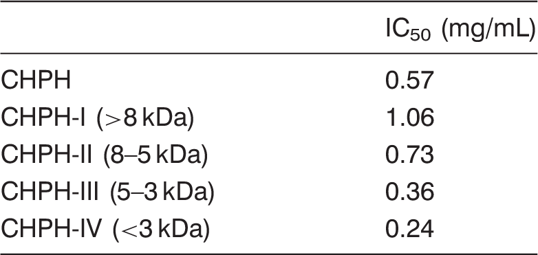
Cobia head protein hydrolysate (CHPH) with angiotensin I converting enzyme (ACE) inhibitory activity was prepared with papain. The 3 kDa ultrafiltration filtrate CHPH-IV of the hydrolysate exerted a potent ACE inhibitory activity with IC50 being 0.24 mg/mL. The fractions with molecular weight located between 1749 Da and 173 Da represented up 66.96% of CHPH-IV, and those between 494 Da and 173 Da represented up 31.37% of CHPH-IV. It was found that the ACE inhibitory activity of CHPH-IV was intensified from IC50 0.24 mg/mL to 0.17 mg/mL after incubation with gastrointestinal proteases. The CHPH-IV significantly decreased the systolic blood pressure in a dose-dependent manner after oral administration to spontaneously hypertensive rats (SHR) at dose of 150 mg/kg, 600 mg/kg and 1200 mg/kg body weight. These results suggested that CHPH-IV from cobia head protein hydrolysate by papain could serve as a source of peptides with antihypertensive activity in functional food industry.
Introduction
Hypertension, which affects 15–20% of all adults, is one of the major risk factors for the development of cardiovascular diseases, stroke and the end stage of renal disease (Zhang et al., 2006). Angiotensin I converting enzyme (peptidyl carboxy peptidase, EC 3.4.15.1, ACE) belongs to the class of zinc proteases that needs zinc and chloride for activation. ACE plays an important and physiological role in regulating blood pressure. It converts an inactive form of decapeptides, angiotensin I, to a potent vasopressor octapeptide, angiotensin II, and inactivates catalytic function of bradykinin. Therefore, inhibition of ACE is considered to be an important therapeutic approach for controlling hypertension. Many synthetic ACE inhibitors such as captopril, enalapril, alacepril and lisinopril were used in the treatment of essential hypertension and heart failure in humans (Ondetti, 1977). However, some undesirable side effects may occur such as cough, lost of taste, renal impairment and angioneurotic edema (Antonios and MacGregor, 1995). The peptides derived from food proteins are considered to be milder and safer compared with synthetic drugs. Many studies have been directed toward enzymatic hydrolysates of different food proteins, including casein (Silva and Malcata, 2005), mushroom (Lee et al., 2005), whey protein (Vermeirssen, 2004), porcine muscle (Arihara, 2001), chicken muscle (Fujita et al., 2000), soybean (Kuba et al., 2005) and fish protein (Ono et al., 2006).
Fish by-products are usually discarded as processing waste or used for animal feed because of its poor functional properties. Recognition of the limited biological resources and the increasing environmental pollution have emphasized the need for better utilization of by-product from the fisheries (Guerard et al., 2005). Fish by-products are considered as a safe material and provide proteins with high nutritional properties and a good pattern of essential amino acid (Shahidi et al., 1995). These by-products are very important bioresources that might be utilized for applications in food, health care products and pharmaceuticals. Therefore, the use of fish by-products has been of increasing interest for years. Many studies especially paid attention to prepare bioactive peptides using enzymatic hydrolysis from fish by-products (Byun and Kim, 2001; Je et al., 2008; Qian et al., 2007). Recent studies have identified a number of bioactive compounds or bioactive peptides with health benefits such as antioxidative activity, antihypertensive activity, from fish skin, fish frame and fish bone (Je et al., 2007; Kim et al., 2001; Lee et al., 2010).
Cobia, Rachycentron canadum, is a large, migratory, coastal pelagic fish of the monotypic family Rachycentridae, which is distributed worldwide in tropical and subtropical seas, with the exception of the eastern Pacific (Ditty and Shaw, 1992). In China, the cobia-farming regions are located in the southern areas, namely Guangdong and Hainan provinces. China’s total output is 19,634 tons in 2006, ranking first in the world (FAO, 2009). Cobia was consumed now mainly as fillets and sashimi. A sizeable amount of by-products including head, skin, viscera and bone is generated during fillets processing and can be as high as 40–50% of the original material. Furthermore, Cobia head takes about 20–25% of the original material. And with the amount of 45.38% protein on a dry weight basis, cobia head can serve as an additional source of protein (Liu et al., 2009). The objective of this study was to prepare papain hydrolysate from cobia head protein with angiotensin I converting enzyme inhibitory activity. We also determined the molecular weight distribution of high activity fraction of the hydrolysate and assessed its antihypertensive action by oral administration in spontaneously hypertensive rats (SHR).
Materials and methods
Materials
Fresh cobia (Rachycentron canadum) head used was provided by ZhanJiang GuoLian Aquatic Products Co., Ltd., (Guangdong China) in November 2008. After washing with water, the cobia heads were immediately grinded and packed (about 100 g per bag) and stored at −20 °C until use.
Papain (650,000 U/g) was purchased from Pangbo Biological Engineering Co., Ltd (Nanning, China), pepsin and pancreatin were purchased from Shanghai Biochemical Company (Shanghai, China). ACE (from rabbit lung), HHL (Hippuryl-L-histidyl-L-leucine), and captopril were purchased from Sigma Chemical Co. (St. Louis, MO, USA). Other reagents used were of analytical grade.
Preparation of cobia head protein hydrolysate
Hydrolysate of cobia head was produced according to pilot-study (Jiang et al., 2010). The cobia head was ground and homogenized with two volumes of distilled water, then poured into a reaction vessel. The pH was adjusted to 6.0 and the system was incubated in a water bath of 73 °C. The enzymatic hydrolysis was started by adding 0.35% (by weight of raw material) papain with stirring. The reaction was terminated after 4 h and the reaction system was heated at 100 °C in a water bath for 15 min to inactivate the protease, followed by cooling to room temperature and pH was adjusted to 8.3. The resulting solution was centrifuged at 4 °C, 5000 × g for 15 min and the supernatant was collected for further analysis. The hydrolysate solution was separated into four fractions (CHPH-I, > 8 kDa; CHPH-II, 8–5 kDa; CHPH-III 5–3 kDa, CHPH-IV, < 3 kDa) using an ultrafiltration membrane bioreactor system (Millipore, USA). The ultrafiltration was performed at 0.25 MPa, 4 °C. CHPH-IV was collected, lyophilized and stored at −20 °C until further analysis within a month.
Determination of ACE inhibitory activity
ACE inhibitory activity was assayed using the spectrophotometric assay of Cushman and Cheung (1976) with some modification. A 50 µL sample solution was mixed with 50 µL of 5 mM HHL as substrate containing 100 mM sodium phosphate buffer (pH 8.3) and 500 mM NaCl and then pre-incubated at 37 °C for 6 min. The reaction was initiated by the addition 50 µL of 25 mU/mL ACE solution in a buffer containing 100 mM sodium phosphate buffer (pH 8.3) and the mixture was incubated at 37 °C for 30 min. The reaction was stopped by adding 150 µL of 1.0 M HCl. The liberated hippuric acid was extracted with 1 mL of ethyl acetate. The mixture was centrifuged and 0.5 mL of ethyl acetate layer was transferred to a fresh test tube and evaporated to dryness on a water bath for 15 min at 100 °C. The residue containing hippuric acid was dissolved in 3 mL deionized water and the solution was measured using a UV visible spectrophotometer (UV-1700, Shimadzu Co., Japan) at 228 nm against deionized water as a blank. The ACE inhibitory activity was also expressed as IC50. The IC50 value was defined as the concentration of inhibitor (soluble protein) required to inhibit 50% of the ACE activity. The soluble protein content of the samples was determined using the Folin-Lowry method (1951).
Simulation of gastrointestinal digestion
The stability of the ACE inhibitory peptide against gastrointestinal proteases was assessed in vitro. The lyophilized sample CHPH-IV (1 mg/mL) was individually digested with pepsin (pH 2.5, 37 °C, 3 h) with stirring, in a ratio of substrate to enzyme (200:1, w/w) as previously described (Hernandez-Ledesma et al., 2004) and the digests were used for ACE inhibitory activity determination. Moreover, the samples were successively hydrolyzed with pepsin (pH 2.5, 37 °C, 3 h) and pancreatin (pH 8.0, 37 °C, 4 h) with stirring in a ratio of substrate to enzyme (200:1, w/w) and the digests were used for ACE inhibitory activity determination also.
Stability of CHPH-IV for ACE
A 50 µL aliquot CHPH-IV was incubated with 50 µL of 50 mU/mL of ACE at 37 °C for 3 h. IC50 values were compared with that of before and after pre-incubation.
Determination of molecular weight distribution
The molecular weight distribution of CHPH-IV was analyzed by high performance size exclusion chromatography (HPSEC) using Waters-PROTEIN-PAK 60 column. The mobile phase was 50 mM Tris-HCl buffer (pH 7.4). Its flow rate was 0.7 mL/min. Detection was performed by UV-light absorption at 214 nm, and the whole system was maintained at constant temperature 25 °C. After the equilibration between mobile phase and packing was established, the aliquot of 10 µL samples was injected into HPSEC apparatus (Waters 600E, Waters Co., Milford, USA) for analysis. A molecular weight calibration curve was obtained from the following standards from Biorad: Triosephosphate-isomerase (MW 26,625 Da), Myoglobin (MW 16,950 Da), Aprotinin (MW 6512 Da), Insulin-B (MW 3496 Da), Bacitracin (MW 1423 Da), HHL (MW 423 Da). This yielded a near linear correlation between the retention time (t) and the log of the molecular mass (Mr) of peptides. The regression equation was: log Mr = - 0.244 t + 6.652(R2 = 0.9691).
Antihypertensive action in spontaneously hypertensive rats
Thirty spontaneously hypertensive rats (SHR) (9-week-old, male, SPF, 279 ± 3 g) with tail systolic blood pressure (SBP) over 170 mm Hg were obtained from Slac Laboratory Animal Co. Ltd. (Shanghai, China). Rats were housed in a room kept at 23 ± 1 °C with a relative humidity of 50% and a 12 h light-dark cycle, and fed with standard laboratory diet. Tap water was freely available. The lyophilized CHPH-IV was dissolved in distilled water and orally administered to rats at a dose of 150 mg/kg, 600 mg/kg and 1200 mg/kg body weight (BW), respectively. The positive control group was administered a commercial antihypertensive drug, captopril, at a dosage of 4 mg/kg BW. The negative group was administered the distilled water only. All of the products were orally administered in 2 mL of water. The SBP of the rats was measured at 2, 4, 6, 8, 10 and 24 h after administration of the peptides using the tail-cuff method with a programmable electro-sphygmomanometer (BP-98A; Softron Co., Ltd., Tokyo, Japan) after warming the rats in a chamber maintained at 37 °C for 15–20 min.
Statistical analysis
The differences in SBP values for animals in the control and experimental groups were analyzed by one-way ANOVA and the magnitude of the differences using software SPSS13.0.
Results and discussion
Effect of ultrafiltration on the ACE inhibitory activity of CHPH
ACE inhibitory activity of CHPH and four UF fractions
Molecular weight distribution of CHPH-IV
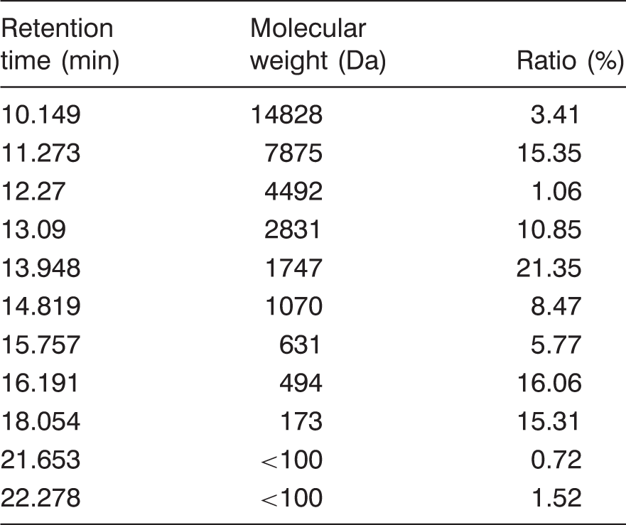
CHPH-IV was separated using HPSEC to analyze peptide size composition. HPSEC of CHPH-IV yielded 11 absorbance peaks at 214 nm (Figure 1). The molecular weight of each peak was shown in Table 2. The molecular weight distribution of CHPH-IV varied in a relatively broad range. The fraction was composed mostly of molecular weight peptides located at 1749–173 Da (66.96%), and low molecular weight peptides located between 494 Da and 173 Da took 31.37%. Bioactive peptides of 3–20 amino acids (Pihlanto, 2000) and molecular masses of less than 6000 Da (Sun et al., 2004) have been reported. Lee et al. (2010) reported an ACE peptide derived from tuna frame protein hydrolysate whose molecular weight was 2480 Da. CHPH-IV contained a considerably proportion of low molecular weight peptides and probably had potential bioactivity.
HPSEC chromatogram of CHPH-IV. Molecular weight distribution of CHPH-IV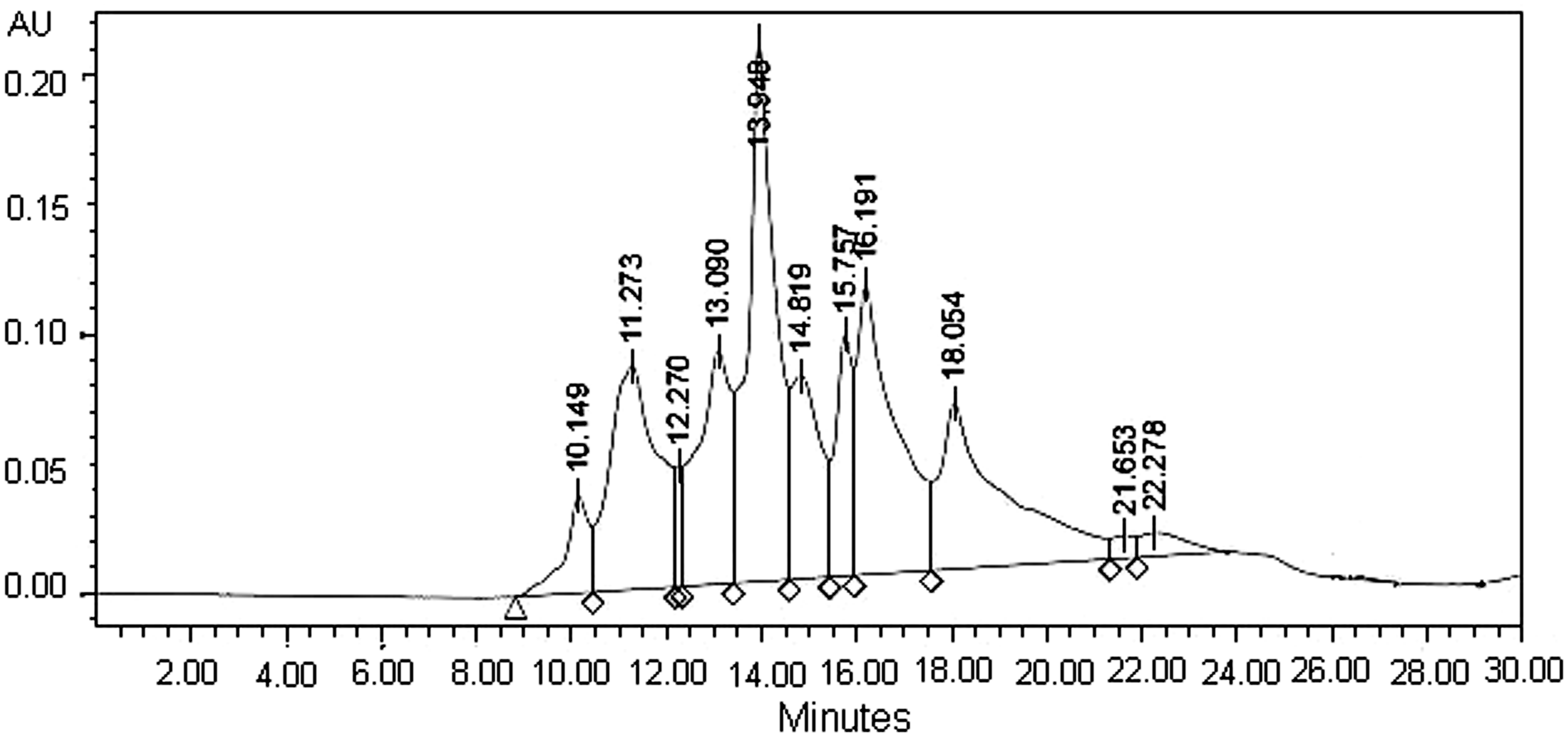
Determination of stability of CHPH-IV for ACE
In order to check the inhibitory character of the isolated peptides, IC50 values of CHPH-IV were determined before and after pre-incubation with ACE. It was found that pre-incubation did not change the inhibitory activity of the CHPH-IV, and IC50 values for the CHPH-IV before and after pre-incubation with ACE were 0.24 and 0.21 mg/mL, respectively. Fujita et al. (2000) classified ACE inhibitory peptides into three types, based on changes in ACE inhibitory activity after the peptides were digested by ACE. Substrate type peptides are those which show a decrease in ACE inhibitory activity after being digested by ACE. The inhibitory activity of the inhibitor type peptide is not significantly affected by ACE digestion. The pro-drug type peptides are those which show an increase in ACE inhibitory activity after ACE digestion. It has also been reported that the substrate type peptides do not affect the blood pressure of SHR, but the inhibitor and pro-drug type peptides produce a reduction in blood pressure values. We would class CHPH-IV as an inhibitor type peptide and we suggest that CHPH-IV has antihypertensive activity in vivo.
Stability of CHPH-IV for gastrointestinal proteases
Not all of the ACE inhibitory activity peptides in vitro have antihypertensive activity in vivo. It is of crucial importance that ACE inhibitory peptides remain active during gastrointestinal digestion. Gastrointestinal enzyme incubation in vitro provided an easy process to imitate the fate of these peptides under oral administration. The stability of CHPH-IV against gastrointestinal proteases in vitro was examined in order to predict the antihypertensive effect in vivo. The ACE inhibitory activity of the peptide showed a change after in vitro incubation with gastrointestinal proteases. The results showed that the ACE inhibitory activity of CHPH-IV was increased from IC50 0.24 to 0.15 mg/mL after pepsin digestion, and from IC50 0.24 to 0.17 mg/mL after a successive digestion with pepsin and pancreatin.
Grimble and Silk (1989) suggested that intact dipeptides or tripeptides could be transported from the intestinal lumen into the blood circulation. Many researchers found that other short peptides with peptide length more than three could also be absorbed into the blood circulation (Langguth et al., 1994). Zhao et al. (2009) reported that the inhibitory activity of the peptide isolated from Acaudina molpadioidea hydrolysate was intensified from IC50 15.9 to IC50 5.3 mM after pepsin digestion and increased from IC50 15.9 to IC50 4.5 mM following further chymotrypsin digestion. This inhibitory peptide from A. molpadioidea showed a clear antihypertensive effect in spontaneously hypertensive rats. So, intensification of the ACE inhibitory activity was found after gastrointestinal proteases treatments, which indicated that CHPH-IV may have a potential antihypertensive effect.
Antihypertensive effect of CHPH-IV in SHR
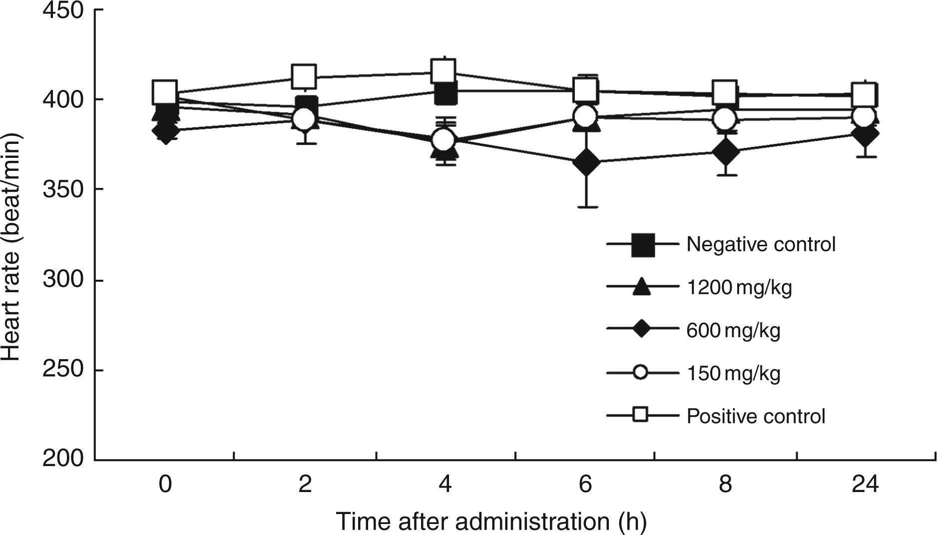
Antihypertensive effect of CHPH-IV and captopril were evaluated by measuring changes in systolic blood pressure (SBP) of SHR at 2, 4, 6, 8 and 24 h after oral administration as shown in Figure 2. There was no change in SBP in the negative control group during the investigation period. The oral administration of captoril, caused significantly decrease in SBP at 2, 4, 6 and 8 h after administration. Three concentrations of CHPH-IV could all significantly decrease the SBP in SHR compared with negative group in a dose-dependent manner after oral administration 2 to 8 h. The decreases in SBP caused by 1200 mg/kg of CHPH-IV at 4, 6 and 8 h after oral administration were even greater than that caused by 4 mg/kg of captopril contrasting with negative control group, and showed significant difference compared with positive group (p < 0.05). The maximum decreases of 57 mmHg in SBP caused by 1200 mg/kg of CHPH-IV was observed at 4 h post-administration. The decreases in SBP caused by 600 mg/kg of CHPH-IV at 6 and 8 h were also greater than that caused by 4 mg/kg of captopril contrasting with negative control group but had insignificant difference compared with positive group. Heart rates of these SHR were also measured after oral administration. As shown in Figure 3, there was no significant change in the heart rate of SHR at 2, 4, 6, 8, and 24 h after oral administration. Moreover, in order to investigated the antihypertensive activity of CHPH-IV to normotensive rats, six wistar-kyoto rats (WKY, 9-week-old, male, SPF, 283 ± 7 g, SBP < 120 mm Hg) were administered CHPH-IV at a dose of 600 mg/kg. The results showed that CHPH-IV did not modify the SBP of WKY (data not shown).
Decrease of SBP in SHR after administration of ACE inhibitor. Distilled water was used as negative control and captopril (4 mg/kg) as positive control Significance different from the negative control *p < 0.05, **p < 0.01, significance different from the positive control #p < 0.05. Effects of CHPH-IV on the heart rate of SHR. Distilled water was used as negative control and captopril (4 mg/kg) as positive control.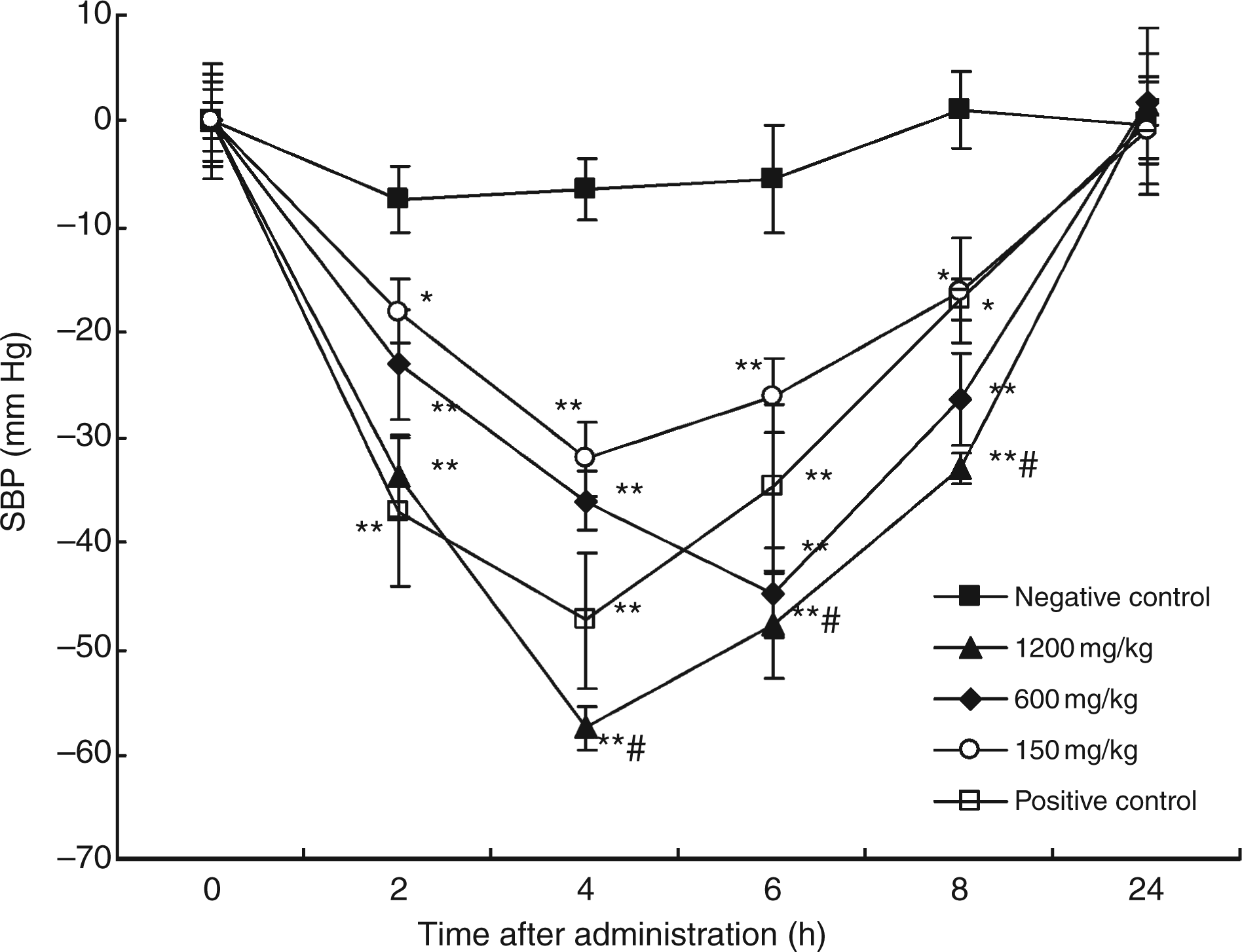
Conclusion
CHPH-IV of Cobia head protein papain hydrolysate exerted a potent inhibitory activity to ACE in vitro and decreased the systolic blood pressure of SHR after oral administration. It is also important that the administration of CHPH-IV did not change the systolic blood pressure of the normotensive WKY rats. Therefore, CHPH-IV from Cobia head protein papain hydrolysate could be very useful in the preparation of antihypertensive peptides. A further research on the purification of ACE inhibitory peptides from CHPH-IV is undergoing.
Footnotes
Funding
This work was supported by National Key Technology R&D Program of China [grant number 2007BAD29B09].
Acknowledgments
The authors are grateful for the technical support of Slac Laboratory Animal Co. Ltd. Shanghai, China.