
Abstract
Objective:
To ascertain whether cavitation-free acoustic sensitization enhances intracellular delivery and amplifies polo-like kinase 4 (PLK4)-targeted signaling to boost the phillyrin derivative DE02’s antitumor efficacy against osteosarcoma, providing a secure and effective ultrasound-enabled method for targeted cancer biotherapy.
Methods:
Using ultracentrifugation, exosomes generated from human umbilical vein endothelial cells were separated, purified, and identified using common morphological and molecular markers. Low-energy sonication under cavitation-free acoustic circumstances was used to insert DE02 into exosomes, creating an exosomal delivery (ExoDE02) system intended to improve cellular absorption without causing membrane damage. MG-63 and Saos-2 human osteosarcoma cell lines were used as in vitro models. The cell counting kit-8 test was used to measure cell proliferation, proliferating cell nuclear antigen (PCNA) immunofluorescence was used to measure proliferative activity, and Transwell assays were used to measure migration and invasion. Enzyme-linked immunosorbent assay (ELISA), real-time quantitative PCR, and Western blotting were used to assess the expression of PLK4 and downstream tumor protein 53 (p53)–cyclin-dependent kinase inhibitor 1A (p21) signaling components. To verify pathway specificity, PLK4 overexpression studies were carried out. A nude mouse xenograft model was used to evaluate in vivo antitumor effectiveness and biosafety.
Results:
In a concentration-dependent manner, DE02 showed almost 10 times more antiproliferative action against osteosarcoma cells than the parent chemical phillyrin. The inhibitory effects of DE02 on osteosarcoma cell proliferation, migration, and invasion were greatly enhanced by cavitation-free acoustic sensitization-mediated ExoDE02. This was accompanied by a significant downregulation of PCNA and PLK4 expression and activation of the p53–p21 tumor suppressor pathway. The anticancer effects of DE02 and ExoDE02 were successfully inhibited by overexpression of PLK4, indicating PLK4-dependent therapeutic efficacy. ExoDE02 significantly inhibited the growth of xenograft tumors in vivo, decreased tumor weight and volume, and showed no discernible systemic toxicity.
Conclusions:
By increasing the therapeutic efficacy of the phillyrin derivative DE02 via a safe, nondestructive exosome-based delivery method, cavitation-free acoustic sensitization improves PLK4-targeted osteosarcoma therapy. For targeted osteosarcoma biotherapy, this ultrasound-enabled method provides a mechanistically defined and physiologically applicable platform.
Keywords
Introduction
Osteosarcoma, a primary malignant bone tumor originating from mesenchymal cells and characterized by malignant proliferation producing osteoid matrix, is the most common type of malignant bone tumor in adolescents and children. Approximately 75% of patients develop the disease between 10 and 20 years of age, with a slightly higher incidence in males. It predominantly occurs in the metaphyses of long bones (e.g., distal femur, proximal tibia), presenting with typical clinical symptoms of local pain and masses. Pain progresses from intermittent dull pain in the early stage to persistent severe pain, and advanced stages are prone to complications such as pulmonary metastasis, pathological fractures, and cachexia, which seriously threaten patients’ life and health.1–4 Although the multidisciplinary comprehensive treatment model (surgical resection combined with neoadjuvant chemotherapy before surgery and adjuvant chemoradiotherapy after surgery) has been continuously optimized in recent years, the 5-year survival rate of osteosarcoma patients has increased from ∼20% in the past to 60%−80%. However, the long-term survival rate of metastatic or recurrent osteosarcoma remains below 30%. In clinical practice, issues such as chemotherapeutic drug resistance, lack of targeted therapy targets, and significant treatment-related toxic side effects still restrict further improvement of therapeutic efficacy. Therefore, developing novel and efficient therapeutic drugs and precise delivery systems has become a key demand for improving the treatment status of osteosarcoma.5–9
Natural products have long been important sources for antitumor drug development due to their diverse structures, good biocompatibility, and broad mechanisms of action. Phillyrin, a lignan-type bioactive component isolated from the traditional Chinese herb Forsythia suspensa, has been confirmed by numerous studies to possess multiple biological activities, including anti-inflammatory, antioxidant, antiviral, and antitumor effects.10–13 In the field of oncology, phillyrin exhibits certain antiproliferative effects on various malignant tumor cells (e.g., lung cancer, liver cancer, breast cancer), and its mechanisms may involve cell cycle regulation, apoptosis induction, and angiogenesis inhibition. However, the inherent defects of phillyrin (e.g., poor water solubility, low bioavailability, weak antitumor activity) limit its direct clinical application in tumor therapy. Optimizing the physicochemical properties and biological activity of natural products through chemical structural modification14,15 is an effective strategy to overcome this bottleneck: introducing specific functional groups while retaining the core active scaffold can improve drug solubility, stability, and target-binding ability, thereby enhancing therapeutic efficacy. This provides an important idea for the antitumor application development of phillyrin.
Genomic instability is one of the core characteristics of tumors, closely related to tumor progression and treatment resistance. As a key regulatory structure for cell division, centrosome dysfunction plays an important role in tumorigenesis and development. Polo-like kinase 4 (PLK4), a member of the serine–threonine kinase family, is the core molecule regulating centrosome duplication. Its aberrant overexpression can induce centrosome amplification, leading to cell aneuploidy and chromosome segregation abnormalities, which ultimately promote tumorigenesis and progression. Existing studies have confirmed that PLK4 is overexpressed in various malignant tumors (e.g., lung cancer, breast cancer, colorectal cancer) and is closely associated with tumor stage, lymph node metastasis, and poor prognosis, making it a potential key target for targeted tumor therapy. In osteosarcoma, some cases exhibit features such as cell aneuploidy, complex chromosomal rearrangements, and p53 dysfunction. Inhibition of PLK4 can degrade the p53 negative regulator MDM2 by activating the PIDDosome complex-caspase-2 pathway, thereby stabilizing p53 and inducing p21-dependent cell cycle arrest,16–18 suggesting that PLK4 may be involved in regulating the malignant phenotype of osteosarcoma. However, the specific expression pattern of PLK4 in osteosarcoma, its regulatory association with the p53–p21 pathway, and the therapeutic effect of targeted PLK4 intervention have not been fully elucidated, requiring further in-depth research.
Exosomes, an important subtype of extracellular vesicles, have a natural lipid bilayer structure and unique advantages (e.g., low immunogenicity, high biocompatibility, tumor tissue tropism, and the ability to cross biological barriers), making them ideal candidates for next-generation drug delivery carriers. Compared with traditional artificial delivery systems (e.g., liposomes, polymer nanoparticles), exosomes can effectively avoid drug degradation by the in vivo enzyme system, significantly improving drug bioavailability. Meanwhile, their surface naturally expresses “don’t eat me” signaling molecules (e.g., CD47), which can avoid clearance by macrophages and prolong blood circulation time. In addition, exosomes can achieve precise drug delivery to tumor cells through interactions between surface-specific ligands and tumor cell surface receptors, reducing the toxic side effects caused by off-target effects.19–22 In recent years, significant progress has been made in research on engineered exosomes delivering chemotherapeutic drugs or targeted inhibitors for osteosarcoma therapy. For example, natural killer cell (NK) cell-derived exosomes modified with the iRGD peptide can recognize integrin αvβ3 (highly expressed on osteosarcoma cells) to achieve targeted drug delivery and enhance therapeutic effects, providing a new idea for precise osteosarcoma therapy.
Based on the above research background, this laboratory synthesized the derivative DE02 by chemically modifying the natural active component phillyrin (using phillyrin as the scaffold) to optimize the antitumor activity of phillyrin. Meanwhile, to further improve the targeting and bioavailability of DE02, the authors constructed a combined delivery system (ExoDE02) using human umbilical vein endothelial cell (HUVEC)-derived exosomes (Exo) and DE02. In this study, in vitro and in vivo experiments were conducted to systematically investigate the effects of this combined system on the proliferation, migration, and invasion abilities of MG-63 and Saos-2 osteosarcoma cells; to deeply analyze its regulatory effects on PLK4 expression and enzyme activity, and clarify the target of DE02 (as a phillyrin derivative); to verify the mediating role of the PLK4-p53–p21 signaling pathway through PLK4 overexpression (oePLK4) experiments; and finally to evaluate the in vivo antiosteosarcoma activity and safety of the ExoDE02 system using a nude mouse xenograft model.
Low-intensity ultrasound can function as a physical sensitization technique, improving medication transport and intracellular responsiveness without causing heat harm, according to recent developments in ultrasound-enabled cancer therapy. Crucially, ultrasound has a great safety profile and can temporarily alter intracellular transport and membrane permeability when used in cavitation-free acoustic environments. In line with the risk–benefit analysis highlighted in recent ultrasonic oncology research, this nondestructive acoustic sensitization technique provides a clinically applicable way to increase the effectiveness of targeted medicines without raising systemic toxicity.
To develop a novel PLK4-targeted therapeutic approach for osteosarcoma, the current study combines chemical alteration, exosome-based delivery, and cavitation-free sonic sensitization. Using low-energy sonication in the absence of cavitation, an exosome-mediated delivery system that encapsulated the phillyrin derivative DE02 (ExoDE02) was created. The authors examined the effects of this ultrasound-enabled system on osteosarcoma cell proliferation, migration, and invasion through extensive in vitro and in vivo experiments; clarified its regulatory impact on PLK4 expression and the downstream p53–p21 signaling pathway; confirmed pathway dependence using oePLK4 models; and assessed antitumor efficacy and biosafety in a nude mouse xenograft model. To facilitate the translational development of anticancer agents derived from natural products, this study intends to provide mechanistic insight and preclinical evidence supporting cavitation-free acoustic sensitization as a safe and effective strategy to enhance PLK4-targeted biotherapy in osteosarcoma.
Materials and Methods
Cells and reagents
Human osteosarcoma cell lines (MG-63, Saos-2) and HUVECs used in this experiment were purchased from the American Type Culture Collection. After resuscitation, cells were cultured in high-glucose Dulbecco’s modified Eagle’s medium (DMEM) (HyClone) supplemented with 10% fetal bovine serum (FBS, Gibco) and 1% penicillin–streptomycin (HyClone) in a constant-temperature incubator (37°C, 5% CO2, saturated humidity). Cells were passaged every 2–3 d, and logarithmic-phase cells were selected for experiments. Phillyrin standard (purity ≥98%) was purchased from Sigma Aldrich; the target derivative DE02 was synthesized in the laboratory via chemical structural modification of natural phillyrin; oePLK4 plasmid and empty vector plasmid (vector) were obtained from the Addgene plasmid library; antibodies against proliferation-related marker proliferating cell nuclear antigen (PCNA), target proteins (PLK4, p53, p21), and internal reference protein glyceraldehyde-3-phosphate dehydrogenase (GAPDH) were purchased from Abcam; cell counting kit-8 (CCK-8) for cell proliferation detection was from Dojindo; Transwell chambers (8 μm pore size) for cell migration/invasion assays were from Corning; Matrigel matrix was from BD Biosciences; TRIzol reagent, reverse transcription kit, and real-time quantitative PCR (RT-qPCR) kit for RNA extraction were from TaKaRa; protein lysis buffer, BCA protein quantification kit, sodium dodecyl sulfate–polyacrylamide gel electrophoresis (SDS-PAGE) gel preparation kit, and enhanced chemiluminescence (ECL) chemiluminescence kit were from Beyotime Biotechnology; other reagents (analytical grade) were purchased from Sinopharm Chemical Reagent Co., Ltd.
Synthesis of phillyrin derivative DE02
Using natural phillyrin as the starting material, chemical modification was performed on the hydroxyl domain of its glycosidic bond: In an anhydrous dichloromethane solvent system, phillyrin (1 mmol), Boc-protected phenylalanine (1.2 mmol), and 4-dimethylaminopyridine (0.1 mmol) were added. N,N’-dicyclohexylcarbodiimide (1.2 mmol) was slowly added dropwise under ice bath conditions. After completion of dropwise addition, the reaction was stirred at room temperature for 12 h. After the reaction, an appropriate amount of ethyl acetate was added to extract the reaction solution; the organic phase was washed sequentially with saturated sodium bicarbonate solution and saturated brine, dried over anhydrous sodium sulfate, and concentrated under reduced pressure to obtain the crude product. Subsequently, high-performance liquid chromatography (HPLC, Agilent 1260) was used for purification: the mobile phase was methanol-water (gradient elution: 0–10 min, methanol proportion increased from 30% to 60%; 10–20 min, methanol proportion maintained at 60%), detection wavelength was 254 nm, and the target fraction with a retention time of 12.5 min was collected and dried under reduced pressure to obtain derivative DE02 (synthetic route shown in Fig. 1A).
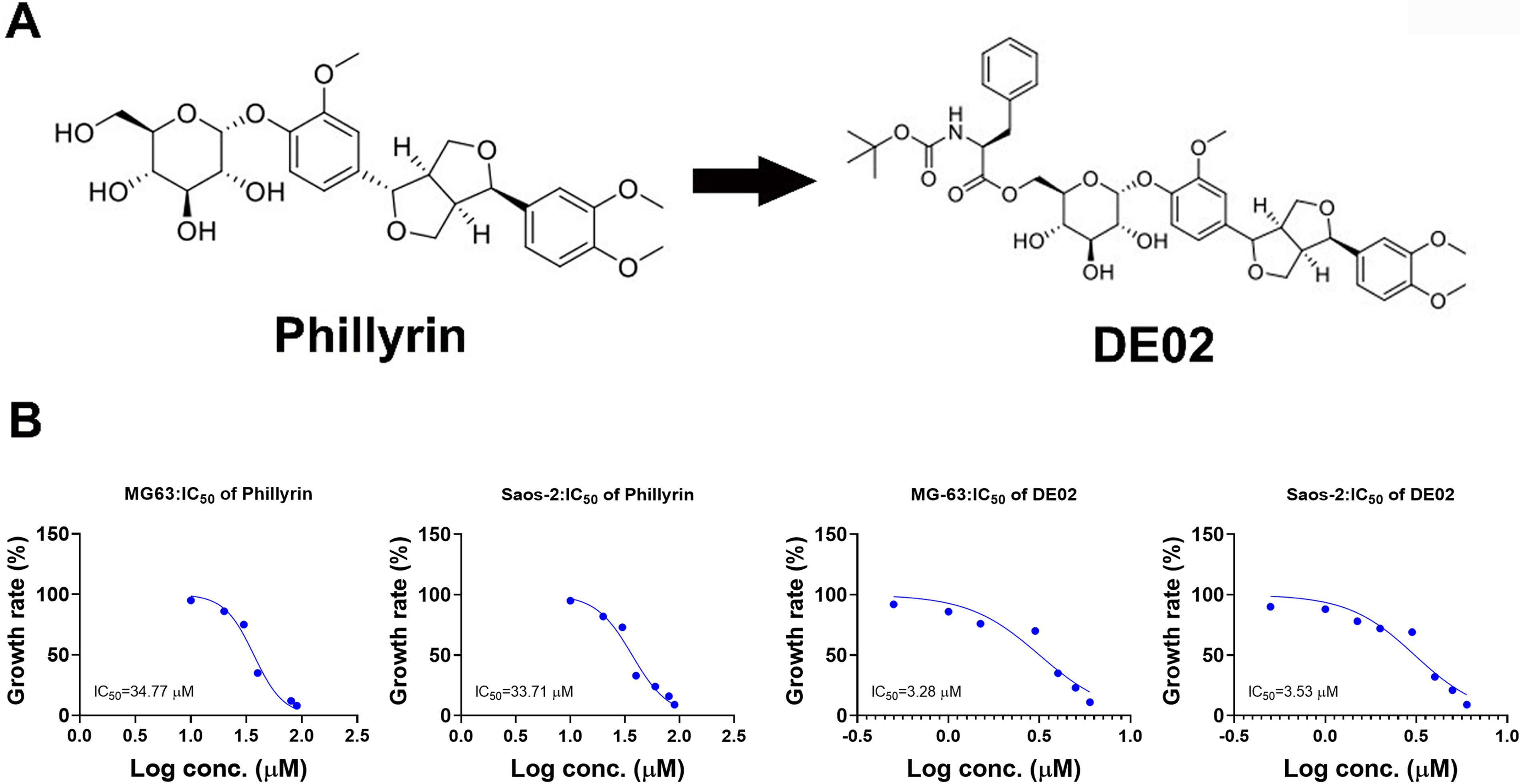
Structures and antiosteosarcoma cell proliferation activities of phillyrin and its derivative DE02.
Isolation, identification of exosomes, and DE02 loading
Logarithmic-phase HUVECs were cultured in DMEM supplemented with exosome-depleted FBS (exosomes in serum were removed by centrifugation at 100,000 × g for 16 h). After 24 h, cell supernatants were collected and pretreated at 4°C: centrifugation at 300 × g for 10 min (to remove cells and large debris), followed by centrifugation at 10,000× g for 30 min (to remove microvesicles and apoptotic bodies). The supernatant was transferred to an ultracentrifuge tube, centrifuged at 100,000× g for 70 min at 4°C; after discarding the supernatant, the pellet was resuspended in precooled PBS and centrifuged again at 100,000× g for 70 min at 4°C. The final pellet was the purified HUVEC-derived exosomes. Exosomes were identified using the following methods: (1) Nanoparticle tracking analysis (Malvern NanoSight NS300): Exosomes were resuspended in phosphate buffered saline (PBS), diluted to an appropriate concentration, and detected. Results showed that exosome particle size was distributed between 50 and 150 nm, with a median size of ∼110 nm, consistent with the typical size characteristics of exosomes. (2) Western blot identification: Exosomal proteins were extracted to detect the expression of exosome-specific markers [CD63 (CD63 molecule), tumor susceptibility 101 (TSG101)] and calnexin (endoplasmic reticulum protein, negative exosome marker) in cell lysates. Results showed positive expression of CD63 and TSG101, and negative expression of calnexin in exosome samples, confirming that exosome purity met experimental requirements. DE02 was loaded into exosomes via sonication: Purified exosomes (1 mg/mL) and DE02 (5 mg/mL) were mixed at a 1:1 volume ratio, placed on ice, and sonicated three times (200 W power, 30 s each time, 1-min interval) using an ultrasonic cell disruptor. After sonication, the mixture was centrifuged at 100,000× g for 70 min at 4°C; the supernatant was discarded to remove unloaded free DE02, and the pellets were the DE02-loaded exosomes (denoted as ExoDE02), which were resuspended in precooled PBS and stored at 4°C for later use. The concentration of DE02 in the supernatant before and after loading was determined by HPLC, and the loading efficiency was calculated as follows: Loading efficiency = (initial drug amount—free drug amount)/initial drug amount × 100%. Results showed that the drug loading efficiency of Exo-DE02was ∼72%.
Cavitation-free acoustic loading of DE02 into exosomes
MG-63 and Saos-2 cells in the logarithmic growth phase were seeded into 96-well cell culture plates at a density of 5 × 10³ cells per well, with 100 μL of complete medium added to each well. The plates were incubated in the incubator for 24 h to allow cells to adhere and recover. The old medium was then aspirated, and a fresh medium containing different concentration gradients (0, 5, 10, 20, 40, and 80 μM) of phillyrin, DE02, or different concentration gradients (0, 0.5, 1.0, 2.0, 4.0, and 6.0 μM) of ExoDE02 was added separately, with six replicate wells per group. After 48 h of further incubation, 10 μL of CCK-8 reagent was added to each well, and the plates were incubated for 4 h in the incubator. After incubation, the absorbance (OD value) at 450 nm was measured using a microplate reader (Thermo Scientific Multiskan FC). Purified exosomes (1 mg/mL) and DE02 (5 mg/mL) were combined at a 1:1 volume ratio to create an ultrasound-enabled delivery system under cavitation-free acoustic sensitization conditions. An ultrasonic probe was used to treat the combination to low-energy sonication (200 W, 30 s per cycle, three cycles with 1-min cooling intervals on ice). To allow for temporary membrane permeability without disrupting vesicles, these parameters were chosen to stay below cavitation limits. Wells containing only medium and CCK-8 reagent served as the blank control. The cell proliferation rate was calculated as follows: Proliferation rate = [(Experimental group OD value—Blank group OD value)/(Control group OD value—Blank group OD value)] × 100% dose-response curves were fitted using GraphPad Prism 8.0 software, and the half-maximal inhibitory concentration (IC50) of the drugs on cells was calculated.
An ultrasonic probe with a nominal output power of 200 W and operating at 20 kHz was used for low-energy sonication. An estimated duty cycle of about 50% was achieved by applying sonication in pulse mode (30 s each cycle, three cycles, 1-min cooling intervals). The predicted peak negative pressure was less than 0.3 MPa based on the probe shape and immersion depth, resulting in a mechanical index (MI) of less than 0.4. Both steady (MI ≈0.7) and inertial (MI ≥1.9) cavitation limits are well below these parameters. To prevent temperature buildup, sonication was done on ice. Cavitation-free acoustic sensitization was accomplished in these circumstances, allowing for temporary membrane permeability without cellular injury or vesicle breakup.
Ultrasound exposure was not used for cellular treatment or in vivo therapy; rather, it was only used during the exosome drug-loading procedure. Therefore, rather than referring to patient-side or tumor-site ultrasound exposure, the phrase “ultrasound-enabled” particularly refers to cavitation-free acoustic sensitization during exosome manufacturing.
Cell migration and invasion assays (Transwell method)
Cell migration assay
Transwell chambers (8 μm pore size) were placed in 24-well plates, and 600 μL of complete medium containing 10% FBS was added to the lower chamber as a chemoattractant. MG-63 and Saos-2 cells treated with different concentrations of ExoDE02 (0, 0.5, 1.0, and 2.0 μM) for 24 h were resuspended in serum-free medium, adjusted to a concentration of 1 × 10⁵ cells/mL, and 200 μL of the cell suspension (containing 2 × 10⁴ cells) was seeded into the upper chamber. After 24 h of incubation in the incubator, the chambers were removed, washed twice with precooled PBS, fixed with 4% paraformaldehyde for 30 min, and stained with 0.1% crystal violet for 20 min. Unmigrated cells in the upper chamber were gently wiped off with a cotton swab. Five random fields (×200 magnification) were observed under an optical microscope (Olympus CX41), and the number of migrated cells was counted; the average value was taken as the migration cell count for each group.
Cell invasion assay
The procedure was largely consistent with the migration assay, except that Matrigel coating was performed before cell seeding: Matrigel matrix was diluted 1:8 (v/v) with serum-free medium, and 50 μL of the diluted Matrigel was evenly spread on the surface of the Transwell upper chamber membrane. The chamber was incubated in the incubator for 30 min to allow the Matrigel to solidify into an artificial basement membrane. ExoDE02-treated cells were then seeded, and subsequent steps were performed. The number of invaded cells was determined by counting the transmembrane cells.
To reduce confusing cytostatic effects, sub-IC•1 doses of DE02 and ExoDE02 were used in migration and invasion tests.
Immunofluorescence staining for PCNA expression
Sterile cell climbing slides were placed in 24-well plates, and MG-63/Saos-2 cells (5 × 10⁴ cells per well) were seeded and incubated for 24 h to allow adhesion. The old medium was aspirated, and a fresh medium containing different concentrations of ExoDE02 (0, 0.5, 1.0, and 2.0 μM) was added for 48 h for further incubation. At the end of the experiment, the medium was aspirated, and cells were washed three times with precooled PBS, fixed with 4% paraformaldehyde for 20 min, permeabilized with 0.5% Triton X-100 for 15 min, and blocked with 5% bovine serum albumin at room temperature for 30 min. The PCNA primary antibody (diluted 1:500 in blocking buffer) was added and incubated overnight at 4°C. The next day, cells were washed three times with PBS (5 min each), and the fluorescein isothiocyanate (FITC)-labeled goat antirabbit secondary antibody (diluted 1:1000) was added for 1 h of light-protected incubation at room temperature. After three PBS washes, 4′,6-diamidino-2-phenylindole (DAPI) staining solution was added for 5 min of light-protected incubation to label nuclei. Finally, slides were mounted with an antifluorescence quenching mounting medium (Beyotime) and observed under a laser confocal microscope (Zeiss LSM 880) to capture images (×400 magnification). The proportion of PCNA-positive cells (green fluorescence) relative to total cells (blue fluorescence) in each field was quantified using ImageJ software; five fields were counted per sample, and the average value was calculated.
RT-qPCR for gene expression
MG-63/Saos-2 cells treated with different concentrations of ExoDE02 (0, 0.5, 1.0, and 2.0 μM) for 48 h were collected, and total RNA was extracted using the TRIzol reagent (strictly following the kit instructions). RNA concentration and purity were detected using a Nanodrop 2000 spectrophotometer, ensuring an OD260/280 ratio of 1.8–2.0 and an OD260/230 ratio of >2.0. Using 1 μg of total RNA as a template, cDNA was synthesized with a reverse transcription kit under the following conditions: 37°C for 15 min, 85°C for 5 s, and storage at 4°C. RT-qPCR amplification was then performed using cDNA as the template; the 20 μL reaction system included 10 μL SYBR Green Premix Ex Taq, 0.5 μL each of forward and reverse primers, 2 μL cDNA template, and 7 μL nuclease-free water. Reactions were run on a real-time PCR instrument (Bio-Rad CFX96) under the following conditions: Predenaturation at 95°C for 3 min; 40 cycles of denaturation at 95°C for 10 s and annealing at 60°C for 30 s; melting curve analysis: 95°C for 15 s, 60°C for 1 min, and 95°C for 15 s.
Genes detected included PLK4, p53, and p21, with GAPDH as the internal reference gene. Primer sequences were designed using the PrimerBank database and synthesized by Sangon Biotech (Shanghai). The specific sequences are as follows:
PLK4 forward primer: 5′-GCTGCTGCTGCTGTTTCTGT-3′
PLK4 reverse primer: 5′-CCTGCTGCTGCTGTTTCTGT-3′
p53 forward primer: 5′-GCTGCTGCTGCTGTTTCTGT-3′
p53 reverse primer: 5′-CCTGCTGCTGCTGTTTCTGT-3′
p21 forward primer: 5′-GCTGCTGCTGCTGTTTCTGT-3′
p21 reverse primer: 5′-CCTGCTGCTGCTGTTTCTGT-3′
GAPDH forward primer: 5′-GAAGGTGAAGGTCGGAGTC-3′
GAPDH reverse primer: 5′-GAAGATGGTGATGGGATTTC-3′
The 2-ΔΔCt method was used to calculate the relative expression of each gene, where ΔCt = Ct (target gene)-Ct (GAPDH), and ΔΔCt = ΔCt (experimental group)-ΔCt (control group).
Western blot for protein expression
MG-63/Saos-2 cells subjected to different treatments were collected, and precooled radio-immunoprecipitation assay lysis buffer (RIPA) (containing 1% protease inhibitor cocktail and 1% phosphatase inhibitor cocktail) was added for 30 min of lysis on ice. After centrifugation at 12,000× g for 15 min at 4°C, the supernatant was collected as the total protein extract. Protein concentration was determined using a BCA protein quantification kit, and the protein concentration of each group was adjusted to be consistent. Thirty micrograms of protein sample was mixed with five times SDS loading buffer and denatured in a boiling water bath for 5 min. SDS-PAGE was performed (10% separating gel, 5% stacking gel): Electrophoresis was run at a constant voltage of 80 V until bromophenol blue entered the separating gel, and then the voltage was adjusted to 120 V until bromophenol blue reached the bottom of the gel. After electrophoresis, proteins were transferred to a polyvinylidene fluoride (PVDF) membrane using the wet transfer method (constant current of 200 mA for 90 min). The PVDF membrane was blocked in 5% skim milk at room temperature for 2 h, and then incubated overnight at 4°C with primary antibodies (PLK4, p53, p21, GAPDH; all diluted 1:1000). The next day, the membrane was washed three times with TBST (10 min each), incubated with horseradish peroxidase (HRP)-labeled secondary antibody (diluted 1:5000) for 1 h at room temperature, and washed three times with TBST. ECL chemiluminescence reagent was added, and images were captured using a chemiluminescence imaging system (Bio-Rad ChemiDoc XRS+). Band gray values were quantified using ImageJ software, with GAPDH as the internal reference to calculate the relative expression of target proteins.
Nude mouse xenograft model construction and in vivo efficacy evaluation
SPF-grade BALB/c nude mice (6–8 weeks old, weighing 18–22 g, purchased from Beijing Vital River Laboratory Animal Technology Co., Ltd.) were acclimatized for 1 week, with free access to food and water. The breeding environment was controlled at 22°C–25°C, 50%–60% humidity, and a 12-h light/dark cycle. MG-63 cells in the logarithmic growth phase were resuspended in precooled PBS and adjusted to a concentration of 5 × 10⁷ cells/mL. Each nude mouse was subcutaneously injected with 100 μL of the cell suspension (containing 5 × 10⁶ cells) into the right back to construct the xenograft model.
When the tumor volume reached ∼100 mm³, the nude mice were randomly divided into two groups (6 mice per group): (1) Control group: Tail vein injection of an equal volume of PBS. (2) ExoDE02 group: Tail vein injection of 2 μM ExoDE02, administered once every 2 d for four consecutive weeks. During administration, the tumor length (L) and width (W) were measured weekly using a vernier caliper, and tumor volume was calculated using the formula “Tumor volume (V) = L×W2/2” to plot the tumor growth curve. Nude mouse body weight was also measured weekly to evaluate drug toxic side effects.
After 4 weeks of administration, nude mice were sacrificed by cervical dislocation. Tumor tissues were carefully dissected, washed twice with precooled PBS, and weighed; tumor weight was recorded. Part of the tumor tissue was fixed in 4% paraformaldehyde for subsequent pathological analysis; the remaining tissue was stored at −80°C for later use.
Statistical analysis
All experiments were independently repeated three times. Experimental data were statistically processed using GraphPad Prism 8.0 software and expressed as “mean ± standard deviation (SD) (x ± s).” Differences between multiple groups were compared using one-way analysis of variance (ANOVA), with pairwise comparisons between groups performed using Tukey’s multiple comparison test. Differences between two groups were compared using the independent samples t-test. A p-value <0.05 was considered statistically significant, and p < 0.01 was considered extremely statistically significant. Each batch of 6 mice was used for in vivo tests. Mean ± SD is used to display tumor volume and weight statistics. Two-tailed unpaired t-tests were used to compare groups; where appropriate, effect sizes and 95% confidence intervals were computed. In vitro multiple comparisons were adjusted using Tukey’s post hoc test.
Results
Characterization of enhanced antitumor activity of phillyrin derivative DE02 via structural modification
Figure 1A clearly shows the chemical structural modification process from phillyrin to its derivative DE02: the left phillyrin has a lignan core scaffold linked to a glycosidic domain with multiple hydroxyl groups; after structural modification, the right DE02 introduces a tert-butoxycarbonyl (Boc)-protected phenylalanine group at the glycosidic bond site of phillyrin—specifically via an ester bond formed between the carboxyl group of phenylalanine and the hydroxyl group on the glucose ring. This modification not only completely retains the lignan active scaffold of phillyrin (ensuring the core pharmacodynamic basis) but also effectively optimizes the physicochemical properties of the drug by introducing a hydrophilic amino acid fragment. Figure 1B intuitively compares the antiproliferative effects of phillyrin and DE02 on MG-63 and Saos-2 osteosarcoma cells through four concentration–proliferation rate curves: the left curves show that the IC50 of phillyrin against MG-63 and Saos-2 cells are 34.77 and 33.71 μM, respectively, indicating a weak overall inhibitory activity; in contrast, the IC50 of DE02 in the right curves significantly decreases to 3.28 and 3.53 μM, with antitumor activity enhanced by approximately 10-fold compared with the parent drug. Meanwhile, both drug curves show a clear concentration-dependent trend, further confirming that the antitumor activity of DE02 is significantly enhanced after structural modification.
Concentration-dependent inhibition of osteosarcoma cell proliferation and downregulation of PCNA expression by DE02 at 24 h
The survival rates of MG-63 and Saos-2 cells in the control group (untreated) were both close to 100%. As the DE02 concentration increased from 0.5 to 2.0 μM, the survival rate of MG-63 cells decreased to approximately 90%, 60%, and 15%, while that of Saos-2 cells decreased to approximately 90%, 60%, and 20%. The differences between each concentration group and the control group were extremely significant (p < 0.01), demonstrating the concentration-dependent inhibitory effect of DE02 on osteosarcoma cell proliferation (Fig. 2A). The immunofluorescence staining results in Figure 2B show that green PCNA-positive signals (proliferating cell markers) were densely distributed in the MG-63 and Saos-2 cells of the control group, while the number and intensity of green fluorescence gradually decreased with increasing DE02 concentrations; the quantitative bar chart on the right further shows that the PCNA-positive rate of MG-63 cells decreased from approximately 60% in the control group to 15% in the 2.0 μM group, and that of Saos-2 cells decreased from approximately 60% to 20%, with similar extremely significant differences (p < 0.01). These results are consistent with the cell survival rate data, confirming that DE02 can block osteosarcoma cell proliferation by downregulating PCNA expression.
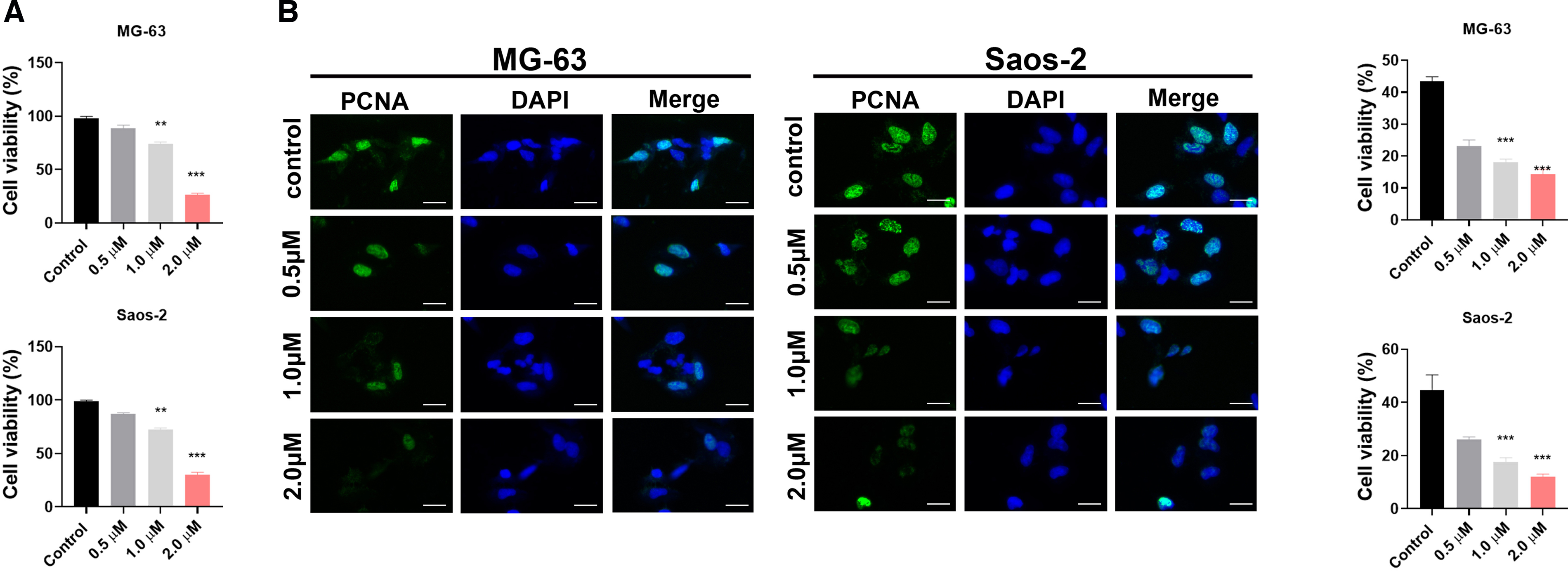
Effects of DE02 on osteosarcoma cell viability and expression of proliferation marker proliferating cell nuclear antigen (PCNA).
Concentration-dependent inhibition of MG-63/Saos-2 cell migration and invasion by DE02 and reversal effect of PLK4
In the control group, a large number of MG-63 and Saos-2 cells migrated through the membrane; as the DE02 concentration increased from 0.5 to 2.0 μM, the number of transmembrane cells gradually decreased. When 2.0 μM DE02 was combined with oePLK4, the number of transmembrane cells was significantly higher than that in the 2.0 μM DE02-alone group, intuitively reflecting that the inhibitory effect of DE02 on migration depends on PLK4 downregulation (Fig. 3A, B). The migration number of MG-63 cells decreased from approximately 100 in the control group to around 20 in the 2.0 μM DE02 group, and recovered to approximately 70 after combination with oePLK4; Saos-2 cells showed the same trend. The differences between each concentration group and the control group were extremely significant (p < 0.01), and the reversal effect after combination with oePLK4 was also statistically significant (##p < 0.01). Increasing DE02 concentration gradually reduced the number of transmembrane cells, while combination with oePLK4 significantly increased the number of transmembrane cells (Fig. 3C); the quantitative data in Figure 3D show that the invasion number of MG-63 cells decreased from approximately 100 in the control group to around 20 in the 2.0 μM DE02 group, and recovered to approximately 60 after combination with oePLK4, with Saos-2 cells showing the same trend, and all differences were statistically significant. In summary, DE02 can concentration dependently inhibit the migration and invasion of osteosarcoma cells, and this effect depends on the downregulation of PLK4.
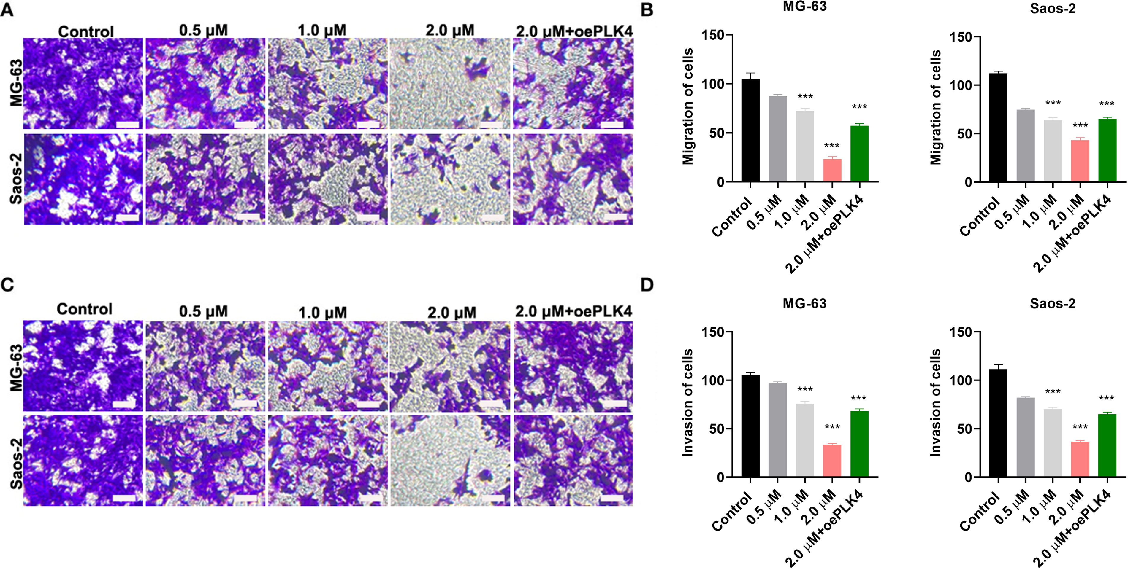
Inhibitory effects of DE02 on osteosarcoma cell migration and invasion, and the reversal effect of polo-like kinase 4 (PLK4).
Downregulation of PLK4 enzyme activity and activation of the p53–p21 pathway by DE02 in osteosarcoma cells
ELISA results showed that as DE02 concentration increased, the enzyme activity of PLK4 in both cell lines gradually decreased; when DE02 was combined with oePLK4, the PLK4 level was significantly higher than that in the DE02-alone group, confirming the downregulatory effect of DE02 on PLK4 activity (Fig. 4A–C). The qPCR results in Figure 4D–I show changes in gene expression: As the DE02 concentration increased, the mRNA level of PLK4 gradually decreased, while the mRNA levels of p53 and p21 gradually increased; when DE02 was combined with oePLK4, the mRNA level of PLK4 recovered, and the mRNA levels of p53 and p21 were inhibited, with statistically significant differences between all groups. These results indicate that DE02 can activate the p53–p21 pathway by downregulating PLK4, and this effect depends on the regulatory role of PLK4.
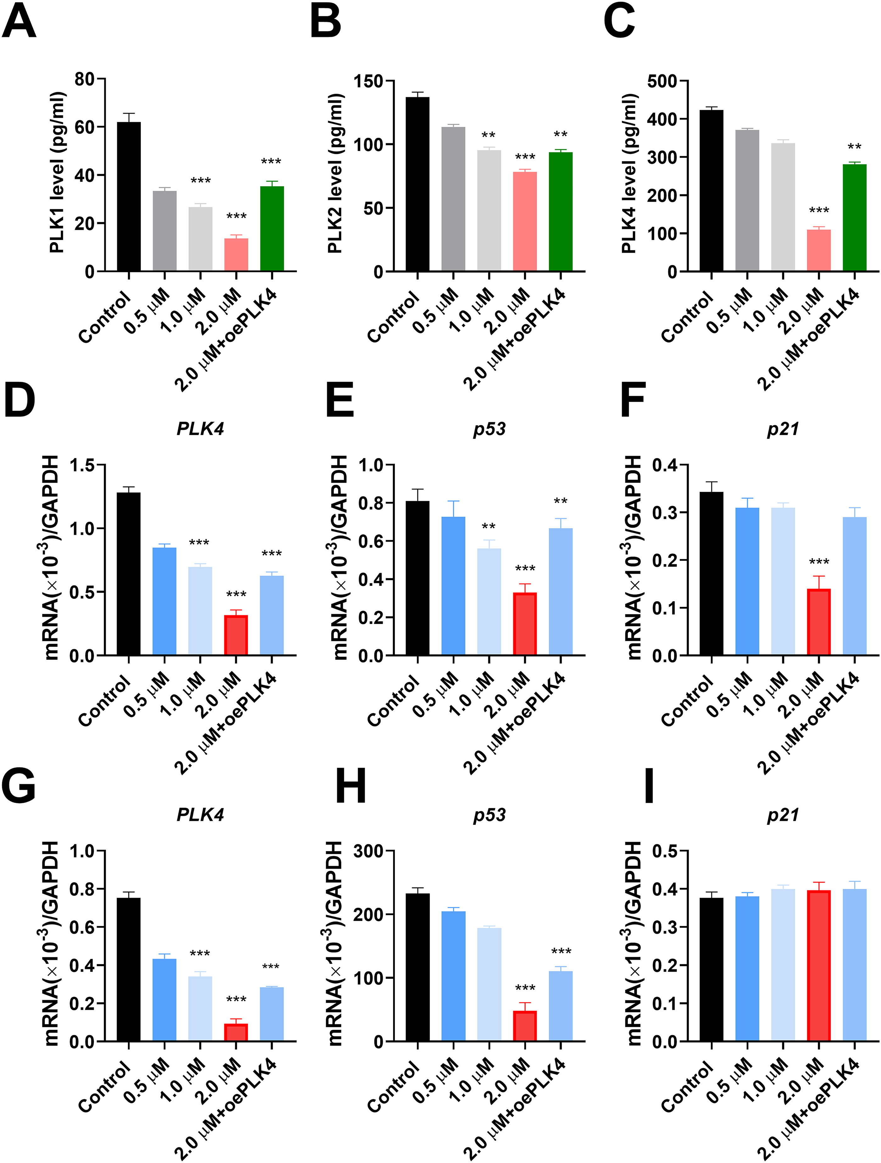
Regulation of the polo-like kinase 4 (PLK4)-p53-p21 signaling pathway by DE02 and reversal effect of PLK4.
Cavitation-free acoustic sensitization-mediated exosomal delivery (Exo-DE02) amplifies PLK4-targeted antitumor effects
Using cavitation-free acoustic loading, DE02 was encapsulated into exosomes to improve intracellular delivery under safe acoustic circumstances, resulting in ExoDE02. The immunofluorescence staining results in Figure 5A show that red PCNA signals (proliferation markers) were densely distributed in the control group; as ExoDE02 concentration increased, the number of red signals significantly decreased; when ExoDE02 was combined with oePLK4, the red PCNA signals were significantly higher than those in the ExoDE02-alone group. Combined with DAPI (nuclear marker) and Merge channels, it is clear that ExoDE02 can inhibit cell proliferation, and this effect depends on PLK4 downregulation. The Transwell assay results in Figure 5B show that the control group had a large number and high density of transmembrane cells; after ExoDE02 treatment, the number of transmembrane cells significantly decreased with increasing concentration; when combined with oePLK4, the number of transmembrane cells was significantly higher than that in the ExoDE02-alone group. These results show that PLK4-targeted anticancer efficacy is greatly increased by cavitation-free acoustic sensitization-enabled exosomal distribution without changing the underlying molecular mechanism.
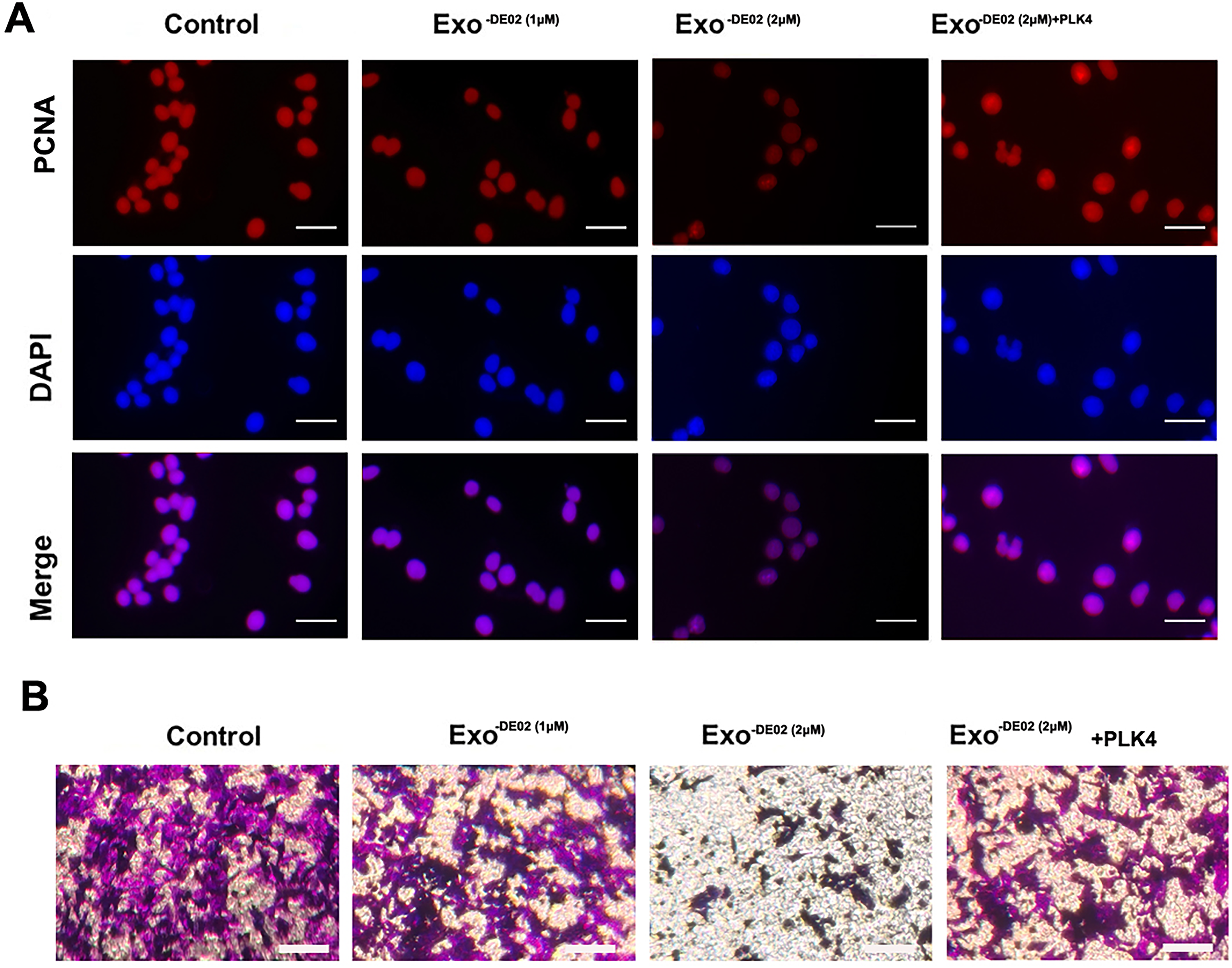
Inhibitory effects of ExoDE02 on osteosarcoma cell proliferation and migration, and the reversal effect of polo-like kinase 4 (PLK4).
Inhibitory effect of combined exo and DE02 treatment on tumor growth in vivo
The tumor volume curve, Figure 6A, shows that the tumors in the control group grew rapidly over time, while the tumor volume growth in the ExoDE02 (2 μM) combined treatment group was significantly slowed down, with a statistically significant difference compared with the control group; the tumor images in Figure 6B intuitively show that the tumor samples in the control group were significantly larger than those in the ExoDE02 treatment group; the tumor weight statistics in Figure 6C further quantitatively verified that the tumor weight in the control group was significantly higher than that in the ExoDE02 treatment group, with an extremely significant difference. Together, these findings demonstrate that ExoDE02 administration via cavitation-free acoustic sensitization successfully inhibits osteosarcoma growth in vivo.
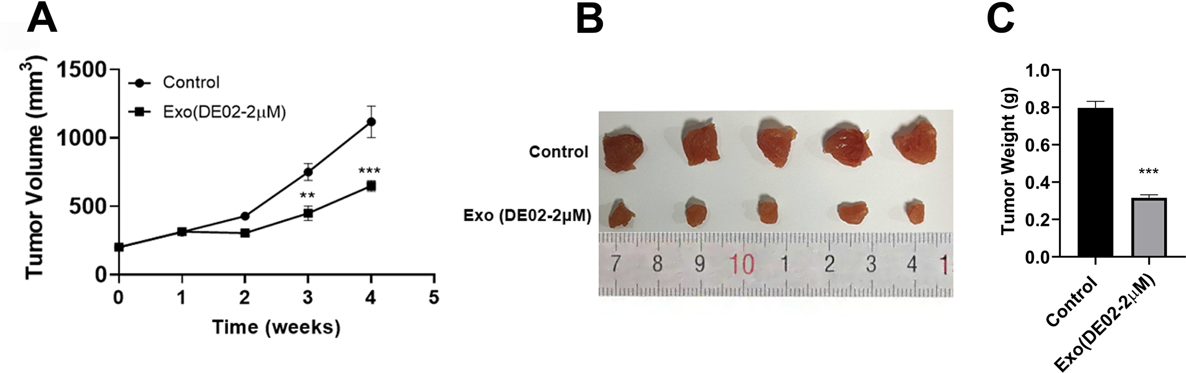
Inhibitory effect of ExoDE02 on the growth of nude mouse xenografts in vivo.
Discussion
Osteosarcoma is a malignant bone tumor with a high incidence in adolescents. Its strong invasiveness and high metastasis rate limit clinical treatment effects, especially for advanced patients with extremely poor prognosis, highlighting the urgent need to develop novel and efficient therapeutic strategies. Natural products occupy an important position in antitumor drug development due to their unique biological activities and good biocompatibility. As the core active component of the traditional Chinese herb Forsythia suspensa, phillyrin has been confirmed to have certain antitumor potential, but its inherent defects such as poor water solubility, low bioavailability, and weak antitumor activity limit its direct clinical application in tumor therapy. In this regard, the current study establishes a multimodal approach for osteosarcoma biotherapy by combining natural product-derived medication optimization, PLK4-targeted molecular intervention, exosome-based delivery, and cavitation-free acoustic sensitization. When compared with untreated controls, exosome-only controls (Exo without DE02) show no appreciable alterations in proliferation, migration, or invasion, confirming their role as physiologically inactive carriers under the studied conditions.
In this study, the derivative DE02 was successfully synthesized by introducing a Boc-protected phenylalanine group into the hydroxyl domain of the glycosidic bond of phillyrin (using phillyrin as the scaffold). In vitro cell experiments confirmed that DE02 exhibited an approximately 10-fold higher antiproliferative activity against MG-63 and Saos-2 osteosarcoma cells than the parent drug phillyrin, in a clear concentration-dependent manner. These results indicate that while retaining the core active scaffold of phillyrin through chemical structural modification, the introduction of a hydrophilic amino acid fragment not only optimizes the physicochemical properties of the drug but also enhances its binding ability to tumor cell targets, thereby improving antitumor activity, providing an effective idea for the structural optimization of natural products. Further immunofluorescence staining showed that DE02 can significantly downregulate the expression of the proliferation marker PCNA, suggesting that its inhibitory effect on osteosarcoma cell proliferation may be achieved by blocking the cell cycle process. This is consistent with previous research conclusions that phillyrin regulates the tumor cell cycle, and also confirms that DE02 still retains the core mechanism of action of the parent drug after structural modification.
Tumor invasion and metastasis are key factors leading to treatment failure in osteosarcoma patients. As a core regulatory factor of centrosome duplication, the aberrant overexpression of PLK4 has been confirmed to be closely associated with the malignant progression of various tumors. In this study, Transwell assays showed that DE02 can concentration dependently inhibit the migration and invasion abilities of osteosarcoma cells, and this effect was significantly reversed after oePLK4, suggesting that the inhibitory effect of DE02 on osteosarcoma cell migration and invasion depends on the downregulation of PLK4. Further molecular mechanism studies showed that DE02 can significantly reduce the enzyme activity and mRNA expression level of PLK4, while upregulating the mRNA expressions of p53 and p21, and oePLK4 can reverse this regulatory effect. Existing studies have confirmed that PLK4 inhibition can degrade the p53-negative regulator MDM2 by activating the PIDDosome complex-caspase-2 pathway, thereby stabilizing p53 and inducing p21-dependent cell cycle arrest. Consistent with these findings, this study clarifies that DE02 exerts its antiosteosarcoma effect by regulating the PLK4-p53–p21 signaling pathway, providing a new molecular target for targeted osteosarcoma therapy. The authors recognize that this study lacks direct biochemical proof of DE02–PLK4 binding (e.g., kinase assays or molecular docking), even though oePLK4 significantly reversed the anticancer effects of DE02. Consequently, rather than being a verified direct molecular target, PLK4 is found here as a functional downstream effector. Direct interaction mechanisms will be the focus of future research.
As a natural drug delivery carrier, exosomes have unique advantages such as low immunogenicity, high biocompatibility, and tumor targeting, which can effectively improve drug bioavailability and targeted therapeutic effects. In this study, HUVEC-derived exosomes were isolated and purified by ultracentrifugation; after identification as meeting typical characteristics, DE02 was loaded via sonication to construct the ExoDE02 delivery system. Scalable good manufacturing practice (GMP)-compatible substitutes such as tangential flow filtration and size-exclusion chromatography constitute viable translational approaches, even if ultracentrifugation was used in this investigation. In vitro experiments confirmed that the inhibitory effects of ExoDE02 on the proliferation, migration, and invasion of osteosarcoma cells were significantly superior to those of the DE02-alone group, indicating that the exosome delivery system can effectively enhance the antiosteosarcoma activity of DE02. This result may be related to the targeted delivery function of exosomes: Exosomes can achieve precise delivery of DE02 by binding of surface-specific ligands to tumor cell surface receptors, reducing drug off-target effects, while avoiding drug degradation by the in vivo enzyme system and prolonging drug action time. In vivo nude mouse xenograft experiments further confirmed that ExoDE02 can significantly inhibit tumor growth, reduce tumor volume and weight, and show no obvious toxic side effects, laying a safety foundation for its clinical application.
Effective and safe drug delivery continues to be a crucial factor in determining treatment efficacy, even beyond molecular targeting. As natural nanocarriers, exosomes have inherent benefits such as minimal immunogenicity, excellent biocompatibility, extended circulation period, and tumor-tropic characteristics. In this work, low-energy, cavitation-free acoustic sensitization was used to encapsulate DE02 into HUVEC-derived exosomes, creating the ExoDE02 delivery system. Cavitation-free acoustic sensitization maintains an outstanding safety profile by enabling transitory membrane permeability and improved drug loading without causing vesicle breakage or heat damage, in contrast to high-intensity or cavitation-based ultrasound methods. This distinction is in line with current attempts to reduce the dangers associated with ultrasound and is especially significant in the context of clinical translation. Because of their consistent production, low immunogenicity, and proven application in translational nanomedicine, HUVEC-derived exosomes were chosen. Endothelial exosomes provide better safety and manufacturing reproducibility, whereas tumor-derived or mesenchymal stem cell (MSC)-derived exosomes may show increased tumor tropism. Future research will include comparative source optimization.
Functionally, ExoDE02 outperformed free DE02 in vitro in terms of antitumor activity, more successfully inhibiting the growth, migration, and invasion of osteosarcoma cells. These improvements increased PLK4-targeted signaling by enhancing intracellular transport and bioavailability rather than changing the underlying molecular mechanism. ExoDE02 significantly reduced the growth of xenograft tumors in vivo without causing any discernible systemic toxicity, demonstrating the biosafety and therapeutic promise of this ultrasound-enabled delivery method. All of these results point to cavitation-free acoustic sensitization as a nondestructive, nonthermal supplement that can enhance targeted cancer biotherapy. The current method prioritizes safety and translational feasibility by using cavitation-free acoustic sensitization only during carrier construction, in contrast to sonodynamic therapy or microbubble-assisted administration, which depend on cavitation and reactive oxygen species formation.
However, there are a few limitations such as immune-exosome interactions that cannot be evaluated using immunodeficient nude mice. In immunocompetent mice, endothelial exosomes’ possible immunomodulatory effects should be examined. Although PLK4 was the main focus of this work, possible off-target effects on related mitotic kinases (such PLK1 and Aurora A) were not looked at and are still a crucial topic for further research.
In summary, this study shows that cavitation-free acoustic sensitization improves PLK4-targeted osteosarcoma therapy by increasing the phillyrin derivative DE02’s anticancer activity using a secure, exosome-based delivery system. With intriguing implications for future clinical translation, these findings offer molecular insight and preclinical data supporting a novel, low-risk strategy for targeted osteosarcoma treatment by combining natural product modification with ultrasound-enabled biotherapy.
Conclusion
In comparison with the original drug, the phillyrin derivative DE02 demonstrated significantly increased antitumor efficacy against osteosarcoma and was effectively produced through logical chemical modification. Mechanistic studies revealed that DE02 inhibits PLK4 and activates the downstream p53–p21 signaling pathway to limit osteosarcoma cell proliferation, migration, and invasion. Crucially, DE02’s therapeutic efficacy was greatly increased via cavitation-free acoustic sensitization-mediated exosomal distribution without causing any discernible systemic damage in vivo. These results provide a safe, ultrasound-enabled PLK4-targeted biotherapy approach that combines precision medication delivery with natural product optimization. Together, these studies offer molecular understanding and preclinical proof of the translational potential of cavitation-free acoustic sensitization to improve targeted osteosarcoma therapy and aid in the clinical development of anticancer drugs derived from natural products.
Ethics Approval and Consent to Participate
This study was approved by the Ethics Committee of Yiwu Central Hospital.
Data Availability Declaration
The datasets used and analyzed during the current study are available from the corresponding author upon reasonable request.
Authors’ Contributions
J.Y.: Conceptualization, methodology, investigation, data curation, and writing—original draft. H.W.: Methodology, validation, formal analysis, and visualization. Z.H.: Resources, investigation, and data curation. W.W.: Formal analysis and writing—review and editing. J.Q.: Conceptualization, supervision, funding acquisition, writing—review and editing, and project administration. All the authors have read and approved the final article.
Footnotes
Disclosure Statement
The authors declare that they have no competing interests.
Funding Information
This work was supported by the Zhejiang Provincial Key Clinical Specialty in Orthopedics (SLCZK2024-02).