






Abstract
Background:
Hepatocellular carcinoma (HCC) is a highly prevalent malignant tumor globally, with low survival rates in advanced stages. Therefore, early diagnosis and precision therapy are pivotal to improving patient prognosis. Glypican-3 (GPC3) acts as a specific molecular target for HCC, which is highly expressed in 70%–85% of HCC tissues but undetectable in normal liver tissues, hepatitis, liver cirrhosis, and other pathological conditions. This stringent expression regulation renders it an ideal tumor-specific target.
Main Content:
Based on the biological properties of GPC3, various specific tumor imaging agents have been developed, covering multiple biomolecular types, including antibodies, peptides, and aptamers. This review systematically summarizes the research advances, application advantages, and existing challenges of three types of molecular imaging probes in the precise diagnosis and treatment of HCC. It focuses on the development strategies of these probes and evaluates their current limitations in light of key characterization parameters.
Conclusion:
This review aims to provide new research ideas for the precision diagnosis of HCC and promote the translation of clinical applications.
Introduction
Hepatocellular carcinoma (HCC) is the main histological subtype of liver cancer, accounting for about 90% of cases, and is recognized as a highly invasive malignant tumor worldwide. Its morbidity and mortality rates are still high, and the clinical prognosis is generally poor. 1 According to reports from the International Agency for Research on Cancer and the World Health Organization, liver cancer became the third leading cause of cancer-related death in the world in 2022, recording 865,000 newly diagnosed cases and 750,000 deaths. 2 Although targeted therapy and immunotherapy have improved the survival results of some patients, the 5-year survival rate of advanced patients is still less than 20%, 3 which highlights the limitations of existing treatments. Early diagnosis and intervention have become the core strategies to improve the survival rate of patients. 4 The accuracy of early tumor diagnosis depends to a large extent on reliable immunohistochemical markers, which makes it an urgent task to identify treatment targets for early HCC. These limitations emphasize the urgent need to develop a new imaging probe for early diagnosis of liver cancer with high specificity and sensitivity.
However, the early and precise diagnosis of HCC presents substantial challenges. In the clinical diagnosis of HCC, serum α-fetoprotein is often used as a traditional biomarker. However, its detection sensitivity is below 40%, resulting in poor overall diagnostic performance. 5 Traditional imaging techniques such as ultrasound and computed tomography (CT) have insufficient specificity in the diagnosis of early small lesions and the distinction between benign and malignant lesions.6,7 Positron emission tomography/computed tomography (PET/CT) is a cutting-edge molecular imaging technology that usually uses the radioactive tracer 18F-fluorodeoxyglucose to detect early primary lesions. 8 However, it also faces some limitations, such as low diagnostic rates and high incidence of false-positive results. 9 In view of the limitations of these traditional detection methods and the failure of early HCC detection to meet the medical needs, the development of highly specific and sensitive HCC-targeted imaging agents has become the key to early diagnosis.
Nuclear medicine molecular imaging uses radionuclide-labeled molecular probes to achieve noninvasive in vivo imaging by detecting emitted positrons or γ-rays, enabling dynamic monitoring of physiological and biochemical processes at the molecular level. 10 The imaging performance of radiotracers depends on radionuclide selection and radiochemical design. The half-life, decay mode, and emission energy of radionuclides should be matched with the pharmacokinetics of the targeting vector. For example, short- to intermediate-half-life radionuclides, such as 68Ga, 18F, and 99mTc, are suitable for fast-clearing small molecules, peptides, or antibody fragments, whereas long-lived radionuclides, such as 89Zr and 124I, are more appropriate for intact antibodies with slow clearance. 11 In addition, factors such as labeling methods, chelators, radiochemical purity, molar activity, hydrophilicity, and in vivo stability can affect probe biodistribution, tumor-to-background contrast, radiation dose, and safety. Unstable labeling may increase nonspecific uptake in nontarget organs and reduce image quality. Therefore, rational radionuclide selection and optimization of radiochemical properties are essential for developing high-performance glypican-3 (GPC3)-targeted imaging agents.
Among many potential molecular targets, GPC3 shows excellent potential. It is highly and specifically expressed in 70%−85% of HCC tissue, but shows almost no expression in normal liver tissue and benign lesions, making it an ideal tumor-specific target.12,13 Based on this, researchers have developed antibodies, peptides, aptamers, and other GPC3-targeted molecular imaging probes, and relevant reviews have been published one after another. Filippi, 14 Grega, 15 and Liu Yaru 4 have systematically reviewed the research progress of GPC3-targeted imaging and therapy. They summarized the research and theranostic applications of antibody, peptide, and aptamer probes and illustrated the clinical value of GPC3 in the diagnosis, prognostic evaluation, and targeted therapy of HCC, laying an important foundation for understanding the significance of the GPC3 target and the research and development of relevant probes.
It should be pointed out that most existing literature merely lists and categorizes probes, with insufficient in-depth analysis of their molecular design strategies, key performance parameters, and inherent limitations. In addition, most studies do not display probe structural diagrams, making it impossible to visually present their molecular architecture and hindering readers from clearly understanding the relationship between probe design rationale and performance. Accordingly, this review focuses on probe optimization strategies as the main thread, systematically summarizes the advantages and applicable scenarios of each strategy, and deeply analyzes probe design approaches and imaging performance. Through comparative analysis, the respective strengths and translational challenges of various probes are clarified, to provide forward-looking references for the optimal design and clinical translation of next-generation high-performance GPC3-targeted imaging agents.
Research Methods
A literature search was conducted in PubMed, Elsevier ScienceDirect, ACS Publications, and China National Knowledge Infrastructure for studies published between 2008 and April 2026. The search terms included “Glypican-3 (GPC3),” “hepatocellular carcinoma (HCC),” “imaging agent,” “molecular probe,” “PET,” “SPECT,” “targeted imaging,” “radiotracer,” and “radiopharmaceutical,” used alone or in combination. Studies were included if they focused on GPC3-targeted imaging agents or molecular probes for HCC and reported relevant experimental, preclinical, or clinical data. Conference abstracts and articles without relevant imaging or probe-performance data were excluded. Finally, 57 articles were included for qualitative analysis.
Molecular Structure and Function of GPC3
GPC3 is a key member of the heparan sulfate proteoglycan family. It consists of 580 amino acid residues, with a theoretical molecular weight of ∼70 kDa and a single furin cleavage site. Following cleavage at this site, GPC3 is processed into N-terminal and C-terminal fragments. 16 Figure 1 shows the structural diagram of GPC3 on the cell membrane.
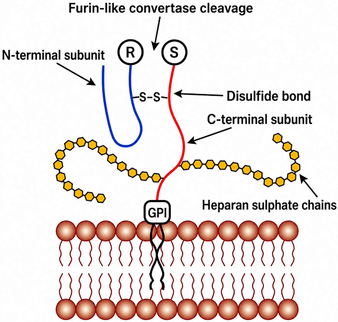
A schematic diagram of the glypican-3 (GPC3) structure on the cell membrane. Adapted from Guo et al. 23
In the signal pathway regulation, GPC3 activates the Wnt/β-catenin protein and Hedgehog signaling pathways related to the development of HCC through its heparan sulfate chain or core protein structure: The former promotes β-catenin to enter the nucleus and initiates proliferation-related gene expression by binding to Wnt ligands and stabilizing the receptor complex,17–19 while the latter fine-tunes the signal conduction efficiency by regulating ligand–receptor interaction.20–22 Concurrently, GPC3 binds to proliferation-related factors such as insulin-like growth factor and fibroblast growth factor to enhance their proliferation-promoting effect on HCC cells. Its expression is positively regulated by the oncogene c-Myc, which ultimately promotes the HCC cell cycle process and inhibits apoptosis.23,24 As shown in Figure 1, GPC3 is anchored to the cell membrane via a glycosylphosphatidylinositol (GPI) anchor.
GPC3-Targeting Imaging Agents
Antibody-based probes
Antibody probes have become the earliest molecular type used in GPC3-targeted imaging due to their high affinity, strong targeting, and long in vivo half-life. Its large modifiable space is also convenient for multimodal imaging. However, the high molecular weight (about 150 kDa) leads to an excessive half-life of blood circulation, resulting in obvious background uptake in organs such as the liver, which limits the application of early diagnosis of HCC. For this reason, the researchers gradually optimized the antibody structure, from complete antibodies to antibody fragments, and then to single-domain antibodies and humanization to improve the pharmacokinetic performance.
Clinical validation and limitations of intact antibodies
In 2018, 124I-codrituzumab became the first clinical-grade probe to demonstrate the tumor-targeting capability of GPC3 antibody probes. Using a humanized full-length IgG (GC33), tumor-specific uptake was detected in 13 out of 14 HCC patients, validating its clinical safety.25–27 However, intact IgG molecules have a large molecular weight and slow blood clearance, requiring an imaging waiting period of several days. Guan developed the probe 131I-scFv using a 30 kDa anti-GPC3 single-chain variable fragment (scFv) as the targeting vector, 28 which preliminarily overcomes poor tumor penetration and immunogenicity limitations of conventional full-length antibodies.
Long-half-life radionuclide matching and optimization of F(ab′)2 fragments
Yang first conjugated the long-half-life radionuclide 89Zr (half-life of 78.4 h) with the murine anti-GPC3 intact antibody 1G12 to construct the probe 89Zr-DFO-1G12. 29 In the HepG2 orthotopic xenograft model, its tumor/liver (T/L) ratio reached 6.65 ± 1.33 at 168 h, enabling clear differentiation between patient-derived xenografts and normal liver tissue. These findings established GPC3 as a promising molecular target for HCC immuno-PET imaging and demonstrated the potential of 89Zr-DFO-1G12 for noninvasive visualization of GPC3-positive HCC lesions. However, the potential immunogenicity of murine antibodies and the slow clearance characteristics of full-length antibodies (blood half-life ≈115 h) limited clinical application. To overcome pharmacokinetic limitations, Sham et al. 30 developed the probe 89Zr-αGPC3-F(ab′)2 by enzymatically removing the Fc fragment, reducing the blood half-life to 11 h, achieving a T/L ratio of 23.3 within 4 h (significantly higher than the T/L ratio of 1.2 for the intact antibody at the same time point), markedly enhancing early imaging contrast, and reducing hepatic background. This advance demonstrates the crucial value of antibody fragmentation in improving the contrast of early-phase imaging, while its murine framework still carries the risk of immunogenicity.
Site-specific labeling and bispecific design
Conventional random lysine labeling methods (89Zr-nHN3) often result in fluctuating affinity (KD = 30 ± 12 nM) and high hepatic uptake. Fayn adopted a sortase-mediated site-specific labeling strategy to construct 89Zr-ssHN3. 31 It exhibited stable affinity with a KD value of 10 ± 4 nM and achieved a significantly enhanced tumor uptake in HepG2 tumor models. Waaijer developed a bispecific antibody ERY974 targeting both GPC3 and CD3. The probe [89Zr]Zr-N-suc-Df-ERY974 can be applied to immuno-PET imaging. 32 This bispecific design not only achieves tumor targeting but also enables visualization of the immune microenvironment through T cell recruitment, thereby expanding the functional dimension of antibody-based probes.
Humanization modification and theranostic integration
From 2021 to 2024, humanization has become the core strategy to meet the needs of clinical transformation. Natarajan retained the GPC3-binding affinity (KD ≈ 3.5–3.9 nM) of the murine 1G12 through Complementarity-Determining Region grafting and developed the probe 89Zr-Df-H3K3. The homology was increased to 94.8% through the humanized framework area, which effectively reduced the immunogenicity. 33 Subsequently, Dickerson developed the probe 89Zr-αGPC3H to further verify the clinical potential of humanized probes. 34 There was no significant difference between the tumor uptake (97% ± 50% ID/mL) and that of the mouse probe, and the T/L ratio was maintained at 12 ± 7.6, confirming that humanization would not affect the imaging performance. On this basis, 89Zr/90Y-αGPC3 broke through the limitation of single-function imaging with 89Zr used for preoperative imaging and 90Y mediating radioimmunotherapy, achieving integrated diagnosis and treatment. 35
Emergence of single-domain antibodies and clinical-grade RDC
The emergence of single-domain antibodies provides a new method for the miniaturization of probes and the enhancement of penetration. Al[18F]F-ssHN3 is a second-generation GPC3-targeted single-domain antibody PET probe developed on the basis of the first-generation 89Zr-ssHN3. It exhibits high affinity for GPC3 (KD = 48 ± 0.7 nM) and rapid blood clearance in vivo. The hepatic orthotopic tumor uptake reaches 16% ± 4% IA/g at 1 h postinjection, with an exceptionally high T/L ratio of 13. This probe enables same-day imaging and accurately identifies residual lesions after ablation, providing a highly specific molecular imaging modality for the diagnosis and therapeutic response monitoring of HCC. 36 The An team constructed [68Ga] Ga-NOTA-G2, [18F] F-G2, and the fusion protein probe [68Ga] Ga-NOTA-ABDG2, 37 based on the GPC3-specific single-domain antibody G2 with high affinity (KD = 1.297 nM). Among them, [68Ga]Ga-NOTA-ABDG2 showed significant advantages, reaching a tumor-to-muscle ratio of 8.7 ± 0.9 in 24 h, which was obviously better than unmodified probes. This breakthrough took advantage of the low molecular weight of single-domain antibodies to improve tumor penetration and blood clearance, providing new insights for optimizing antibody-based probes. In 2025, GPN02006 became the world’s first GPC3-targeted radioligand (RL) to enter clinical trials. Utilizing antibody-68Ga conjugation technology, this RL exhibits an affinity for GPC3 of 0.21 nM. Among 80 HCC patients, PET/CT imaging was completed within 30 min after administration. The tumor/normal liver SUVmax reached 8.5, and the detection rate of microfoci with a diameter of less than 1 cm was 92%. At present, the phase II clinical trial has been completed.
From the early clinical research of complete antibodies to engineering transformation, humanization, and diagnosis–treatment integration, the development of GPC3-targeted antibody probes has gradually improved the performance. This brings hope for the creation of greater value for the accurate diagnosis of HCC. Table 1 summarizes the RLs mentioned in this article.
Currently Reported Radiolabeled Antibody-Based Probes Targeting Glypican-3
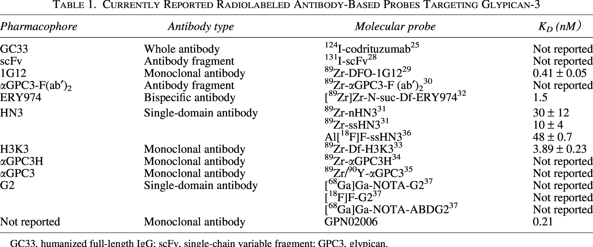
GC33, humanized full-length IgG; scFv, single-chain variable fragment; GPC3, glypican.
Peptide-based probes
The molecular weight of peptide probes is usually 1–5 kDa. Compared with antibodies, they have higher tumor penetration, lower immunogenicity, faster in vivo clearance rate, and more flexible radioactive labeling ability. They are highly adapted to the clinical imaging process. It is the core direction of the evolution of GPC3-targeted probes from macromolecules to small molecules. Its development process revolves around solving the problems of insufficient affinity and high intake of nontarget organs in early probes. Through chemical modification, screening strategy upgrade, multitarget coupling, and other means, its performance is gradually improved. Some high-affinity macrocyclic peptide probes have entered the clinical research stage.
Limitations of early linear peptides and basic chemical modification
The early research of GPC3-targeted linear peptides mainly focused on chemical modification of single sequences. However, restricted by the intrinsic binding potency and tissue selectivity of peptides themselves, their clinical translation faces numerous challenges. In 2016, Wang Z took the lead in developing PET probes 18F-NFP-L5 and 18F-NOTA-L5 using L5 peptide (sequence: RLNVGGTYFLTTRQ) as the target core. However, excessive intra-abdominal radioactive deposition seriously interfered with the detection of liver tumors. 38 Subsequently, in 2018, further modifications of chelation chemistry led to the preparation of 18F-AlF-NOTA-MP-6-Aoc-L5. 39 Although this probe shows good stability and potential for GPC3 in vitro, its T/L uptake ratio (0.93 ± 0.16) is still insufficient due to the high physiological uptake of the liver. In 2020, the GP series of PET probes entered the field of research. The probes Al[18F]F-GP2076 and [18F]F-GP2633 are based on TP peptide (sequence: RLNVGGTYGLTTRQ). The latter shows enhanced hydrophilicity due to the introduction of linkers and has a stronger binding affinity for GPC3 (KD = 63.3 vs. 101 nM). Its renal excretion route also reduces the interference from the background signal of high liver, 40 which shows that the distribution and background signals in the body can be effectively optimized through hydrophilic modification of the peptide skeleton, providing a reference for subsequent probe optimization. Studies conducted at this stage have illustrated that a single chemical modification strategy is insufficient to completely address the issue of nonspecific uptake of linear peptides, necessitating fundamental optimization from the perspective of peptide sequence screening.
Upgraded screening strategies and multitarget synergistic design
Traditional peptide probes obtained via a single in vitro phage display screening generally suffer from insufficient in vivo specificity. Accordingly, the upgrading of screening strategies has become critical to improving the performance of peptide probes. In 2019, the Berman team confirmed that the early GPC3-targeted peptide TJ12P1 (sequence: DHLASLWWGTEL, KD = 280.4 ± 33.51 nM) lacked binding selectivity and effectiveness and failed to meet the imaging requirements. 41 In 2019, Qin et al. 42 developed the 18F-AlF-NOTA-TJ12P2 probe (sequence: SNDRPPNILQKR, KD = 158.2 ± 26.25 nM) based on a two-step in vivo–in vitro phage display screening strategy, which showed high specificity, low background signal, and rapid blood clearance characteristics. This effectively solves the inherent limitations of TJ12P1, indicating that the transition from “single in vitro screening” to “in vivo–in vitro combined screening” is the key way to improve the affinity and specificity of polypeptide probes. In 2024, Chen et al. 43 further coupled TJ12P2 with PSMA inhibitors to construct dual-targeted probes [68Ga]Ga-T2P and [18F]AlF-T2P. Compared with monomer probes, these probes show higher tumor uptake and longer residence time, realizing the functional expansion from single-target to multitarget synergies.
Functional group modification and expansion of imaging modalities
Beyond the optimization of peptide sequences themselves, functional group modification can further modulate the pharmacokinetics, internalization efficiency, and imaging modalities of probes, serving as a key strategy to enhance the performance of peptide probes. Mo et al. 44 developed a new type of PET probe, [18F] AlF-NOTA-IPB-GPC3P, due to the presence of the IPB group, that showed higher tumor cell uptake (15.13% vs. 5.96%) and internalization rate (80.63% vs. 35.93%) at 120 min, compared with [18F]AlF-GP2633, reduced kidney accumulation, and improved imaging performance. This highlights the significant advantage of enhancing the effectiveness of peptide probes through functional group modifications. In terms of imaging modalities, Xu 45 developed the SPECT radiotracer 99mTc-HPG based on the GBP peptide (sequence: THVSPNQGGLPS). This compound combined excellent GPC3 binding specificity with rapid clearance from normal organs, filling the technical gap in GPC3-targeted SPECT imaging. In 2024, breakthroughs were achieved in developing GBP-based PET probes. 68Ga-PEG2-GBP and 68Ga-ALB-GBP incorporated polyethylene glycol and 4-methylphenylbutyric acid (an albumin-binding group), 46 respectively, where 68Ga-ALB-GBP significantly extended circulation time and enhanced bioavailability through efficient binding to serum albumin, ultimately enabling high-contrast PET imaging. This achievement marks the cross-modal application of GBP peptides from SPECT to PET and further validates the value of albumin-binding strategies in improving the imaging contrast and bioavailability of peptide probes.
Macrocyclic peptides: High affinity and clinical translation prospects
Linear peptides are susceptible to degradation by endogenous proteases and exhibit flexible conformations, resulting in limited affinity and stability. In contrast, macrocyclic peptides adopt rigid and stable conformations through intramolecular cyclization, which markedly enhances target binding affinity and in vivo stability, making them a research hotspot in the development of peptide probes in recent years. The cyclic tumor target peptide F3 (KD = 0.7 nM) was radiolabeled to produce 68Ga-DOTA-F3. The tracer can effectively distinguish GPC3-positive tumors in different cancer types, improving the tumor-specific diagnostic value of peptide imaging agents. 47 Chen modified the cyclic peptide 10YX (sequence: CLNHELFQTC) via polyethylene glycol conjugation and albumin-binding engineering to develop the probe 10P3Me (KD = 93.8 ± 49.9 nM). In GPC3-positive HCC models, it achieves a high T/L ratio of 8.28 at 1 h postinjection and enables accurate lesion localization. 48 Nevertheless, it still suffers from nonspecific uptake in blood and kidneys, as well as insufficient sensitivity for subcentimeter lesions, leaving room for further optimization.
The cyclic peptide RYZ-GPC3 reported in 2024 exhibits markedly higher affinity than other reported GPC3-binding agents, with KD values as low as 0.35 and 0.42 nM for human and murine GPC3, respectively. 49 The Poot team conducted the first-in-human study of the probe [68Ga]Ga-RAYZ-8009. The study enrolled 24 patients with suspected/confirmed HCC and pediatric hepatoblastoma patients. No adverse events were reported postimaging, with clear lesion differentiation achieved. 50 This study laid the foundation for subsequent clinical applications. Building on this, the 177Lu/225Ac-RYZ-GPC3 probe realized an integrated “targeted imaging-radiation therapy” design, while ensuring efficient binding and rapid internalization on the surface of tumor cells. 51 It therefore holds great potential for further development as a targeted therapeutic agent against HCC.
Reviewing the development of GPC3-targeted peptide probes, their evolution has centered on the core peptide backbone. Through the modification of functional groups, optimized screening strategies, dual-target coupling, and diagnosis-treatment integration, these probes gradually address early challenges, such as high background signals, low specificity, and limited imaging modalities. This progress provides key support for the accurate diagnosis and treatment of HCC. The current research results are only based on animal experiments and lack large-scale clinical verification. In the future, comprehensive research is needed to objectively evaluate the clinical application potential of the probe. Figure 2 shows the structural diagrams of selected radiolabeled peptide probes, while Table 2 summarizes the radiolabeled peptide probes mentioned in this article. (Note: In the 68Ga-DOTA-F3 structural diagram, X represents the seven amino acids of the F3 peptide, which were not disclosed in the reference.)
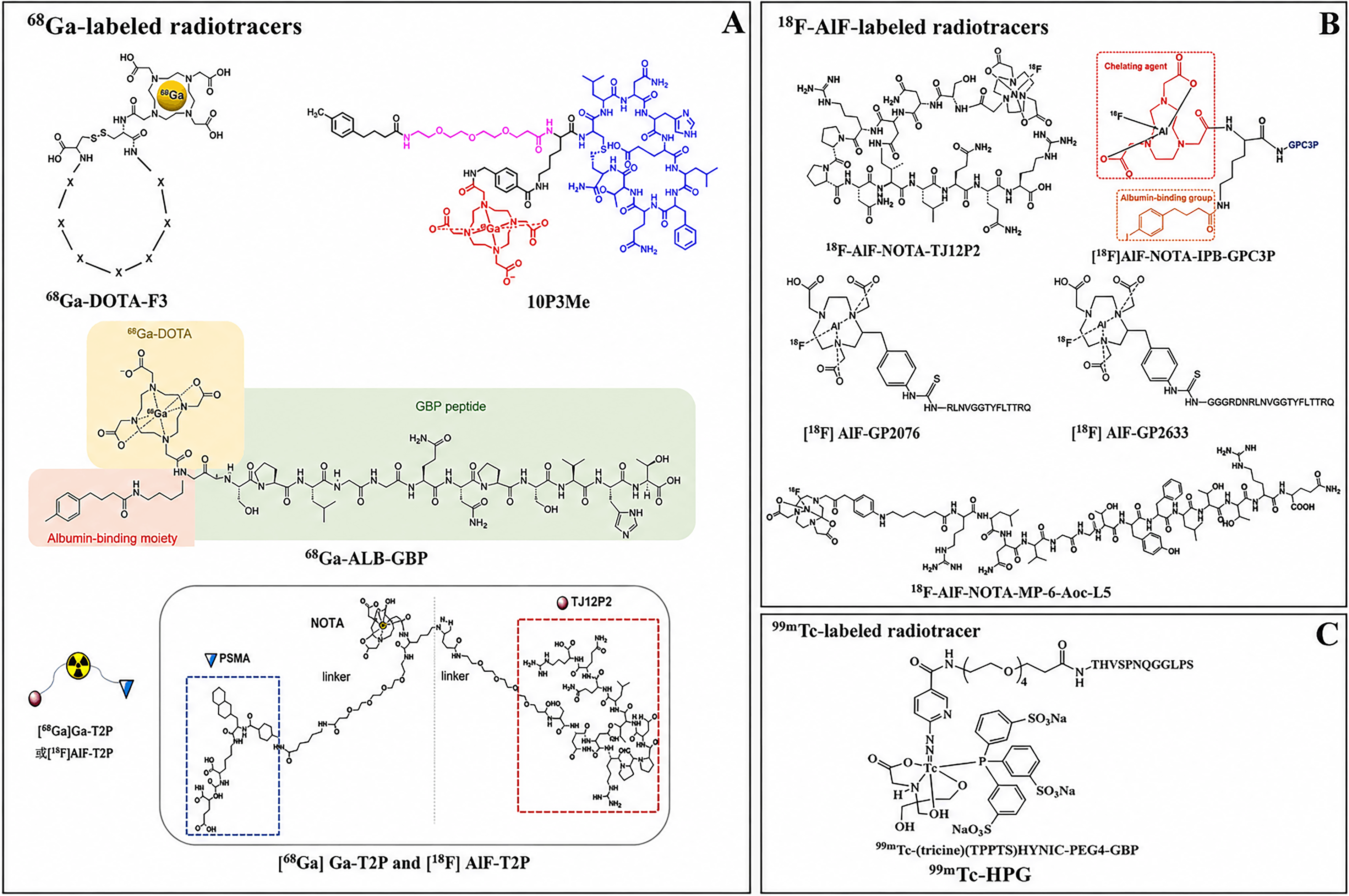
Schematic diagram of the chemical structures of selected radioactive peptide probes.
Currently Reported Radiolabeled Peptide Probes Targeting Glypican-3
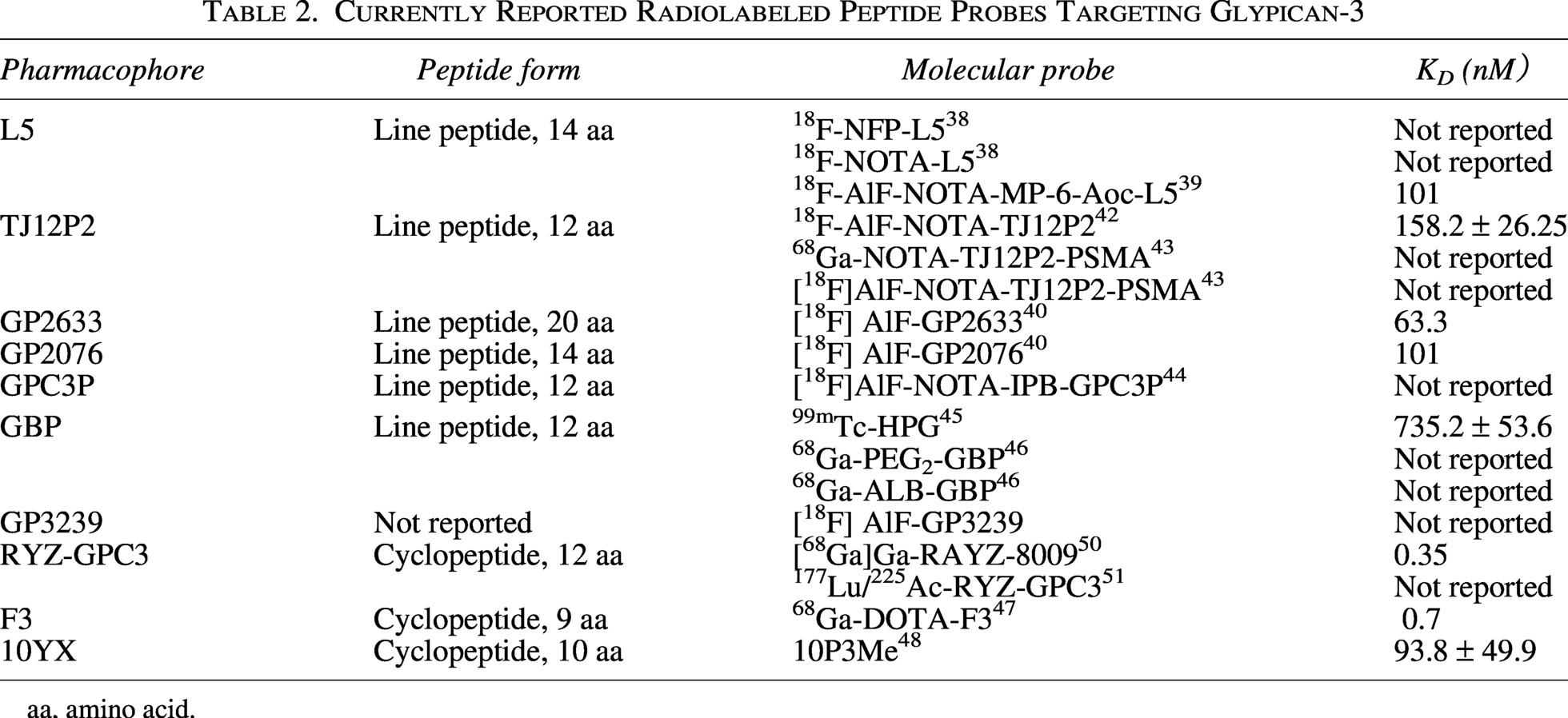
aa, amino acid.
Aptamers and other probes
Aptamers are single-stranded RNA or DNA oligonucleotides with secondary or tertiary structures, and multiple intramolecular interactions among their sequence components enable them to specifically recognize and bind to targets after proper folding. 52 The expression frequency of GPC3 mRNA is significantly higher in HCC than in normal tissues, providing a molecular basis for aptamer-based targeting of GPC3 to achieve tumor imaging. Compared with antibodies and peptides, aptamers feature the low immunogenicity and facile synthesis of peptides while achieving antibody-like high affinity. As such, they represent a promising class of GPC3-targeted probes, although further clinical validation is still required. In addition, fluorescent probes also exhibit unique advantages for this target.
Significant progress has been made in the development of ligands and fluorescent probes for GPC3 for accurate imaging of liver cancer. In 2018, Zhao et al. 53 used CE-SELEX technology to screen and truncate the sequence, and obtained the single-stranded DNA ligand AP613-1 that specifically targeted GPC3. After phosphorothioate modification, its affinity is almost quadrupled (KD = 15.48 nM). When coupled with Alexa Fluor 750, it can accurately display GPC3-positive tumors in the nude mouse HCC xenotransplantation model while overcoming the inherent degradation problem of natural nucleic acids. 54 The IRDye800 fluorescent probe based on the IRDye800-labeled 12-peptide (sequence: ALLANHEELFQT) showed a binding dissociation constant of 39.5 nM in vitro. In the in situ Hep3B liver cancer mouse model, the probe reached the peak of tumor uptake within 1.5 h and was cleared within 24 h. It can effectively distinguish between HCC and non-HCC and cirrhosis in human HCC samples. The ligand exhibits a serum half-life of 3.5 h and is nontoxic, 55 which meets the pharmacokinetic requirements of rapid ingestion and removal. GPC-ICG is a humanized anti-GPC3 monoclonal antibody conjugated with indocyanine green (ICG), an NIR-II (1000–1700 nm) probe. It specifically binds GPC3-positive HCC cells in vitro and can maintain stable fluorescence for 5 d. In the subcutaneous tumor model, it reached a peak fluorescence intensity within 48 h, and the tumor-to-background ratio was 3 at 96 h. It clearly depicts GPC3-positive tumors through specific signals, providing a multifunctional technical path for HCC-targeted imaging. 56 In 2025, Wu developed fluorescent magnetic resonance dual-modal imaging and photothermal/photodynamic therapy. This multifunctional diagnostic-treatment probe accurately depicts the tumor boundary and realizes real-time surgical navigation and prognosis evaluation. 57
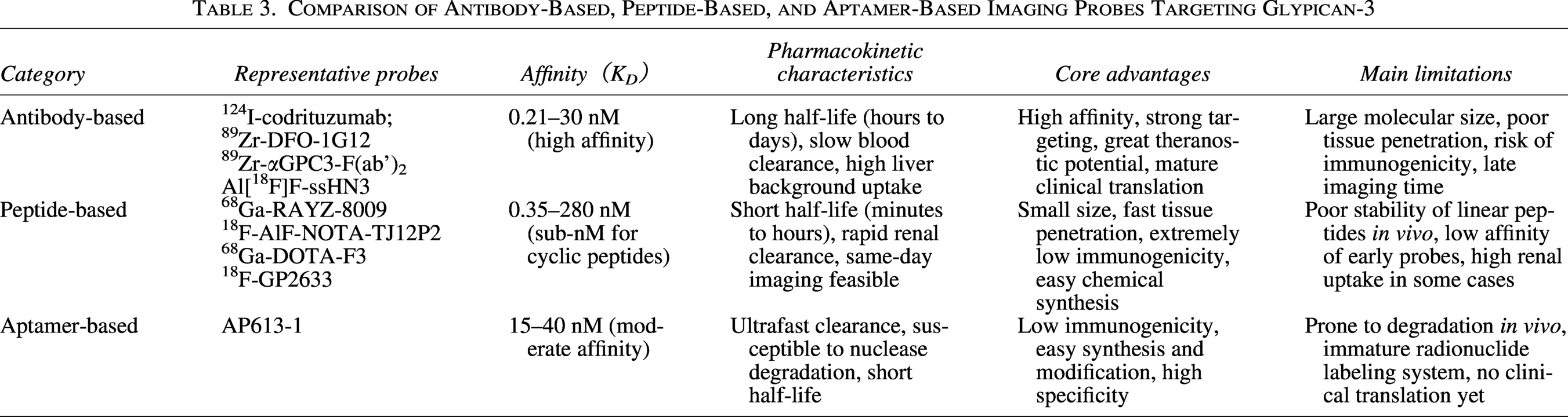
Overall, antibody-based, peptide-based, and aptamer-based probes each have distinct advantages and limitations in GPC3-targeted imaging. In Table 3, the authors summarize their representative probes, reported affinity values (KD), pharmacokinetic characteristics, core advantages, and main limitations, thereby facilitating a direct comparison among these probe categories.
Comparison of Antibody-Based, Peptide-Based, and Aptamer-Based Imaging Probes Targeting Glypican-3
Discussion
This review systematically summarizes the research progress of GPC3-targeted molecular imaging probes, including antibody-based, peptide-based, and aptamer-based probes. A horizontal comparison shows that antibody-based probes have high affinity and substantial potential for theranostic applications. However, their large molecular size is often associated with high hepatic background uptake and a certain risk of immunogenicity. Peptide-based probes exhibit rapid tissue penetration, fast in vivo clearance, and low immunogenicity, making them more suitable for same-day rapid imaging. Nevertheless, they commonly suffer from insufficient affinity and poor in vivo stability. Aptamer-based probes are easy to synthesize and have low immunogenicity; however, they are prone to degradation in vivo, their radionuclide labeling systems remain immature, and their clinical translation has been relatively slow. Overall, these three classes of probes face common challenges, including high nonspecific hepatic uptake, insufficient tumor-to-liver contrast, and a mismatch between pharmacokinetics and radionuclide half-life. These issues have become major bottlenecks limiting their clinical application.
From the perspective of clinical translation, only a small number of antibody fragments and macrocyclic peptide probes have entered early-stage clinical trials, whereas most GPC3-targeted probes remain at the preclinical stage. Future studies should further focus on improving probe stability, optimizing target-to-background contrast, and reducing nonspecific uptake. For example, cyclization, hydrophilic modification, site-specific labeling, and albumin-binding strategies may be used to improve their in vivo behavior. For antibody-based probes, continued efforts toward humanization and miniaturization are needed, and radionuclides should be rationally matched according to their pharmacokinetic characteristics to balance the imaging time window and immunogenicity control. In addition, the development of dual-target, multimodal, and theranostic probes may further expand the application value of GPC3-targeted imaging in preoperative precise imaging, intraoperative navigation, and postoperative therapeutic response monitoring. Future prospective studies in real-world clinical settings, such as cirrhosis, are also required to further evaluate their ability to detect small lesions and to promote the use of GPC3-targeted imaging in the early diagnosis and precision treatment decision-making of HCC.
Authors’ Contributions
W.L. was responsible for the literature search, data extraction, and drafting of the original article. L.L. contributed to the conceptualization, critical review, and revision of the article. All authors read and approved the final version of the submitted review.
Footnotes
Disclosure Statement
The authors declared no potential conflicts of interest with respect to the research, authorship, and/or publication of this article.
Funding Information
The authors received no financial support for the research, authorship, and/or publication of this article.