
Abstract
Background:
Bevacizumab, a humanized monoclonal antibody targeting vascular endothelial growth factor (VEGF), was lyophilized and radiolabeled with 99mTc for tumor-targeted imaging. In this study, a freeze-dried kit was developed, the formulation was optimized, and the radiolabeled bevacizumab was evaluated in vitro and in vivo for preclinical evaluation.
Methods:
The preparation process was optimized by investigating pH, temperature, time, and stabilizers. The radiochemical purity (RCP) and immunoreactivity of the lyophilized antibody were determined and confirmed by specific binding to VEGF immobilized on Ni–NTA agarose. Preclinical evaluations included binding assays on three human cancer cell lines (A-549, MCF-7, and HT-29), in which binding affinity (Kd) and maximum binding capacity (Bmax) were calculated. Biodistribution studies were performed in normal mice.
Results:
The optimized kit contained 2.0 mg bevacizumab per vial in phosphate buffer at pH 7.5, ensuring reproducible reconstitution and high labeling efficiency. Quality control demonstrated RCP >97% and in vitro stability of 99mTc-bevacizumab for at least 6 h post-labeling, with the freeze-dried form stable for 12 months. The 99mTc-bevacizumab retained >90% immunoreactivity. Binding assays revealed approximately 80% specific binding, with a Kd of 2.8–9.9 nM and a Bmax of 1.6–2.0 amol/cell, corresponding to about one million molecules per cell. Biodistribution revealed high initial blood retention, which was moderate in the liver, with a blood clearance half-life of 76.97 min, primarily via renal excretion.
Conclusions:
The freeze-dried bevacizumab enables convenient 99mTc radiolabeling, providing a preclinical proof-of-concept supporting further development as a tumor imaging-agent.
Introduction
With the rapid advancement of positron emission tomography/computed tomography and single-photon emission computed tomography (SPECT) technologies, combined with the influence of artificial intelligence in accelerating radiotracer development, molecular imaging of various cancers, cardiovascular diseases, neurodegenerative disorders, and inflammatory conditions has become increasingly accurate and of high quality. Importantly, these innovations have enabled earlier assessments and more detailed molecular-level evaluations of genetic abnormalities, metabolic alterations, and functional status.1,2 Recent statistics reported that 54 out of 67 radiopharmaceuticals approved by the U.S. Food and Drug Administration (US FDA) are used for diagnostic purposes, accounting for approximately 80% of all radiopharmaceuticals manufactured and marketed worldwide. Among them, 99mTc-labeled preparations account for approximately 44%, comprising mostly diagnostic agents related to oncological disorders of the bone, brain, kidney, lung, heart, liver, spleen, thyroid, and sentinel lymph nodes.2–4 Particularly notable are radiopharmaceuticals based on monoclonal antibodies that specifically target pathological lesions for the diagnosis of malignant tumors because of their high target affinity, minimal damage to normal tissues, and improved safety and efficacy. For instance, 99mTc-arcitumomab for colorectal and other CEA-expressing carcinomas, 5 99mTc-sulesomab for musculoskeletal infections and osteomyelitis, 6 99mTc-fanolesomab for acute inflammatory lesions, 7 and 99mTc-besilesomab (Scintimun) for osteomyelitis and deep infections 8 have been used. Specifically, 99mTc-bevacizumab binds to vascular endothelial growth factor (VEGF), a key angiogenic factor involved in tumor angiogenesis, microenvironment remodeling, tumor growth, invasion, and metastasis, making it an attractive molecular target for theranostic applications and supporting further investigation as a tumor imaging agent.9–12
Bevacizumab, which is commercially known as Avastin, was approved by the FDA in 2004 and remains widely used to treat angiogenesis-dependent cancers such as colorectal, renal, ovarian, and cervical carcinomas. Bevacizumab is a humanized anti-VEGF-A IgG1 monoclonal antibody. It binds VEGF-A isoforms through its Fab regions at the receptor-binding domain, thereby preventing interaction with VEGFR-1 and VEGFR-2. Bevacizumab does not bind VEGFR directly. By blocking the binding of antigen to its endothelial receptors, this antibody effectively cuts off the tumor’s blood supply, thereby inhibiting growth and metastasis.11–14 Because of its stable structure and high affinity for vascular growth mediator, it has been radiolabeled with several isotopes, such as 99mTc, 89Zr, and 131I, for diagnostic and therapeutic applications in cancers, including renal cell carcinoma, primary breast cancer, atherosclerosis imaging, pancreatic neuroendocrine tumors, and hemangioblastomas.15–18
99mTc, with a γ energy of 140 keV and a physical half-life of 6.01 h, is well-suited for radioimmunoscintigraphy. 99mTc was first used in 1960 and was approved by the FDA for clinical use in 1973. It has since become the most widely used radionuclide for SPECT imaging and is incorporated into numerous diagnostic compounds for imaging diseases of major organs, including the brain, lungs, and heart.4,19 Owing to its short half-life, reduced radiation exposure to patients, and cost effectiveness, 99mTc remains the most frequently used radionuclide in nuclear medicine. Recent market analyses show that 99mTc-based agents accounted for approximately 42% of the global radiopharmaceutical revenue in 2024, underscoring their dominant role in diagnostic imaging. The development of radiolabeled antibody represents a novel approach in molecular imaging, enabling early tumor detection, disease staging, treatment planning, and monitoring of therapeutic response in VEGF-rich tumors.20,21
To date, although several research groups have developed freeze-dried kits for antibody-based radiolabeled monoclonal antibodies, no widely marketed commercial product is available.22,23 To enable long-term storage, transport, and on-demand radiolabeling with 99mTc, monoclonal antibody was formulated in a freeze-dried form. On the basis of studies by Taniwaki et al., 24 who demonstrated that lyophilization did not affect its biological activity when it was used for corneal neovascularization therapy, we developed a lyophilized kit and performed comprehensive quality assessments. These assessments included radiochemical purity (RCP), stability, and immunoreactivity; preclinical evaluations, such as in vitro binding to A-549, MCF-7, and HT-29 cancer cell lines; and in vivo biodistribution studies in mice. In this report, we present the formulation development and freeze–drying process, the radiolabeling efficiency with 99mTc, stability, and the immunoreactive binding with purified antigen. Furthermore, we describe the affinity and maximum binding capacity of labeled compound to VEGF-expressing tumor cell lines and the biodistribution and pharmacokinetic profile in mice. These results provide an experimental basis for the future clinical application of this imaging agent in molecular tumor imaging and VEGF-targeted radioimmunoscintigraphy.
A schematic of the preparation and evaluation of the kit is shown in Figure 1.
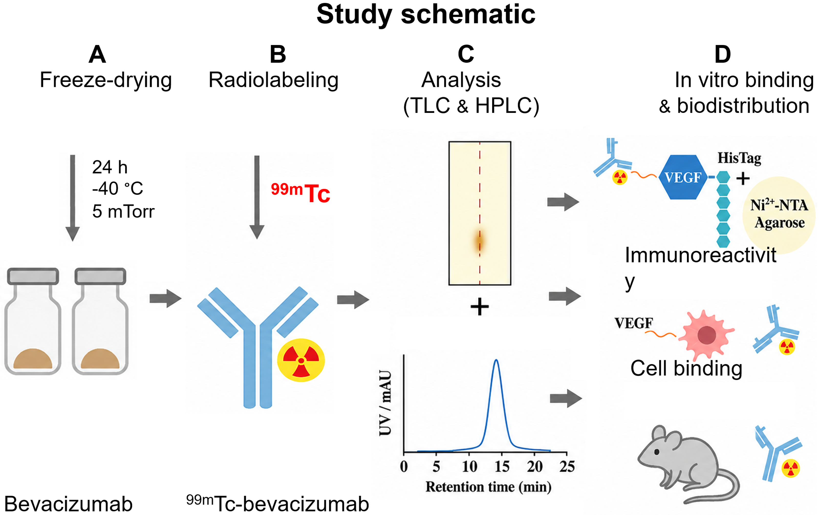
Schematic workflow of kit preparation. The workflow includes
Materials and Methods
Reagents
Bevacizumab (25 mg/mL, 149 kDa; Roche, Switzerland) was used for kit lyophilization and radiolabeling. The human cancer cell lines A-549 (alveolar basal epithelial carcinoma, CCL-185), MCF-7 (Michigan Cancer Foundation-7, breast adenocarcinoma, HTB-22), and HT-29 (colorectal adenocarcinoma, HTB-38) were obtained from the American Type Culture Collection (ATCC, USA). The cell culture media included Eagle’s minimum essential medium (EMEM), RPMI-1640, McCoy’s 5A modified medium (ATCC 30–2007), and fetal bovine serum (FBS; ATCC 30–2020). Recombinant human His-tag VEGF-A/VEGF121 (Elabscience, USA) and HisPur Ni-NTA resin (Thermo Scientific, USA) were used for the affinity binding assays. Carrier-free sodium pertechnetate (Na99mTcO4, 1.85–3.70 GBq/mL) was freshly eluted from a 99Mo/99mTc generator at the Dalat Nuclear Research Institute in Vietnam. MultiScreen 96-well plates, filtration units (Millipore, USA), Fiolax glass vials (Schott, Germany), and rubber stoppers (West Pharmaceutical Services, USA) were used. An LC-20AD system equipped with UV (280 nm) and radiometric detectors coupled to a Shodex OHpak SB-803 HQ size exclusion column (Shimadzu, Japan) were used for the Size Exclusion Chromatography–High Performance Liquid Chromatography (SEC-HPLC) analysis. A cyclone phosphor storage system (PerkinElmer, USA) and a dose calibrator (CRC-127, Capintec, USA) were used for the radioactivity measurements.
Optimization and preparation of lyophilizate
The antibody was radiolabeled with 99mTc under optimized conditions that were previously established in our laboratory (see Supplementary Data S1). The optimized labeling mixture consisted of 2.0 mg of bevacizumab, 3700 MBq of Na99mTcO4, 0.5 M phosphate-buffered saline (PBS) (pH 7.5), and 0.4 mg of stannous chloride as the reducing agent. To optimize the freeze-dried formulation, the solutions were prepared in different buffer systems, including 0.5 M PBS (pH 7.5) and 0.5 M acetate buffer (pH 6.0). The effects of pH (4, 5, 6, 7, and 8) and duration (8, 16, 24, 32, and 48 h) were systematically evaluated (Freeze dryer, IlShinBioBase, Korea). The freeze–drying process consisted of four main stages. The composition was first cooled to −40°C for 10–12 h to ensure complete freezing, followed by primary drying at −20°C for 20 h under a vacuum. Secondary drying was performed at 0°C for 2 h, and the samples were finally dried at 24°C for an additional 2 h to remove residual moisture. The total number of freeze–drying cycles was approximately 36 h, yielding a uniform, white, cake-like product with good mechanical integrity.
Characterization of the freeze-dried kit
The labeling efficacy and RCP were determined by thin-layer chromatography (TLC) using TLC-SG strips (Silicagel 60 F254, Merck) with three mobile phases: acetone, 0.9% NaCl, and ammonium hydroxide:ethanol:water (1:2:5, v/v/v), together with SEC-HPLC analysis. Free 99mTcO4− migrated with the solvent front, whereas reduced-hydrolyzed 99mTc and 99mTc-colloids remained at the origin. The percentage of 99mTc-bevacizumab was calculated by subtracting the proportions of free 99mTcO4− and reduced-hydrolyzed/colloidal 99mTc impurities from the total radioactivity. The lyophilized vial was reconstituted with sterile water, followed by the addition of freshly eluted Na99mTcO4 solution (1850–3700 MBq). The mixture was incubated at room temperature (25°C) for 15 min, after which the RCP was determined using different chromatography systems described above. The stability of the radiolabeled kit was evaluated at multiple time points (0, 3, 6, 9, 12, and 24 h) following labeling with 99mTc under storage conditions of 4°C, 24°C, and 37°C. The stability studies of 99mTc-bevacizumab in human serum at 37°C were performed as previously reported, with minor modifications from Ramos-Membrive et al., 33 and RCPs were evaluated at 0, 3, 6, and 24 h (Supplementary Data S2). In addition, the long-term stability of the freeze-dried kit prepared in different buffer systems, pH conditions, and durations was assessed at 0, 1, 2, 3, 6, and 12 months after production.
VEGF–His-tag in vitro affinity assay
The immunoreactivity of 99mTc-bevacizumab was evaluated using an in vitro VEGF–His-tag binding model based on Ni–NTA affinity chromatography. One hundred microliters of Ni–NTA agarose was loaded into 2-mL Piercespin columns (Thermo Scientific) equipped with a polyethylene frit and sequentially washed with double distilled water and 20 mM Tris–HCl buffer (pH 8.0), followed by centrifugation at 2000 × g for 3 min. His-tagged antigen was dissolved in 200 µL of PBS and incubated with the resin at the indicated concentrations for 60 min at room temperature with gentle agitation. After incubation, the resin was centrifuged, the supernatant was discarded, and the pellet was washed twice with PBS to remove unbound fractions. The conjugation was prepared at a final concentration of 0.66 nmol in PBS. The antibody solution was added to the growth factor-coated resin at VEGF: 99mTc-bevacizumab molar ratios of 0.5:1, 1:1, 1.5:1, 2:1, 3:1, and 4:1 in a final volume of 500 µL. The samples were incubated for 120 min at room temperature and then washed three times with PBS. Bound and unbound fractions were collected separately, and the percentage of antibody bound to antigen was calculated from the measured radioactivity.
Cell culture
A-549 cells were cultured in RPMI-1640, MCF-7 in EMEM, and HT-29 cells in McCoy’s 5A medium, each supplemented with 10% FBS and 0.2% kanamycin. Cells were seeded at 0.15–0.2 × 10⁶ cells per 25-cm2 flask and incubated at 37°C with 5% CO2. When the cultures reached 80%–90% confluence, the cells were passaged for expansion. After 8–20 d (depending on the cell line), the cells were harvested using trypsin, counted with a hemocytometer, washed by centrifugation, and resuspended in sterile PBS.
Specific binding and saturation binding assays
Specific binding and saturation binding were evaluated in A-549, MCF-7, and HT-29 cells to determine the immunoreactive fraction (IRF) or r value. For specific binding according to Lindmo, increasing numbers of cells (1–8 × 10⁶) were incubated in duplicate with a fixed amount of radioligand (6.6 µCi/0.66 µg in 1.0 mL of 10 mM PBS, pH 7) at 37°C for 120 min. Nonspecific binding tubes were preincubated with 50 µg of unlabeled antibody for 60 min at 37°C before the addition of radioactivity.25,26 The two total activity tubes contained only the tracer. The cell-bound and cell-free fractions were separated by Millipore filtration and then were washed four times with phosphate buffer, after which the cells were counted on a γ counter (Caprac 13, Capintec) to calculate the % bound activity. For saturation binding, ∼5 × 10⁵ cells/tube were incubated with 99mTc-bevacizumab (10 µCi/µg) at 0.75–50 nmol/L in the presence or absence of excess cold antibody (50 µg) under the same conditions. Specific binding was analyzed by Scatchard plots using GraphPad Prism 8 to determine the Kd and Bmax.27,28
Biodistribution studies in mice
A total of 48 ICR mice (6 weeks old; Biotechnology Center, Vietnam) were allocated into eight groups (n = 6 per group) and intravenously injected with 3.7 MBq of 99mTc-bevacizumab in 100 µL. The mice were sacrificed at 1, 5, 30, 60, 120, 180, 360, and 1440 min post-injection. Blood and major organs/tissues (heart, liver, kidneys, spleen, lung, muscle, bone, stomach, intestine, and tail) were collected, weighed, and measured for radioactivity using a γ counter. Biodistribution data are expressed as %ID/g, with blood, muscle, and bone masses estimated as 7%, 40%, and 10% of body weight, respectively. 29
Ethics statement
All animal procedures were reviewed and approved by the Animal Ethics Committee of the Dalat Nuclear Research Institute and were conducted in accordance with all relevant institutional and national guidelines for the care and use of laboratory animals.
Statistics
Data are reported as the mean ± SD, and statistical significance was defined as p < 0.05. Saturation binding data were fitted using a one-site binding model to obtain the Kd and Bmax, and Scatchard analysis was performed to confirm single-site interactions. The results of the stability studies, buffer comparison experiments, and freeze-dried time evaluations were analyzed using two-factor Analysis of Variance (ANOVA). Biodistribution data were analyzed using one-way ANOVA followed by Dunnett’s test (*p < 0.05, **p < 0.01). Differences between the two groups were assessed using the Student’s t-test. RCPs were analyzed using OptiQuant 5 software.
Results
Optimization and preparation of the lyophilized formulation
The kit was formulated with 2.0 mg of bevacizumab in 0.5 M phosphate buffer (pH 7.5) with 0.4 mg of stannous chloride as the reducing agent and stabilizers, including 0.5 mg of ascorbic acid, 0.05% HSA, and 0.9% NaCl, before freeze drying. The kit composition was selected according to the radiolabeling efficiency obtained in the optimization studies (Supplementary Data), which revealed 20 µg of stannous chloride (for labeling 100 µg antibody with 185 MBq 99mTc), an incubation time of 5–15 min at room temperature, an optimal pH of 7.5, and good stability (Supplementary Fig. S1A–E). As shown in Figure 2A, the radiolabeling efficiency did not significantly differ before and after lyophilization. The radiolabeling efficiencies of the kit in phosphate buffer, 0.9% NaCl, and 0.5 M acetate buffer were 99.55 ± 0.21%, 99.60 ± 0.28%, and 99.65 ± 0.21%, respectively, which were comparable to those of the liquid form (99.70 ± 0.14%, 99.30 ± 0.28%, and 99.68 ± 0.04%, respectively). At pH 8, the radiolabeling efficiencies before and after drying were 95.41 ± 0.79% and 94.70 ± 0.56%, respectively. No significant differences were observed before or after lyophilization across freeze–drying durations of 8, 16, 24, 32, and 48 h, with all radiolabeling efficiencies exceeding 99%, as shown in Figure 2B. We chose a time profile with a lyophilization cycle consisting of four stages: freezing, primary drying, and two-step secondary drying. The formulation was first cooled to −40°C for 10–12 h, followed by primary drying at −20°C for approximately 20 h under a 5 mTorr vacuum and secondary drying at 0°C for 2 h and 24°C for 2 h (Fig. 2C). The entire process lasted approximately 36 h, resulting in the production of a stable, uniform, white cake suitable for radiolabeling with 99mTc (Fig. 2D).
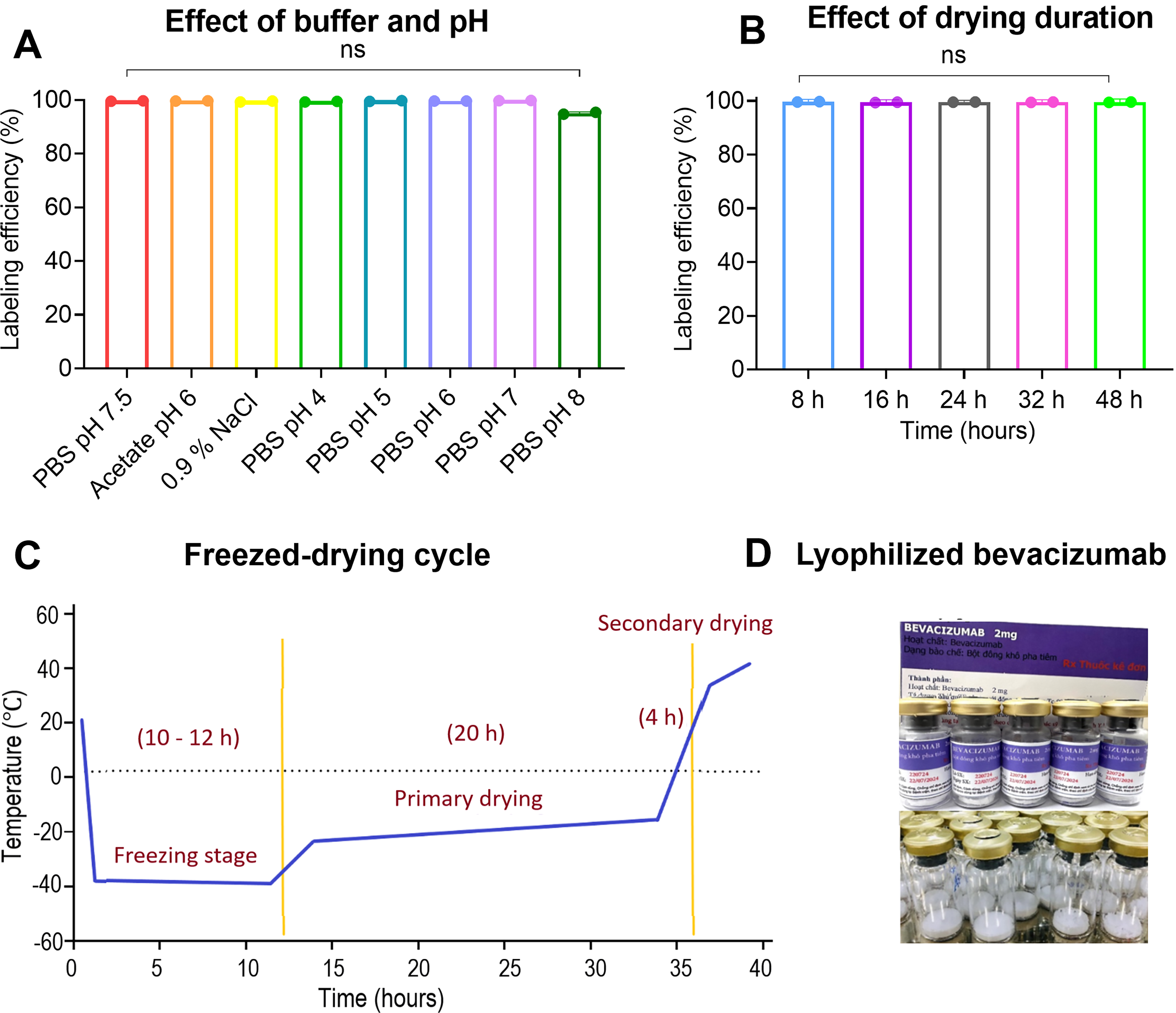
Freeze–drying process of the bevacizumab kit.
Characterization of the freeze-dried kit
The Sn2+ in the formulation acts as a reducing reagent during the radiolabeling process. Sn2+ first reduces 99mTcO4− to a lower oxidation-state technetium intermediate, likely Tc(V)/Tc(IV), with the transient formation of intermediate Sn–Tc species.30,31 Separately, Sn2+ also reduces accessible interchain disulfide linkages (–S–S–) within the IgG1 structure to generate free thiol groups (–SH) that provide reactive coordination sites for stable binding of 99mTc to the antibody. Subsequently, the reduced technetium intermediate coordinates with the newly generated thiols and adjacent donor atoms, such as amide nitrogen and carbonyl oxygen atoms, leading to the formation of the 99mTc-IgG complex8,32 (Fig. 3).
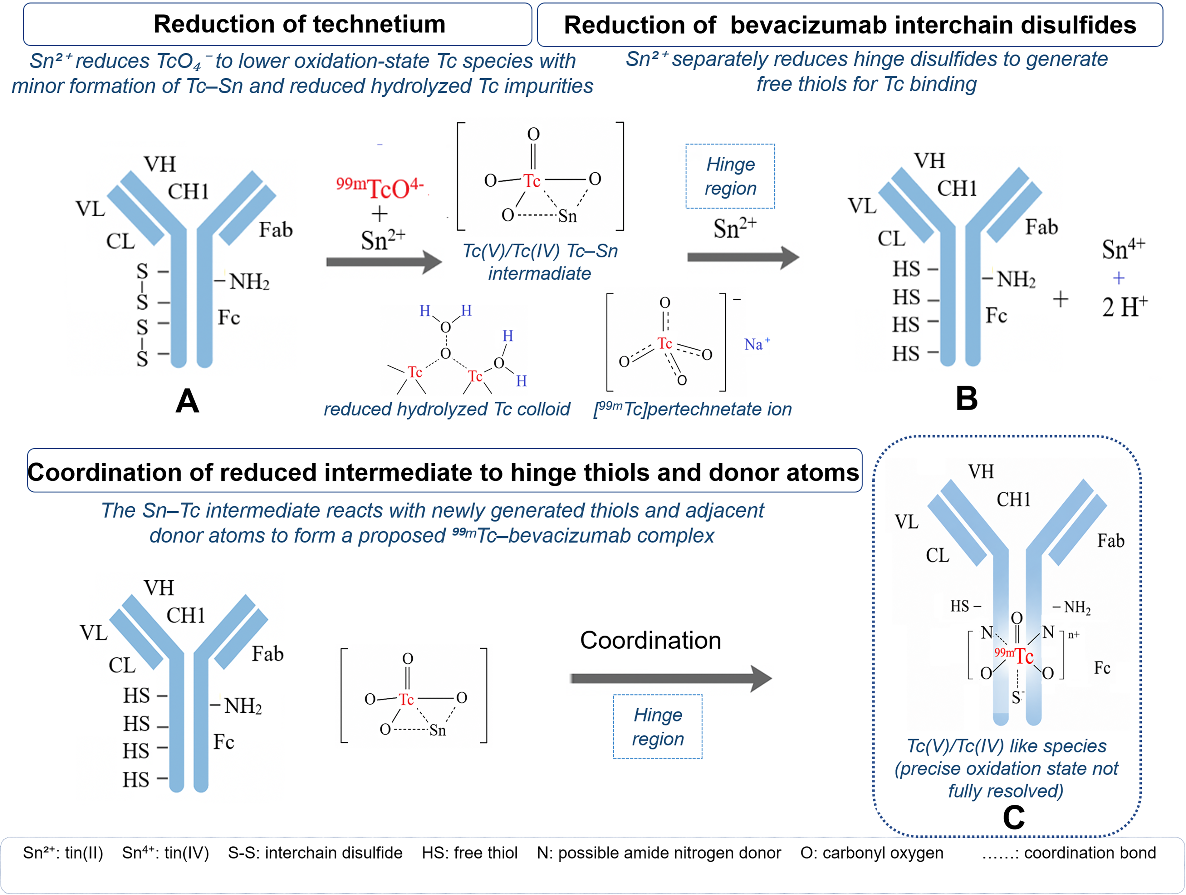
Proposed radiolabeling mechanism of 99mTc-bevacizumab.
Radiochemical purity
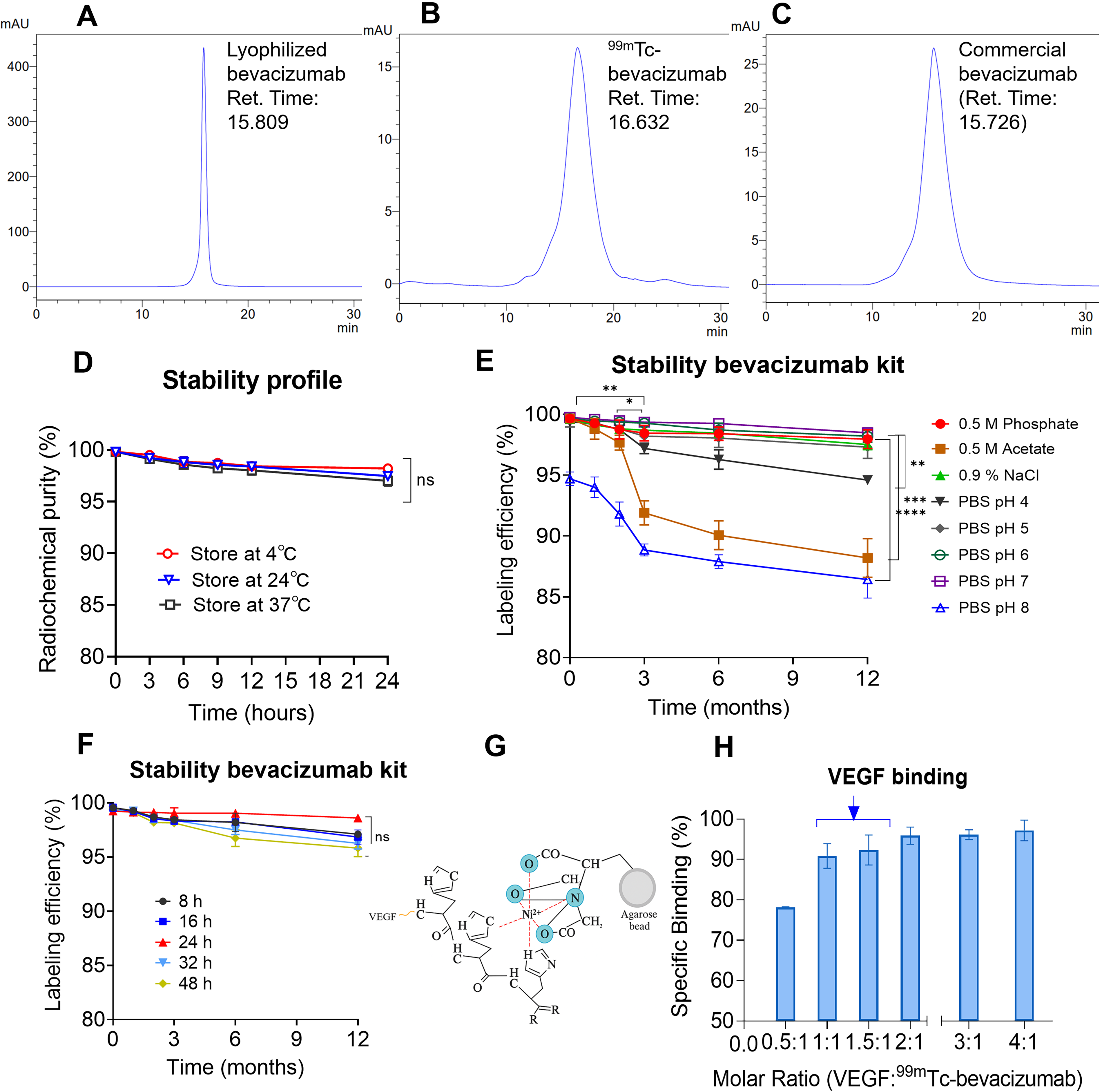
SEC-HPLC analysis showed that lyophilized bevacizumab, 99mTc-bevacizumab, and commercial bevacizumab exhibited major peaks at retention times of 15.809, 16.632, and 15.726 min, respectively. Based on the analysis of three randomly selected samples from three independent production batches, TLC analysis revealed a radiolabeling efficiency of 97.77 ± 0.67% for 99mTc-bevacizumab (Fig. 4A–C). For comparison, Na99mTcO4− moved to the solvent front, with Rf values ranging from 0.8 to 1.0 (Supplementary Fig. S2A), and the radiolabeled yields before lyophilization are shown in Supplementary Fig. S2B.18,34
Characterization and quality control of lyophilized kit
Stability
The short-term stability of the freshly reconstituted formulation was evaluated from 1 to 24 h at 4°C, 24°C, and 37°C. The labeling remained >97% at all temperatures throughout the study (Fig. 4D). Stability results in human serum are shown in Supplementary Figure S2G. The long-term stability of the freeze-dried preparation under different buffer pH conditions was also assessed (Fig. 4E). Kits formulated in 0.5 M PBS (pH 7.5) or PBS (pH 5–7) showed no significant loss of RCP over 12 months (p > 0.05), and the labeling efficiency was maintained above 97%. In contrast, formulations with 0.5 M acetate (pH 6), PBS (pH 4), and PBS (pH 8) significantly decreased in a time-dependent manner. The acetate concentration at a pH of 6 showed the earliest and most pronounced decrease, followed by those at PBS pH values of 4 and 8. The long-term stability kits at different durations were assessed on RCP with time. In all the batches at 8, 16, 24, 32, and 48 h, no significant differences were detected among the batches under storage conditions of 2°C–8°C (Fig. 4F).
Immunoreactivity
VEGF immobilized on Ni2+ NTA agarose beads on the basis of the Ni2+ ion in the NTA–agarose complex formed six coordinate covalent bonds. Four of these are coordinated by oxygen and nitrogen atoms from NTA, while the remaining two were available for coordination with the imidazole nitrogens of the histidine residues in the His-tagged protein (Fig. 4G). Specific binding increased with an increasing VEGF-to-antibody molar ratio, reaching >90% at a 1:1 ratio and exceeding 95% at a 2:1 ratio. The binding followed a one-site specific binding model, yielding a Kd of 0.1405 nM and a Bmax of 101.4% (Fig. 4H). The characteristics are summarized in Table 1.
Physicochemical Characteristics of 99mTc, Bevacizumab (mAb), and Radiolabeled mAb
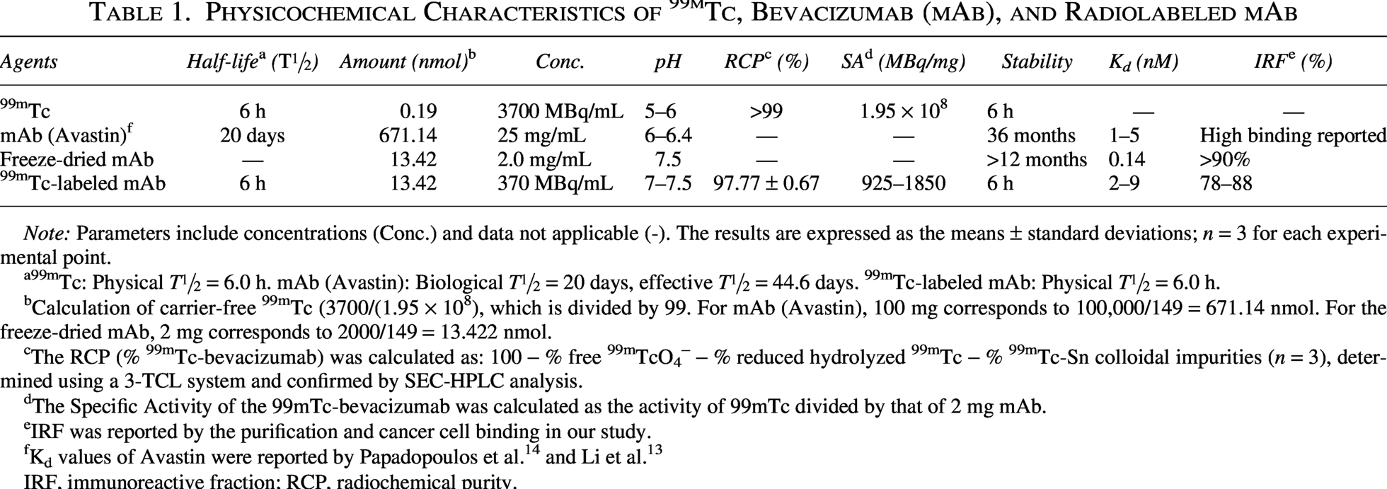
Note: Parameters include concentrations (Conc.) and data not applicable (-). The results are expressed as the means ± standard deviations; n = 3 for each experimental point.
99mTc: Physical T½ = 6.0 h. mAb (Avastin): Biological T½ = 20 days, effective T½ = 44.6 days. 99mTc-labeled mAb: Physical T½ = 6.0 h.
Calculation of carrier-free 99mTc (3700/(1.95 × 108), which is divided by 99. For mAb (Avastin), 100 mg corresponds to 100,000/149 = 671.14 nmol. For the freeze-dried mAb, 2 mg corresponds to 2000/149 = 13.422 nmol.
The RCP (% 99mTc-bevacizumab) was calculated as: 100 − % free 99mTcO4− − % reduced hydrolyzed 99mTc − % 99mTc-Sn colloidal impurities (n = 3), determined using a 3-TCL system and confirmed by SEC-HPLC analysis.
The Specific Activity of the 99mTc-bevacizumab was calculated as the activity of 99mTc divided by that of 2 mg mAb.
IRF was reported by the purification and cancer cell binding in our study.
IRF, immunoreactive fraction; RCP, radiochemical purity.
In vitro specific binding and saturation binding analysis
After 48–72 h of culture, the cells formed a well-attached monolayer and were harvested at 85%–90% confluence. The cells were washed with sterile PBS and adjusted to a concentration of 107 cells/mL. Representative morphologies of A-549, HT-29, and MCF-7 cells are shown in Figure 5A. In A-549 cells, the binding curve exhibited a typical hyperbolic saturation profile, with a maximal specific binding of 89.52% (Fig. 5B). Scatchard transformation yielded a linear plot and the IRF calculated from the slope was approximately 88% (Fig. 5C). In a separate saturation binding experiment, 99mTc-bevacizumab showed binding to A-549 cells, with a Kd of 2.629 nM and a Bmax of 1.659 amol/cell, corresponding to ∼1.0 × 10⁶ binding sites per cell (Fig. 5D). In a similar manner, the binding curve for the HT-29 cells displayed a comparable saturation profile, with a binding of 80.51% (Fig. 5E) and an IRF of approximately 84%, as determined from the Scatchard plot (Fig. 5F). Saturation binding analysis in HT-29 colorectal cancer cells yielded a Kd of 9.969 nM and a Bmax of 2.062 amol/cell, corresponding to ∼1.24 × 10⁶ binding sites per cell (Fig. 5G). Similarly, MCF-7 cells also exhibited a clear saturation profile, with a binding of 71.41% (Fig. 5H). The IRF was approximately 80% (Fig. 5I). Nonlinear regression analysis yielded a Kd = 7.078 nM and a Bmax = 1.672 amol/cell, corresponding to approximately 1.0 × 10⁶ binding sites per cell on the basis of Avogadro’s number (Fig. 5J).
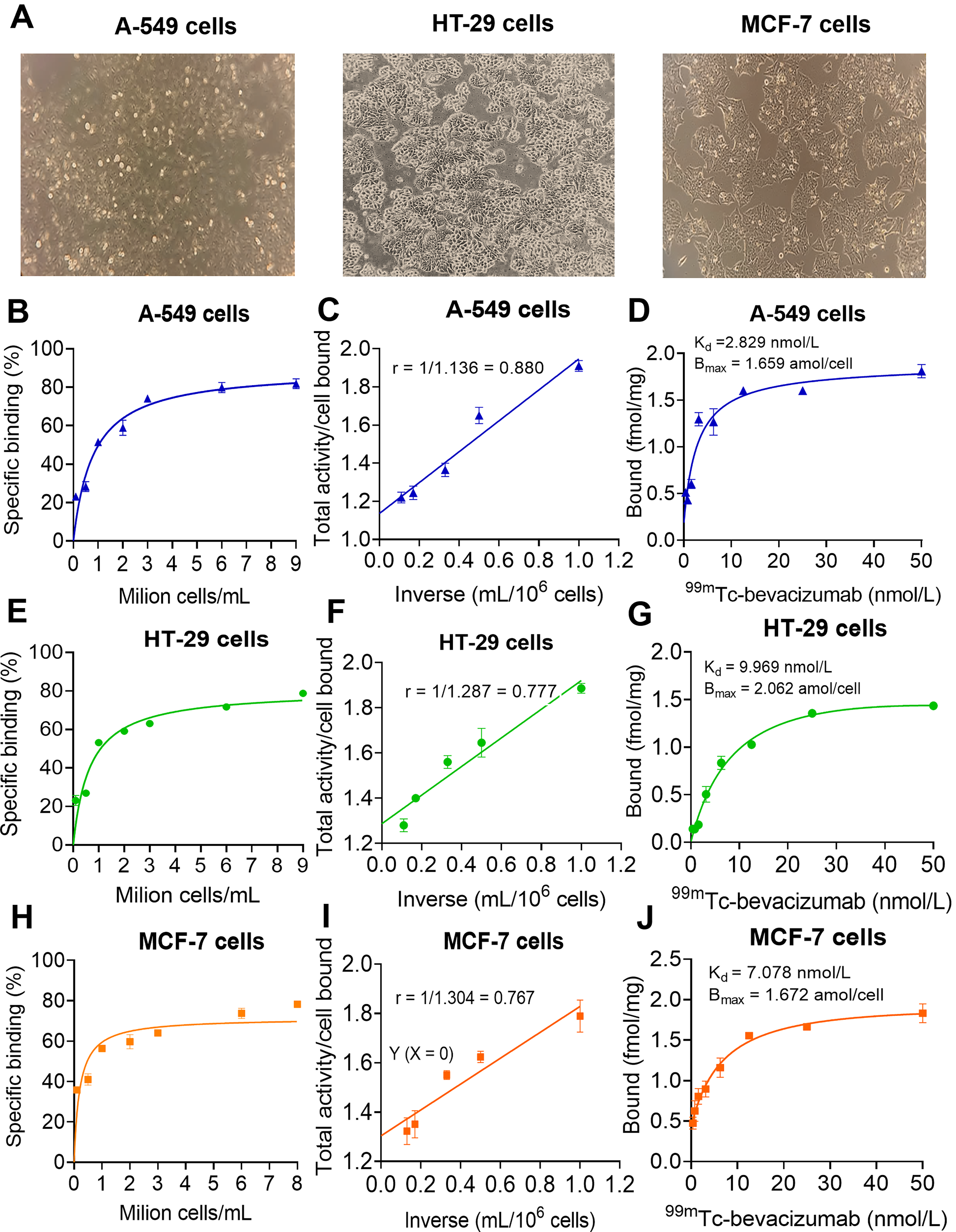
Specific binding of 99mTc-bevacizumab to the cancer cell lines A-549, HT-29, and MCF-7.
Biodistribution results
After injection, the activity of 99mTc-bevacizumab (in %ID/g) was initially high in blood 11.70 ± 0.53, followed by 5.20 ± 0.45 in the heart, 3.34 ± 0.92 in the liver, 8.39 ± 0.55 in the kidneys, and 6.61 ± 2.78 in the lungs. At 60, 360, and 1440 min, respectively, the activity in blood was 4.49 ± 1.76, 2.27 ± 0.39, and 0.76 ± 0.30; 2.55 ± 2.15, 1.23 ± 0.21, and 0.39 ± 0.21 in the heart; 1.65 ± 1.39, 3.22 ± 1.76, and 1.42 ± 0.74 in the liver; 5.59 ± 1.71, 4.61 ± 0.83, and 3.79 ± 1.41 in the kidneys; and 3.00 ± 2.20, 1.87 ± 0.70, and 1.40 ± 1.03 in the lungs (Fig. 6A). Blood clearance half-life was 76.97 min and R squared = 0.7353 (Fig. 6A–B). Spleen uptake decreased slowly (Fig. 6C). Bone uptake increased slightly up to 60 min and then declined, whereas muscle, intestine, and stomach showed minimal activity (Fig. 6D–E). Blood-to-liver ratios (Fig. 6F) and the blood-to-kidney ratios were high during the early period (1–30 min) (Fig. 6J).
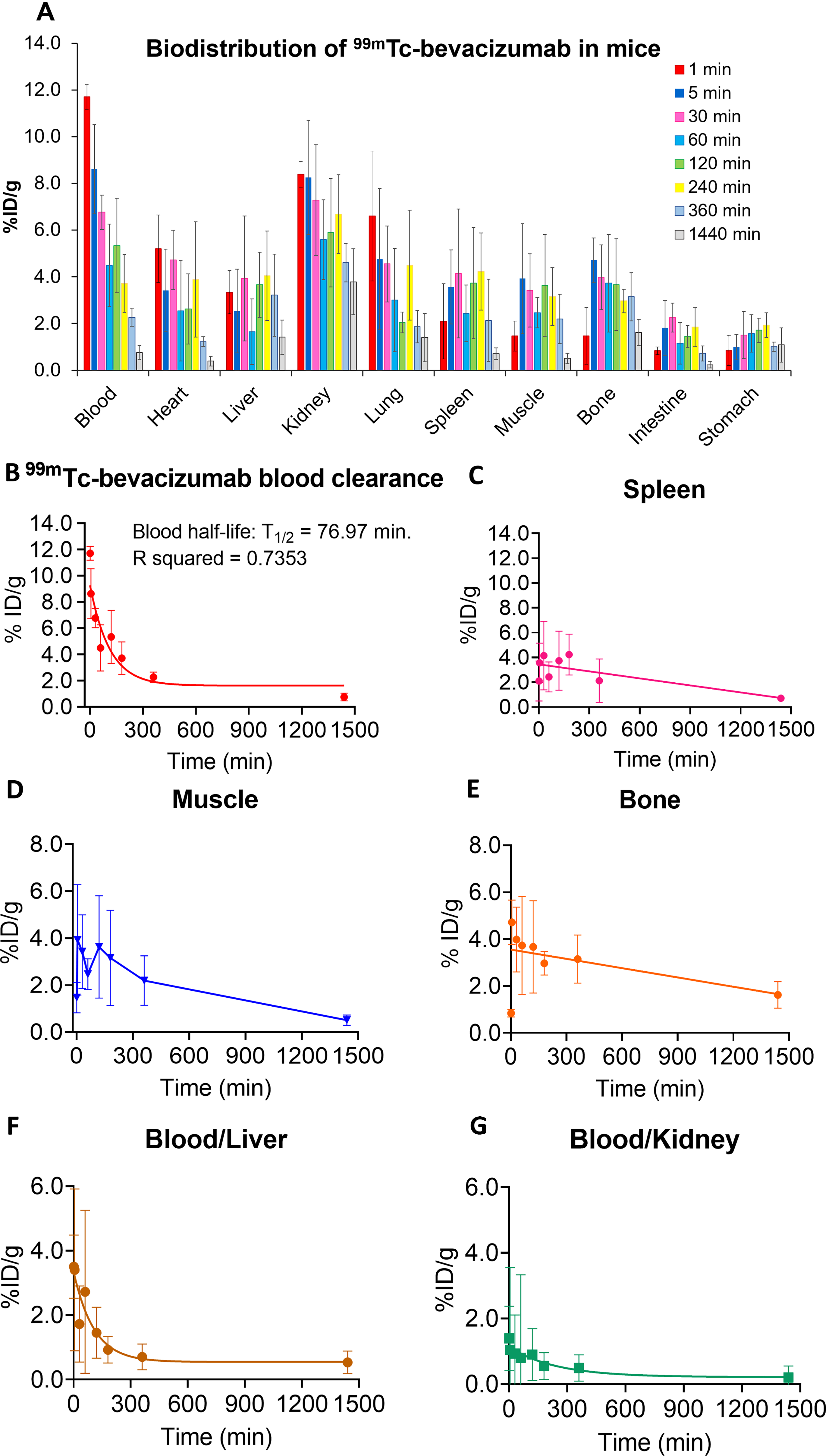
Biodistribution, pharmacokinetic, and organ-specific ratios of 99mTc-bevacizumab in normal mice over time.
Discussion
In this study, we developed a freeze-dried bevacizumab kit suitable for instant radiolabeling with 99mTc. The kit was designed to improved stability, shelf-life, transportation, storage, and hospital use, requiring only simple reconstitution with 99mTc-pertechnetate before administration. 35 The final formulation contained three essential components: bevacizumab, a reducing agent, and an optimized phosphate buffer with stabilizers. This composition is comparable to previously reported lyophilized monoclonal antibody kits, including rituximab at 1.0 mg/vial, nimotuzumab at 3.0 mg/vial, and trastuzumab at 10.0 mg/vial. All of these shared similar requirements regarding buffer conditions and stabilizers to maintain antibody structure during lyophilization.22,23,36,37 The 24-h lyophilization, high pressure protocol in this study was also consistent with previously described freeze–drying procedures for monoclonal antibody.23,36,38
Bevacizumab was selected because it is a recombinant humanized IgG1 monoclonal with a stable molecular architecture, including inter–heavy-chain disulfide bonds in the hinge region and multiple intrachain disulfide bonds within each domain.32,39 Compared with IgG2 or IgG4, IgG1 has fewer free thiol groups and is less prone to disulfide scrambling and aggregation, allowing controlled partial reduction by Sn2+ to generate thiol groups for 99mTc coordination while preserving antibody integrity. 39 Previous studies reported approximately two potential 99mTc binding sites per IgG molecule, 40 providing a useful benchmark for formulation development. This structural stability supports the preservation of functional binding capacity after reduction, lyophilization, and radiolabeling.
The optimized kit showed high RCP, with an RCP value above 97% (Fig. 4, Table 1). This performance is comparable to other 99mTc-labeled monoclonal antibody preparation, including besilesomab, sulesomab, rituximab, nimotuzumab, and trastuzumab, which typically show RCPs in the range of 97%–99%.6,8,23,36,37 Each vial was designed to be labeled with 1850–3700 MBq of 99mTc, corresponding to an administered activity of 370–740 MBq and approximately 0.4 mg of antibody per patient. This amount is far below therapeutic bevacizumab doses (5–15 mg/kg) and is consistent with diagnostic antibody preparations such as 99mTc-besilesomab (300–800 MBq with 0.25–1.0 mg). 8 The Tc-to-antibody molar ratio of 0.014 was also compared to reported values for 99mTc-besilesomab (0.01–0.03), sulesomab (0.02), and rituximab (0.01–0.02).6,41 These findings indicate that only a limited fraction of hinge disulfide bonds was reduced during Sn2+-mediated labeling, allowing incorporation of 99mTc while preserving the overall structure. Regarding the radiolabeling mechanism, Sn2+ reduces 99mTcO4− to lower oxidation-state technetium intermediates and partially reduces accessible interchain disulfide bonds to generate thiol groups for coordination. Minor impurities may include free 99mTcO4−, reduced hydrolyzed 99mTc colloids, and Tc–Sn colloidal species. Therefore, RCP was evaluated using complementary TLC systems and further confirmed by SEC-HPLC.30,31 Comparable HPLC retention profiles between lyophilized bevacizumab, 99mTc-bevacizumab, and commercial bevacizumab indicated that lyophilization and radiolabeling did not markedly alter the overall molecular profile of the antibody. 38
Short-term stability assessment revealed that 99mTc-bevacizumab retained RCP above >97% for up to 24 h at 4°C–37°C, without temperature-dependent degradation. Long-term stability studies also demonstrated that PBS-based kits at pH 6–7.5 maintained high radiolabeling efficiency over 12 months, whereas acetate buffer and extreme pH conditions, particularly pH 4 and pH 8, reduced labeling efficiency. These results indicated that near-physiological pH conditions best preserved the physicochemical stability of the freeze-dried formulation during storage.38,42
The VEGF–His-tag/Ni–NTA assay was used as an in vitro affinity model rather than a physiological VEGFR-mediated receptor-binding system. This functional affinity assay showed specific binding above 90%, indicating that antigen recognition was largely preserved after lyophilization and radiolabeling. This high affinity is consistent with previously reported affinities of bevacizumab for VEGF-A of VEGF165, which typically fall within the 0.1–2 nM range.14,15,43
In vitro cellular binding studies using A-549, HT-29, and MCF-7 cells further supported preservation of the functional binding capability of 99mTc-bevacizumab following radiolabeling. Binding profiles were concentration-dependent and saturable, consistent with specific binding interactions. 14 Among the three lines, A-549 lung adenocarcinoma cells exhibited the highest affinity, with a Kd of 2.629 nM and approximately 1.0 × 10⁶ binding sites per cell. Although all three cell lines showed broadly comparable binding site estimates (∼1–1.24 × 10⁶ sites/cell), the Kd values differed by approximately fourfold (2.6 vs. 7.1 vs. 9.97 nM), consistent with heterogeneity in VEGF-A signaling and presentation among tumor cell types.12,43 HT-29 colorectal cancer cells showed the highest Bmax but lower affinity, whereas MCF-7 breast cancer cells showed intermediate binding characteristics. 44 These in vitro findings demonstrate that lyophilization and radiolabeling did not compromise the ability of bevacizumab to recognize cell-associated antigen.38,42
The affinity values obtained in the three cell lines (Kd = 2.6–9.9 nM) were within the range expected for antibody-antigen binding characteristics. Previous studies reported bevacizumab dissociation constants of 1.33 nM by surface plasmon resonance and 2.3 nM by isothermal titration calorimetry, 43 whereas Papadopoulos et al. described sub-nanomolar binding interactions with VEGF165 (58–1100 pM). 14 The single-site binding profiles observed in Scatchard analyses by Lindmo further support preservation of specific binding behavior after radiolabeling. 25
The in vivo biodistribution study in normal mice shows high initial blood activity, followed by gradual clearance and moderate uptake in the heart, kidneys, lungs, and liver (Fig. 6). This pattern reflects prolonged vascular retention, which is characteristic of antibody-based radiopharmaceuticals. Early renal activity may be related to renal perfusion and possible minor contributions from circulating low-molecular-weight or radiochemical species, including free 99mTcO4−, reduced-hydrolyzed 99mTc, 99mTc-colloid, and 99mTc-Sn species.31,45
The calculated blood half-life of 76.97 min, which is similar to the value of 106.7 min reported by Tan et al. for 99mTc-MAG3-bevacizumab, suggesting a clearance profile comparable to that of other technetium-labeled antibodies. However, this reflects the 99mTc decay-limited measurement (T½ = 6 h) not native IgG pharmacokinetics. Therefore, the true biological half-life of bevacizumab (23–26 days) cannot be determined using 99mTc.18,45
Hepatic uptake observed in our study was low, decreasing from approximately 4% at 3 h to nearly 2% at 24 h. This relatively low liver retention may represent an advantage of direct 99mTc labeling compared with some antibody derivatives labeled through bifunctional chelators. For example, Tan et al. reported hepatic retention of 8.19 ± 3.54% ID/g at 2 h and >2% ID/g at 12 h for 99mTc-MAG3-bevacizumab, while Kameswaran et al. reported >10% ID/g liver activity at 24 h.18,34 Other 99mTc-labeled antibodies, such as rituximab and nimotuzumab, also showed higher liver uptake at early time points and retained >4%–6% ID/g at 24 h.41,46 A comparable decreasing pattern in the blood-to-kidney ratio reflects early renal handling followed by slow clearance, which is consistent with the pharmacokinetic profiles of bevacizumab.45,47
Certain limitations in the present in vitro evaluation should be interpreted as follows: the VEGF–Ni–NTA assay does not reproduce physiological VEGF/VEGFR interactions in vivo, and the cellular binding assays do not directly quantify physiological receptor density in tumor tissues. In addition, the biodistribution study in normal mice did not evaluate tumor uptake or imaging performance in tumor-bearing models. Future studies should focus on GMP-compliant kit production, evaluation in additional tumor cell lines with different VEGF expression levels, and biodistribution and imaging studies in angiogenic tumor-bearing animal models.
In conclusion, the development of a freeze-dried bevacizumab kit containing an optimized combination of reducing agent, buffer, and stabilizers enabled efficient 99mTc radiolabeling while maintaining long-term physicochemical stability and preserving functional binding capability. The radiolabeled product also demonstrated favorable biodistribution characteristics in normal mice, including prolonged blood circulation and low hepatic uptake. These findings support further preclinical evaluation of 99mTc-bevacizumab as a potential tumor imaging agent. Future studies should evaluate 99mTc-bevacizumab in tumor-bearing animal models with defined VEGF expression, including in vivo imaging. These studies will be necessary before clinical translation or application in tumor radioimmunoscintigraphy can be considered.
Authors’ Contributions
T.K.G.N.: Investigation, methodology, validation, software, data curation, and quality control analyses. T.N.N.: Investigation, methodology, software, radiolabeling procedures, and quality control analyses. T.B.N. and H.H.Q.D.: Radioisotope production, data curation, and formal analysis. T.M.C.N.: Investigation, data processing, software, and animal studies. D.K.N. and N.B.N.D.: Investigation, data curation, and biodistribution studies. T.N.N. (Thanh Nhan Nguyen): Radioisotope production, software, and methodology. T.Q.T.V. and T.T.D.: Data curation and formal analysis. T.M.P.: Conceptualization and supervision. T.T.N.: Conceptualization, investigation, methodology, formal analysis, writing—original draft, and writing—review and editing.
Data Availability Statement
All data generated or analyzed during this study are available from the corresponding author upon reasonable request.
Footnotes
Acknowledgments
The authors gratefully acknowledge the Dalat Nuclear Research Institute, Vietnam, for providing laboratory facilities and administrative support.
Disclosure Statement
No competing financial interests exist.
Funding Information
This study was funded by the Ministry of Science and Technology of Vietnam under the Ministry-level research project ĐTCB.11/24/VNCHN.
Supplemental Material
References
Supplementary Material
Please find the following supplemental material available below.
For Open Access articles published under a Creative Commons License, all supplemental material carries the same license as the article it is associated with.
For non-Open Access articles published, all supplemental material carries a non-exclusive license, and permission requests for re-use of supplemental material or any part of supplemental material shall be sent directly to the copyright owner as specified in the copyright notice associated with the article.