
Abstract
War and terrorism demonstrate the need for an improved antidote against nerve agents that cause brain damage following inhibition of acetylcholinesterase (AChE). Our laboratories invented substituted phenoxyalkyl pyridinium oxime AChE reactivators (US patent 9,227,937) that demonstrate the ability to cross the blood–brain barrier in in vivo rat tests with a sarin surrogate by reducing time to cessation of seizure‐like behaviors, glial fibrillary acidic protein levels, and neuropathology, compared to the FDA-approved oxime, 2‐pyridine aldoxime methyl chloride (2‐PAM). This study investigated the safety profile of novel oximes through evaluation of their cytotoxicity and genotoxicity. Cytotoxicity was evaluated in mouse areolar fibroblasts using a lactate dehydrogenase (LDH) assay and an MTS assay. All novel oximes showed no toxicity in the LDH assay from 1.25 nM to 5 mM, whereas 2-PAM did; novel oximes were significantly different from 2-PAM (P < .00001). The MTS assay demonstrated that novel oximes at 2 µM to 500 µM significantly reduced the metabolic activity of fibroblasts compared to 2-PAM (P = .00001). A modified Ames assay using 5 bacterial strains with and without S9 assessed the genotoxicity of the lead compound, Oxime 20, at concentrations from 100 µM to 10 mM. No concentration, whether with S9 or without, showed any genotoxicity. The LDH and Ames results indicated no cytotoxicity or genotoxicity was caused by the novel oximes.
Introduction
Organophosphate (OP) compounds include acutely toxic insecticides such as parathion and chemical warfare agents such as sarin. The large quantities of OPs present worldwide pose one of the greatest chemical threats to both civilians and the military. Civilians were killed by deliberate sarin exposure in the Kurdish-inhabited area in 1988 1 and Japan during 1994 and 1995. 2 Sarin was also the cause of death for both Syrian war fighters and civilians during the 2013 to 2017 civil war. 3 Sarin has been described as the terrorist’s “nerve agent of choice” 4 because its volatility and rapid uptake allow for mass casualties. In developing agricultural countries, the availability of OP insecticides combined with poor safety practices leads to both accidental exposures and suicidal ingestions. OPs are the leading cause of acute poisoning and currently account for 20% of global suicides.5,6
The toxicity of OPs stems from their persistent inhibition of acetylcholinesterase (AChE) causing excess synaptic acetylcholine accumulation, followed by overstimulation of postsynaptic nicotinic and muscarinic receptors. This causes cholinergic effects and seizures, which, if not stopped, elicit excitatory amino acid release, followed by excessive calcium influx and stimulation of N‐methyl‐D‐aspartate (NMDA) receptors, causing neuropathology in rodents 7 plus somatic and psychological symptoms that last for years after exposure in humans. 8
At present, in addition to atropine, the approved therapy to counteract OP toxicity includes pyridinium oxime administration to reactivate the inhibited AChE. The present FDA-approved oxime is 2‐pyridine aldoxime methyl chloride (2‐PAM), which cannot effectively cross the blood–brain barrier (BBB). 9 As a result of this inefficiency, brain excitotoxicity is not successfully treated and brain damage occurs. To meet this critical need, the search continues for oximes which can better cross the BBB and reactivate brain AChE.
Our lab designed a platform of novel phenoxyalkyl pyridinium oximes (US patent 9,277,937) that were tested with the sarin surrogate nitrophenyl isopropyl methylphosphonate (NIMP), first described in 2006.
10
Novel oximes 15, 20, and 55 (Figure 1) demonstrated the ability to cross the BBB in in vivo rat tests with NIMP by reducing the time to cessation of seizure‐like behavior,
11
glial fibrillary acidic protein (GFAP) accumulation,
12
and hippocampal neuropathology
13
when compared with 2‐PAM. Structures of the novel substituted phenoxyalkyl pyridinium oximes examined in the study. The number of groups in the alkyl chain is represented by “n” and “R” is the substitution on the phenoxy moiety.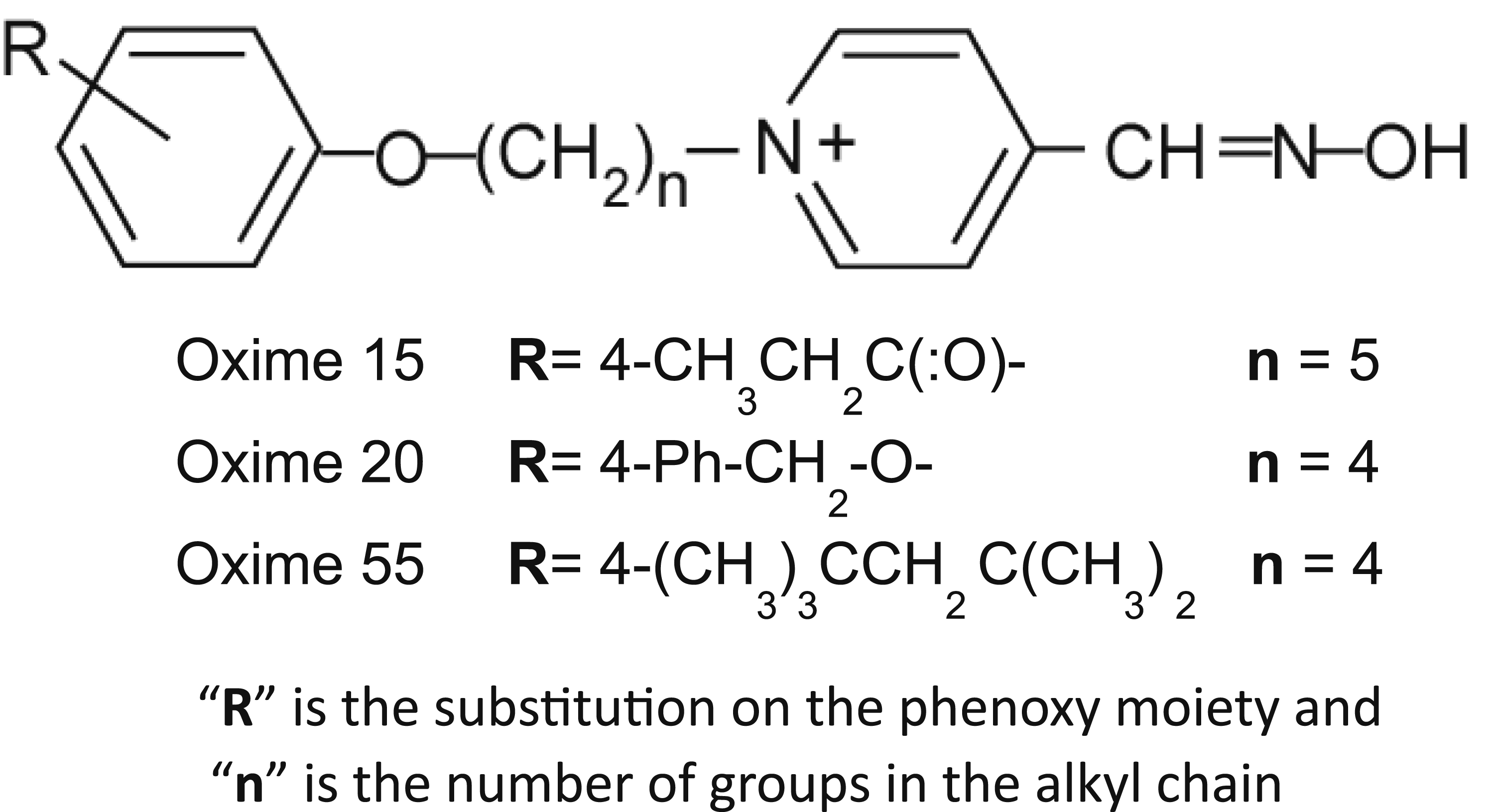
The present study aims to demonstrate acceptable safety of the novel oximes by cell-based cytotoxicity tests using the lactate dehydrogenase (LDH) release and the MTS [3-(4,5-dimethylthiazol-2-yl)-5-(3-carboxymethoxyphenyl)-2-(4-sulfophenyl)-2H-tetrazolium, inner salt] cell proliferation tests. Acceptable safety in this situation means that at least one of the novel oximes will display no greater cytotoxicity than the current FDA-approved oxime, 2-PAM, in either the cell proliferation assay (MTS) or the LDH release assay. After the cytotoxicity testing, Oxime 20 was selected as the lead candidate and tested for genotoxic effects by the in vitro bacterial reverse mutation (Ames) test. Acceptable safety in this situation means that Oxime 20 will not show evidence of genotoxicity in more than 1 bacterial strain when tested at multiple concentrations using standard conditions for bacterial growth.
Materials and Methods
Oxime Reactivators
Novel oximes 15, 20, and 55 (Figure 1), originally described in 2013, 11 were our leading candidates at the start of this study. The mesylate salt forms of the novel oximes were synthesized by the late Dr Howard Chambers following standard procedures from commercially available intermediates at Mississippi State University. 2-PAM was purchased from Sigma-Aldrich (St. Louis, MO, USA).
Cell Culture
The cell line used for cytotoxicity testing was L929 mouse (Mus musculus) areolar fibroblasts purchased from ATCC, Manassas, VA, USA (ATCC® CCL-1 NCTC clone 929 (L cell, L929, derivative of Strain L)). Areolar (ie, loose) connective tissue has fibroblasts, macrophages, collagen fibers, and elastic fibers. The fibroblasts used here secrete the fibers which make a tough, yet flexible, tissue comprising membranes. If approved for human use, the oxime will be delivered via an autoinjector into the thigh muscle and travel through the body prior to reaching the neuronal synapse. Since fibroblasts play a critical role in tissue repair and wound healing, they are relevant for assessing the biological response to an injectable antidote such as our oximes. The L929 cells were grown at 37°C, 5% CO2 in Eagle’s Minimum Essential Medium (EMEM) from ATCC (Manassas, VA, USA) supplemented with 10% heat-inactivated HyClone Donor Equine Serum (U.S.) (Thermo Fisher Scientific, Pittsburgh, PA, USA). Only confluent cells below 11 passages were harvested for experiments. From 4 to 6 individual L929 cultures were grown to confluence to test each oxime (biological replicates) and from 9 to 48 technical replicates (individual wells) were used to test each concentration.
Lactate Dehydrogenase Cytotoxicity Testing
The presence of the cytosolic enzyme lactate dehydrogenase (LDH) in cell media is a well-defined and reliable indicator of cytotoxicity. The CyQUANT LDH Cytotoxicity Assay Kit from Invitrogen (Carlsbad, CA, USA) uses a colorimetric method to quantify cellular cytotoxicity via LDH measurement. The assay was completed following the manufacturer’s instructions. The experimental wells received 10 uls of a novel oxime (oximes 15, 20, or 55) in the biocompatible vehicle Multisol (48.5% water, 40% propylene glycol, 10% ethanol, 1.5% benzyl alcohol). The Spontaneous Lysis Control was 10 uls of Multisol. Absorbance was measured at 490 and 680 nm using a Sunrise microplate reader (Tecan US, Research Triangle Park, NC, USA). To determine LDH activity, the 680-nm absorbance value (background signal from the instrument) was subtracted from the 490-nm absorbance value and the averages were calculated. Data were analyzed for outliers using an interquartile range test.
After outlier removal, the next calculation was as follows:

MTS Cell Proliferation Cytotoxicity Testing
The CellTiter 96 AQueous One Solution Cell Proliferation Assay from Promega (Madison, WI, USA) uses a colorimetric method for determining the number of viable cells in a proliferation cytotoxicity assay. The assay uses MTS [3-(4,5-dimethylthiazol-2-yl)-5-(3-carboxymethoxyphenyl)-2-(4-sulfophenyl)-2H-tetrazolium, inner salt] and PES (phenazine ethosulfate) which is an electron coupling reagent. MTS is reduced by cells to a colored formazan product which is soluble in the tissue culture medium. The quantity of formazan product measured by absorbance at 490 nm is directly proportional to the number of living, metabolically active cells in culture.
The MTS assay utilized the same microplates as the LDH assay with 50 µl of each control or sample placed into 3 wells. The negative control was 1M Na azide, the blank was 0.5% Triton X, and the positive control was complete media. The remaining 87 wells received a novel oxime (oximes 15, 20, or 55) in Multisol. L929 cells were washed twice with EMEM plus 10% heat-inactivated horse serum and resuspended in the same to a concentration of 100 cells per µl. All 96 wells received 50 uls of this mixture prior to incubation at 37°C, with 5% CO2 for 66 h.
After this incubation, the assay was completed as per the manufacturer’s instructions, and the absorbance at 490 nm was immediately read on a SpectraMax M5 Molecular Devices plate reader (San Jose, CA, USA). Outliers were removed using an interquartile range test. The 3 well values (technical replicates) for each control or sample were averaged. The 0.5% Triton X average value was subtracted from the average positive control value and from each averaged value for the tested oxime. The adjusted oxime value was divided by the adjusted positive control value and the result multiplied by 100% to obtain the percent viability. The L929 cells were grown 5 separate times (5 biological replicates) with 3 wells (3 technical replicates) run for each oxime each time at the uM concentrations of 500, 450, 400, 380, 360, 350, 340, 320, 300, 280, 260, 250, 240, 220, 210, 200, 180, 160, 150, 140, 120, 100, 80, 60, 40, 20, 10, 5, and 2.
Genotoxicity Testing (Ames Bacterial Reverse Mutation Testing)
Following completion of the cytotoxicity testing, Oxime 20 was selected to be the lead candidate and moved on to genotoxicity testing by measuring its ability to induce reverse mutations in several bacterial strains using an updated Ames test. 14 Under the OECD 471 Guidelines, 15 at least five strains of bacteria should be used, and we chose S. typhimurium TA97a, TA98, TA100, and TA1535 which require histidine plus E. coli WP2uvrA (pKM101) which requires tryptophan. TA97a and TA98 require histidine due to frameshift mutations which can revert to the wild genotype by exposure to the mutagens 9-aminoacridine (9-AA) and 2-nitrofluorene (2-NF), respectively. TA100 and TA1535 require histidine due to oxidative base-pair substitutions which can revert to the wild genotype by exposure to the mutagen sodium azide (NaN3). E. coli WP2uvrA (pKM101) requires tryptophan due to a base-pair substitution which can revert to the wild genotype by exposure to either 4-nitroquinoline oxide (4-NQO) or methyl methane-sulfonate (MMS). S. typhimurium TA97a, TA98, TA100, and E. coli WP2uvrA (pKM101) all contain the plasmid pKM101 which confers ampicillin resistance and enhances mutagenesis by using a recombinational DNA repair pathway that is highly error prone. 16 If S9 activation is being used an indirect positive control mutagen, 2-aminoanthracene (2-AA) must be included. 2-AA is primarily a frameshift mutagen but can cause base pair mutations when activated by S9.
Environmental Bio-detection Products Inc. (EBPI) (Ontario, CA) supplies a kit which uses a modification of the Ames ISO fluctuation assay to analyze samples in liquid without using agar plates. The MOD-ISO OECD kit with S9 (catalog number: 5041S9-OECD-1S) meets the 1997 OECD 471 guidelines and provides pre-measured reagents for testing with and without S9 metabolic activation, in triplicate and using 5 concentrations (range: 10 µM-10 mM) of Oxime 20 which were shown to be non-cytotoxic in the LDH assay.
All the S. typhimurium bacterial strains, S9, ampicillin, 9-AA, 2-NF, NaN3, and media were supplied by EBPI. E. coli WP2uvrA (pKM101) and 4-NQO were obtained from both EBPI and Molecular Toxicology, Inc. (Boone, NC, USA). MMS was obtained from Molecular Toxicology, Inc.
On the day prior to the assay, 20 μL of ampicillin and a lyophilized bacteria strain (S. typhimurium TA97a, TA98, and TA100 or E. coli WP2uvrA (pKM101)) were added to nutrient broth and placed in a 37°C incubator shaking at 300 rpm overnight (16 h). Since TA1535 does not contain the pKM101 plasmid, no ampicillin was added to the nutrient broth prior to its inoculation.
On the next day, a dilution series (undiluted, 1/5, 1/10, 1/50, 1/100, 1/1,000) of Oxime 20 was made in the Multisol vehicle. The OD at 600 nm of the overnight culture was measured using a Spectronic 1001 Plus spectrophotometer (Milton Roy, Rochester, NY, USA). Based on the OD600 measurement, the overnight bacterial culture was diluted with 1X Exposure Buffer to get a final volume of 6 mL at the working OD600 value given for each strain: TA97a 0.1, TA98 0.05, TA100 0.05, TA1535 0.08, and E. coli WP2uvrA (pKM101) 0.035 using the following calculation: volume of overnight bacteria (in mL) used for dilution = working concentration divided by the overnight OD600 concentration X 6 mL. The assay was performed according to the manufacturer’s instructions.
After incubating at 37°C for 3 days, the plates were removed and placed on white paper towels on the bench and, without lifting the plate lids, the number of wells that had changed from purple to yellow were determined visually by manual counting. Yellow and partial yellow were scored as revertants (positive for reversion) and purple wells were scored as negative since the pH sensitive indicator, bromocresol purple, changes to yellow when the pH is decreased by bacterial growth. The test was considered valid when negative controls had zero or a small numbers of revertants, positive controls had revertants, and the reagent blank/sterility control well is purple (no revertants) and not turbid. A 2-fold increase in the mean number of revertants/plate accompanied by a dose–response to Oxime 20 was considered indicative of mutagenesis/genotoxicity.
Statistics
Outliers were removed from the cytotoxicity data using the Interquartile Range (IQR) method. The remaining data were tested for normal distribution using the Kolmogorov–Smirnov test and homogeneity of variances using the Levene’s test. Since the data failed these tests, the nonparametric Kruskal–Wallis test was used to determine statistically significant differences among the groups at P ≤ .05. When a difference was found, the Mann–Whitney U test was used to determine which of the groups differed. Multiple comparisons were corrected for by using the Benjamini–Hochberg method 17 to rule out false discoveries.
Results
Lactate Dehydrogenase Cytotoxicity Testing
L929 LDH Release Assay Results for the Novel Oximes 15, 20, and 55 and How They Compare to the FDA-Approved AChE Reactivator, 2-PAM.
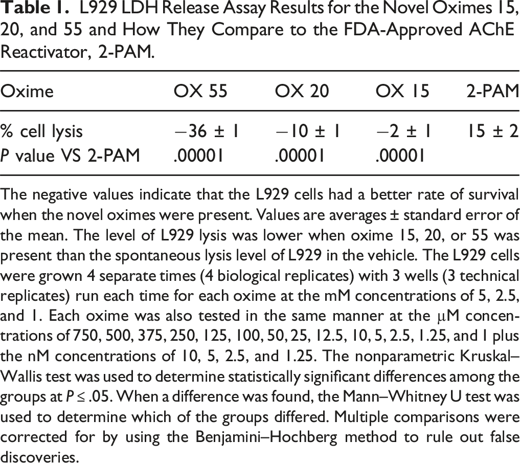
The negative values indicate that the L929 cells had a better rate of survival when the novel oximes were present. Values are averages ± standard error of the mean. The level of L929 lysis was lower when oxime 15, 20, or 55 was present than the spontaneous lysis level of L929 in the vehicle. The L929 cells were grown 4 separate times (4 biological replicates) with 3 wells (3 technical replicates) run each time for each oxime at the mM concentrations of 5, 2.5, and 1. Each oxime was also tested in the same manner at the µM concentrations of 750, 500, 375, 250, 125, 100, 50, 25, 12.5, 10, 5, 2.5, 1.25, and 1 plus the nM concentrations of 10, 5, 2.5, and 1.25. The nonparametric Kruskal–Wallis test was used to determine statistically significant differences among the groups at P ≤ .05. When a difference was found, the Mann–Whitney U test was used to determine which of the groups differed. Multiple comparisons were corrected for by using the Benjamini–Hochberg method to rule out false discoveries.
MTS Cell Proliferation Cytotoxicity Testing
In the MTS test, all 3 novel oximes significantly reduced the metabolic activity of the L929 cells compared to 2-PAM (Figure 2). Oximes 15, 20, and 55 significantly (P = .00001) reduced the metabolic activity of the cells compared to 2-PAM according to the results of the MTS cell proliferation cytotoxicity assay. The negative control was 1M Na azide, the blank was 0.5% Triton X, and the positive control was complete media. The L929 cells were grown 5 separate times (5 biological replicates) with 3 wells (3 technical replicates) run each time for each oxime at the uM concentrations of 500, 450, 400, 380, 360, 350, 340, 320, 300, 280, 260, 250, 240, 220, 210, 200, 180, 160, 150, 140, 120, 100, 80, 60, 40, 20, 10, 5, and 2. Error bars indicate standard error of the mean. The nonparametric Kruskal–Wallis test was used to determine statistically significant differences among the groups of oximes (15, 20, 55, and 2-PAM) at P ≤ .05. When a difference was found, the Mann–Whitney U test was used to determine which of the groups differed. Multiple comparisons were corrected for by using the Benjamini–Hochberg method to rule out false discoveries.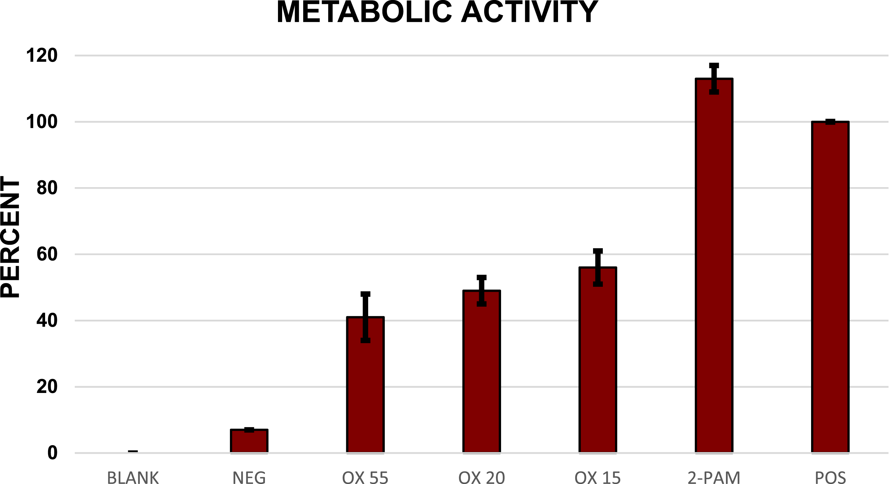
Genotoxicity Testing (Ames Bacterial Reverse Mutation Testing)
Ames Results Showing That Oxime 20 Caused No Reversion in Any of the Test Strains at Any Concentration Whether With or Without S9 Metabolic Activation.
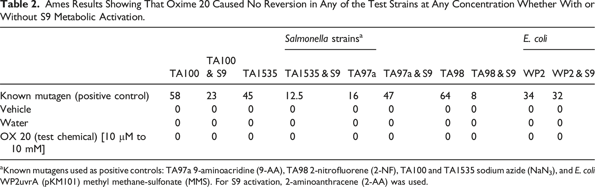
aKnown mutagens used as positive controls: TA97a 9-aminoacridine (9-AA), TA98 2-nitrofluorene (2-NF), TA100 and TA1535 sodium azide (NaN3), and E. coli WP2uvrA (pKM101) methyl methane-sulfonate (MMS). For S9 activation, 2-aminoanthracene (2-AA) was used.
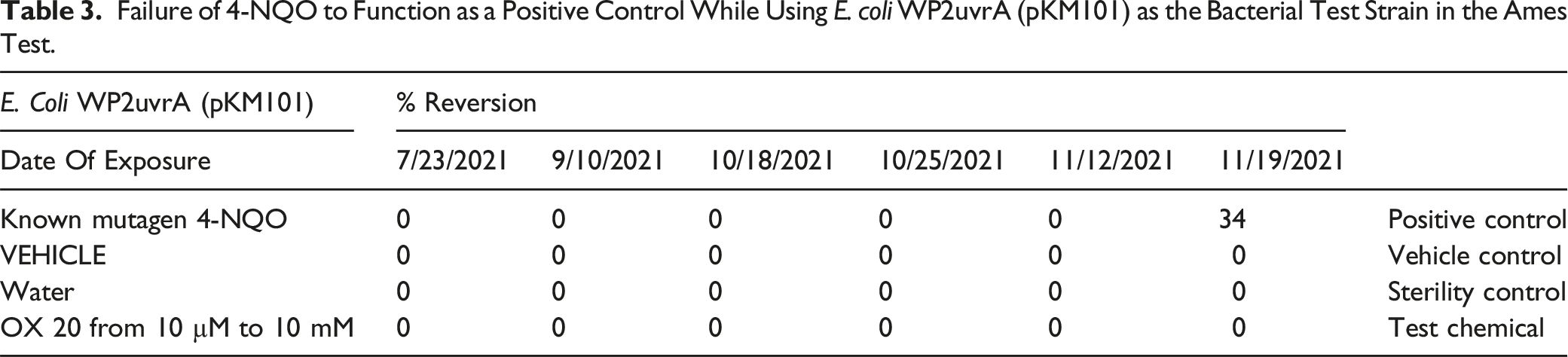
Failure of 4-NQO to Function as a Positive Control While Using E. coli WP2uvrA (pKM101) as the Bacterial Test Strain in the Ames Test.
Discussion
Our lab’s novel oxime acetylcholinesterase reactivators were designed to cross the BBB more efficiently and better prevent OP-induced brain damage than the present FDA-approved oxime acetylcholinesterase reactivator, 2-PAM. The novel oximes are intended to be injectable antidotes for emergency treatment following exposure to nerve agents or other OP compounds such as some insecticides.
Since these novel oximes were invented in our laboratory, no other cytotoxicity or genotoxicity data existed prior to the present work. In light of this, concentration ranges used were based on prior pharmacokinetic analysis done in our laboratory which showed that a 115 µmole/kg intramuscular injection of Oxime 20 in a rat resulted in a maximum concentration of 188 ng/g in its brain. 18 The concentration ranges used were designed to both bracket the 115 µM value and greatly exceed the highest possible dose.
The known mutagen 4-NQO caused no reversion in the E. coli WP2uvrA (pKM101) section of the Ames test. This failure of the EBPI-supplied 4-NQO positive control required the test to be repeated with MMS, which gave the expected results. MMS is a synthetic alkylating agent which transfers a methyl group to DNA upon contact resulting in strand breakage. 19 This triggers the DNA repair process which is highly error prone in E. coli WP2uvrA (pKM101). 16 The mutagenicity/genotoxicity of 4-NQO, however, occurs through the formation of DNA adducts and the generation of reactive oxygen species. 20
Since 10 µg of 4-NQO produced more mutations in Salmonella typhimurium than 570 µg of MMS,14,16 it would appear to be the stronger mutagen. This was not evident in the present work with E. coli WP2uvrA (pKM101). Brüsehafer et al. reported similar data in 2016. 21 They saw little to no statistical mutagenicity in 4-NQO-exposed human lymphoblastoid cells but did see dose–response mutagenicity in mouse lymphoma cells. They suggested that 4NQO mainly induces gene point mutations instead of DNA strand breaks. Possibly, the strand breaks caused by MMS are less able to be repaired by E. coli WP2uvrA (pKM101) than 4NQO point mutations. Brüsehafer et al. suggested that use of 4-NQO as a positive genotoxicity control has to be evaluated for each experiment. 21
Oxime 20 gave no evidence of genotoxicity in any of the Ames bacterial reverse mutation tests. Since TA1535 is uniquely sensitive to mutagenic oximes, 22 its failure to respond is supportive evidence for Oxime 20’s lack of genotoxicity.
Lactate dehydrogenase testing using cultured fibroblasts gave no evidence of cytotoxicity for any of the novel oximes. The data suggested that likelihood of cell lysis was reduced compared to 2-PAM when the novel oximes were present. This may be explained by previous research showing that in the rat hippocampus, in vivo OP exposure decreased the mRNA expression of the antiapoptotic gene Bcl2l1. 23 When administered after OP exposure, Oxime 20 returned Bcl2l1 expression to the control level. 23 Lowered levels of Bcl2l1 expression should increase apoptosis because the BCL2L1 protein removes the apoptosis regulator protein, BAX, from the mitochondrial surface to the cytosol, thereby preventing it from permeabilizing the mitochondrial membrane and initiating apoptosis. 24 In vivo exposure to just Oxime 20 resulted in large, significant decreases of Bcl-2 binding component 3 (Bbc3) gene expression in the rat piriform cortex (unpublished data). Since the protein product of Bbc3 is pro-apoptotic, the decreased expression is likely to reduce the apoptosis, supporting the concept that the novel oximes reduce cellular lysis.
In spite of the LDH data suggesting decreased cytotoxicity in the presence of the novel oximes, the CellTiter 96 AQueous One Solution cell proliferation (MTS) assay indicated that the novel oximes significantly reduced the number of living, metabolically active cells in culture compared to 2-PAM.
Berridge et al 25 suggested that MTS assays do not actually measure the number of viable cells or their growth, but rather the plasma membrane redox system which involves several different electron transport pathways, each capable of reducing the dye. Since the MTS assay is measuring cellular metabolism as a proxy for cell viability and proliferation, when a test chemical alters cellular metabolism this could yield inaccurate results.
In spite of some difficulties with these initial safety tests, the results of the present studies demonstrate that Oxime 20 is not genotoxic, mutagenic, or cytotoxic at clinical dose levels. Overall, the data are highly supportive of the further development of Oxime 20 as a countermeasure against OP anticholinesterases.
Footnotes
Ethical Considerations
This article does not contain any studies with human or animal participants. The Mississippi State University Institutional Biosafety Committee granted approval to “AMES Testing of Oximes 15 and 20” (IBC # 020-21).
Author Contributions
Chambers, J. E. contributed to conception, critically revised manuscript, gave final approval, and agrees to be accountable for all aspects of work ensuring integrity and accuracy.
Funding
The author disclosed receipt of the following financial support for the research, authorship, and/or publication of this article: This work was supported by National Institutes of Health CounterACT grants U01 NS107127 and U01 NS123255.
Declaration of Conflicting Interests
The author declared the following potential conflicts of interest with respect to the research, authorship, and/or publication of this article: The novel oximes are under patent protection to Mississippi State University (US: 9,227,937; Germany: 60 2011 049 991.2; France: 11780924.4; UK: 11780924.4 UK; Italy: 11780924.4 IT) and licensed by Defender Pharmaceuticals, Inc., which had no input into the design of the experiments or the interpretation of the results.
Data Availability Statement
The data that support the findings of this study are available from the corresponding author upon reasonable request.