






Abstract
The Expert Panel for Cosmetic Ingredient Safety (Panel) assessed the safety of 8 Melaleuca alternifolia (tea tree)-derived ingredients as used in cosmetic formulations; 5 of these ingredients are reported to function in cosmetics as skin-conditioning agents. Because final product formulations may contain multiple botanicals, each containing the same constituents of concern, formulators are advised to be aware of these constituents and to avoid reaching levels that may be hazardous to consumers. Industry should use good manufacturing practices to minimize impurities that could be present in botanical ingredients. The Panel noted that oxidized tea tree oil could be a sensitizer, and stated that industry should employ methods to minimize oxidation of the oil in the final cosmetic product. The Panel considered all the data and concluded that these ingredients are safe in cosmetics in the present practices of use and concentration described in this safety assessment when formulated to be non-sensitizing.
Keywords
Introduction
This assessment reviews the safety of the following 8 Melaleuca alternifolia (tea tree)-derived ingredients as used in cosmetic formulations:
Melaleuca Alternifolia (Tea Tree) Extract
Melaleuca Alternifolia (Tea Tree) Flower/Leaf/Stem Extract
Melaleuca Alternifolia (Tea Tree) Flower/Leaf/Stem Oil
Melaleuca Alternifolia (Tea Tree) Leaf
Melaleuca Alternifolia (Tea Tree) Leaf Extract
Melaleuca Alternifolia (Tea Tree) Leaf Oil
Melaleuca Alternifolia (Tea Tree) Leaf Powder
Melaleuca Alternifolia (Tea Tree) Leaf Water
Definitions and Reported Cosmetic Functions 1
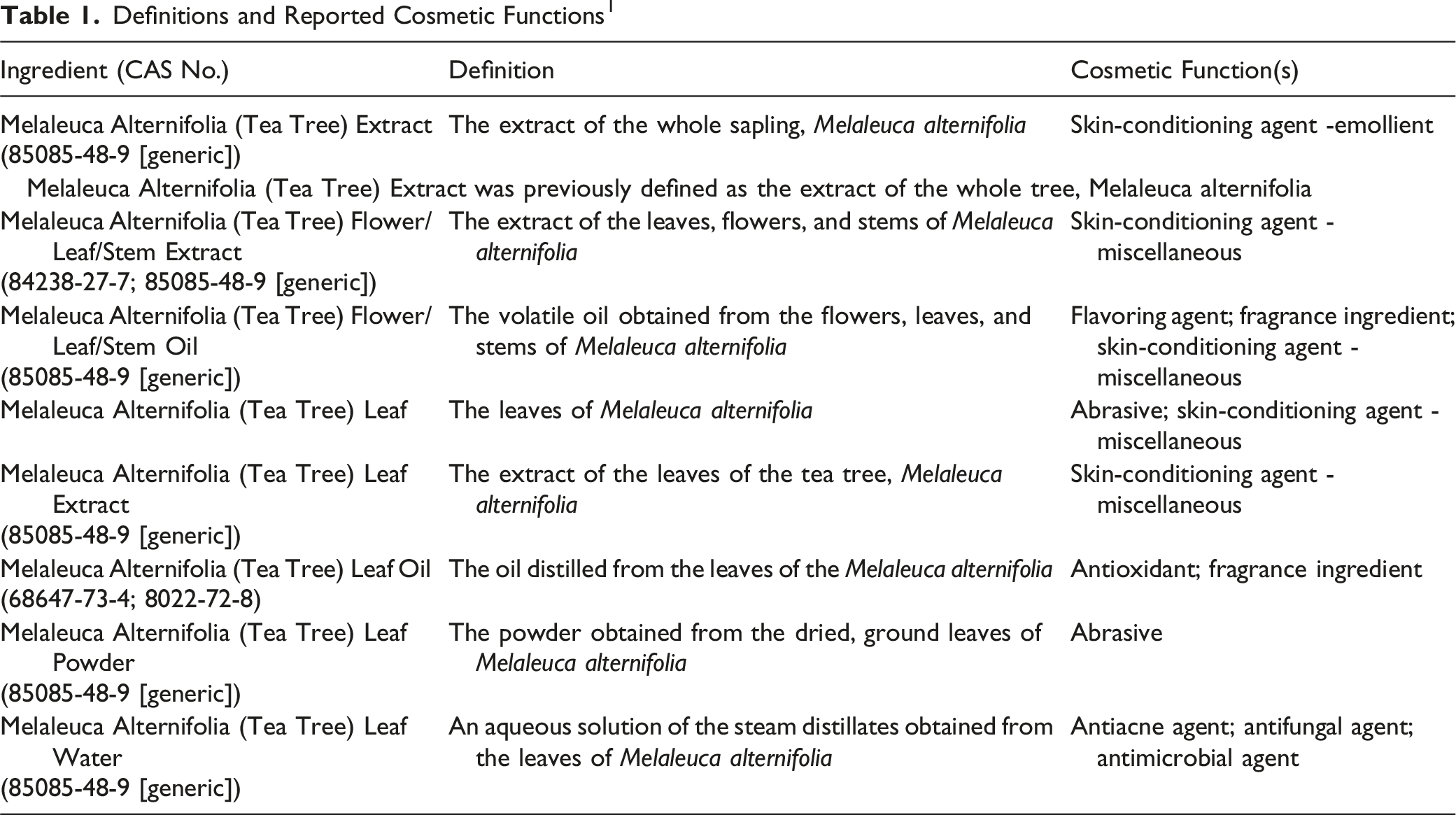
Melaleuca alternifolia contains over 100 constituents, some of which have the potential to cause adverse effects. For example, 1,8-cineole (also known as eucalyptol 2 ) can be an allergen, 3 and terpinolene, α-terpinene, α-phellandrene, limonene, ascaridole (a product of tea tree oil oxidation), and 1,2,4-trihydroxymenthane (a product that might be found in aged tea tree oil) are sensitizers.4,5 In this assessment, the Panel is evaluating the potential toxicity of each of the Melaleuca alternifolia (tea tree)-derived ingredients as a whole, complex substance; toxicity from single components may not predict the potential toxicity of botanical ingredients.
This safety assessment includes relevant published and unpublished data that are available for each endpoint that is evaluated. Published data are identified by conducting an exhaustive search of the world’s literature. A listing of the search engines and websites that are used and the sources that are typically explored, as well as the endpoints that the Panel typically evaluates, is provided on the Cosmetic Ingredient Review (CIR) website (https://www.cir-safety.org/supplementaldoc/preliminary-search-engines-and-websites; https://www.cir-safety.org/supplementaldoc/cir-report-format-outline). Unpublished data are provided by the cosmetics industry, as well as by other interested parties.
Some of the data included in this safety assessment were obtained from reviews (such as those issued by the European Commission (EC) Scientific Committee on Consumer Products (SCCP), 6 European Chemicals Agency (ECHA), 7 and European Medicines Agency (EMA)3,8,9). These data summaries are available on the respective websites, and when deemed appropriate, information from the summaries has been included in this report.
The cosmetic ingredient names, according to the Dictionary, are written as listed above, without italics and without abbreviations. When referring to the plant from which these ingredients are derived, the standard scientific practice of using italics will be followed (i.e., Melaleuca alternifolia). Often in the published literature, the general name “tea tree” is used, especially, tea tree oil. If it is not known whether the substance being discussed is equivalent to the cosmetic ingredient, the test substance will be identified by the name used in the publication that is being cited; it is possible that the oil may be obtained from more than one species of Melaleuca, or from parts other than the leaves. However, if it is known that the substance is a cosmetic ingredient, the Dictionary nomenclature (e.g., Melaleuca Alternifolia (Tea Tree) Leaf Oil) will be used.
Chemistry
Definition and Plant Identification
According to the Dictionary, the most recent definition of Melaleuca Alternifolia (Tea Tree) Extract is the extract of the whole sapling, Melaleuca alternifolia; in the past, this ingredient was defined as the extract of the whole tree (Table 1). 1 Each of the other Melaleuca alternifolia (tea tree)-derived ingredients is named based on the plant part(s) from which they are obtained. Several of these ingredients have the generic CAS No. 85085-48-9; however, Melaleuca Alternifolia (Tea Tree) Leaf Oil has CAS Nos. (68647-73-4; 8022-72-8) that are specific to that ingredient.
Correspondence received from a representative of the Australian Tea Tree Industry Association (ATTIA) stated they are of the opinion that several of the Melaleuca alternifolia-derived ingredients (i.e., the Extract, Flower/Leaf/Stem Extract, Flower/Leaf/Stem Oil, and Leaf Oil) are essentially identical because the definitions for these ingredients describe, in various ways, the essential oil that is steam distilled from the plant (personal communication; T. Larkman, Feb 17, 2021). Additionally, the representative of ATTIA stated that the Melaleuca Alternifolia (Tea Tree) Leaf and Melaleuca Alternifolia (Tea Tree) Leaf Powder both describe the dried leaf.
The Melaleuca genus belongs to the Myrtaceae family, within the Myrtales order. 10 Melaleuca alternifolia occurs in riparian zones of freshwater and swamps. It is a commercially-grown plant that is indigenous to Australia, 11 and plants with the genetic make-up necessary to produce the oil are native to northern New South Wales. 12 However, Melaleuca alternifolia has been introduced and cultivated in China, Indonesia, Kenya, Madagascar, Malaysia, South Africa, Tanzania, Thailand, the US, and Zimbabwe.13,14
Melaleuca alternifolia is a tall shrub or small tree that typically grows up to 7 m high, with a bushy crown and papery bark. 15 The total biomass (above-ground growth) of the tea tree can be subdivided into 3 components: leaves, fine stems, and main stems. 16 The fine stems are defined as stems of less than 2.5 mm in diameter, and they carry virtually all the leaves; the leaves and fine stems, together, are referred to as twigs. The main stems make up the remainder. The hairless leaves are scattered to whorled, and are 10–35 mm long by about 1 mm wide. 15 The leaves, which have prominent oil glands and are rich in aromatic oil, are borne on a petiole (leaf stalk) that is approximately 1 mm long. Tea tree oil is only found in the leaves; it is stored in the subepidermal glands that are adjacent to the epidermis, and the glands are equally distributed on both sides of the leaf. 16 The oil glands first appear in immature leaves, and the number per leaf increases as the leaf expands, reaching a maximum just prior to the leaf fully expanding.
The inflorescences are many-flowered spikes, 3-5 cm long, with axes bearing short hairs. 15 The white flowers are solitary, each within a bract, and have petals 2–3 mm long. There are 30–60 stamens per bundle and the style is 3–4 mm long. The fruit is cup-shaped and 2–3 mm in diameter, with a hole 1.5–2.5 mm in diameter that enables release and dispersal of the seeds by wind. Fruits are usually sparsely spaced along the branches.
Chemical Properties
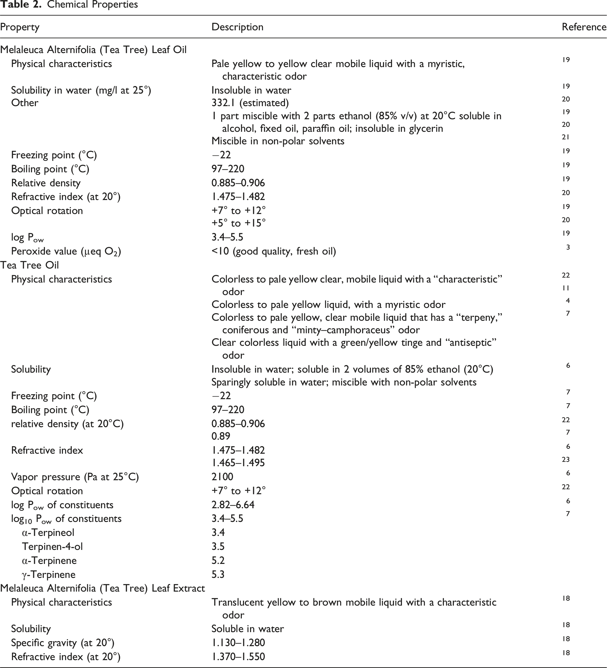
Chemical Properties
Stability
Tea Tree Oil
Because of the possibility for degradation, a supplier of tea tree oil recommends that the use-by date for tea tree oil sold in commercially-available, small (up to 100 ml), dark, glass bottles stored at ambient temperature be set at 12 months from when first opened, or 24 months in unopened bottles. 24 They also recommend that, wherever possible, tea tree oil should be stored at or below 25°C. The supplier also stated that when stored correctly, tea tree oil can retain its quality for periods of up to 10 years.
In a 3-month trial examining stability in accelerated (40°C) and real-time shelf conditions, including exposure to fluorescent light, no discernible difference was demonstrated in the tea tree oil quality based on constituent values in either amber or clear glass bottles. 24 In a 12-month study designed to replicate normal consumer use conditions, there was no appreciable oxidation or degradation of tea tree oil.12,25 No significant change in the level of terpinen-4-ol was reported. A downward trend in α-terpinene and γ-terpinene, and an upward trend in p-cymene, were observed, and peroxide levels increased. The amber glass bottles of tea tree oil were regularly opened, exposed to air and light for short periods of time, and a small amount of oil was removed; when not in use, the bottles were stored away from heat and light.
A supplier also provided some data on the stability of tea tree oil in formulated products, using solvent extraction and gas chromatography/flame ionization detection (GC/FID). 26 The rates of degradation of the oil varied with the medium. Degradation in a cream was faster than seen in a gel or a solution. For the tea tree cream, solution, and gel, the constituents were extremely stable over a period of 1.5, 3, and 5 years, respectively.
Method of Manufacture
The majority of the methods below are general to the processing of Melaleuca alternifolia (tea tree)-derived ingredients, and it is unknown if they apply to cosmetic ingredient manufacturing. In some cases, the definition of the ingredients, as given in the Dictionary, provides insight as to the method of manufacture. 1
Melaleuca Alternifolia (Tea Tree) Leaf Extract
A supplier submitted information describing production of a concentrate; details were not provided regarding raw material or solvents, however, the data were provided for Melaleuca Alternifolia (Tea Tree) Leaf Extract. 27 The supplier indicated that raw material is packed into the extraction system and sealed, liquid extractant is added to the vessel, which is then closed and sealed, and the raw material is extracted under pressure in the closed system. The resulting extract is reported to be a pure extract of the raw material used (e.g., plant, bark, fruit).
Melaleuca Alternifolia (Tea Tree) Leaf Water
Melaleuca Alternifolia (Tea Tree) Leaf Water is an aqueous solution of the steam distillates obtained from the leaves of Melaleuca alternifolia. 1
Tea Tree Oil
Tea tree oil is defined by International Organization for Standardization (ISO) standard 4730:2017 as the essential oil obtained by steam of the leaves and terminal branchlets of Melaleuca alternifolia (Maiden et Betche) Cheel or of Melaleuca linariifolia Sm. 22 ; steam distillation is required to conform to ISO standards. 28 Tea tree oil also can be prepared by hydrodistillation in a laboratory, usually with a Clevenger-type apparatus. 4
More than 80% of the world’s tea tree oil is produced in Australia. 12 Minor quantities come from China, South Africa and Vietnam. Tea tree oil produced in, and exported from, Australia conforms to the ISO standard (personal communication; T. Larkman, Aug 31, 2020).
According to a supplier of Australian tea tree oil, Melaleuca alternifolia tea trees are harvested and mulched into biomass, from which the oil is extracted using low-temperature pressurized steam distillation. 29 Oil from glands in the leaves is vaporized with the steam, and the steam is then condensed with cold water. The oil is separated out, and cooled for 16 h. Following cooling, the oil is filtered to remove any organic debris, sampled for quality assurance, and then bottled.
A researcher extracted tea tree oil from the leaf, twig (<0.3 cm in diameter), and branch (0.3–0.7 cm in diameter) of Melaleuca alternifolia using a Clevenger-type apparatus. 30 After 7 h, the yield of tea tree oil was 2.02% from the leaves, 0.59% from twigs, and 0.01% from branches.
Another possible method for obtaining tea tree oil is solvent extraction. 28 It was reported that solvent extraction methods, including ethanol extraction, have been found to avoid the loss of certain terpenes that occurs during steam distillation, use less leaf material, and are quicker than steam distillation. Total leaf oil content can range from 0.5% to 3%, but yield via “traditional design water distillation” is 1%. 31 A study compared recovery from tea tree leaves by ethanol extraction (3 days) and steam distillation (2–6 h) using both dry and fresh leaves from a low- and a high-oil concentration trees. 32 Ethanol extraction gave 48 and 77 mg of oil/g of leaf for the low- and high-oil concentration trees, respectively; with steam distillation, 42 and 63 mg of oil/g of leaf were obtained after 2 h, and 42 and 66 mg of oil/g of leaf were obtained after 6 h for the same low- and high-oil concentration trees, respectively. Absolute amounts of monoterpenoids and sesquiterpenoids extracted with ethanol were higher than those recovered from the 2-h, and most of the 6-h, steam distillations. As a percent of total oil, the oil obtained by steam distillation for 2 h had a higher percentage of total monoterpenoids.
Oil yield is considered to be more affected by environmental conditions than oil composition, and has been shown to fluctuate diurnally, seasonally and in response to environmental conditions, particularly moisture levels. 28 However, in the study described above, no significant difference in the quantity or quality of oil extracted from fresh (approximately 50% dry matter) and air-dried leaves (approximately 90% dry matter) sampled from either low- or high-oil concentration trees was found. 32
Composition/Impurities
Melaleuca Alternifolia (Tea Tree) Leaf Extract
According to one supplier, Melaleuca Alternifolia (Tea Tree) Leaf Extract is a cellular extraction of the Melaleuca alternifolia leaf that comprises 20%–50% Melaleuca alternifolia leaf, 34–55% glycerin, and 14–24% water, and is preserved with ≤0.5% sodium benzoate, ≤0.4% citric acid, and ≤0.3% potassium sorbate. 18 SCCNFP allergens listed in Annex III of the European Union (EU) Cosmetics Regulation (2003/15/EC) were not detected in the extract (limit of detection, 0.001%). Additionally, according to certificates of analysis provided by another source, specifications for Melaleuca Alternifolia (Tea Tree) Leaf Extract (at ≥ 0.001% leave-on and ≥0.01% w/w rinse-off) indicate that none of the 26 potential fragrance allergens, which according to the EU Cosmetics Regulation are required to be listed on the label, were detected (limit of detection of 0.001%). 33 High-performance liquid chromatography (HPLC) - mass spectrometry (MS) of a test sample of Melaleuca Alternifolia (Tea Tree) Leaf Extract identified a range of phenolic and flavonoid derivatives, based on available ultraviolet (UV)-visible (Vis) and MS spectra. 34
Information was also provided for a cellular extraction comprising <98% Vitis vinifera (grape) seed oil, <1.0%–5.0% Melaleuca Alternifolia (Tea Tree) Leaf Extract, and <0.5% mixed tocopherols (low α-type). 35 According to this submission, as well as certificates of analysis provided by another source, 36 specifications for the mixture (at ≥ 0.001% leave-on and ≥0.01% w/w rinse-off) indicate that none of the 26 potential fragrance allergens requiring labeling (according to the EU Cosmetics Regulation) were detected (limit of detection of 0.001%). Fatty acid analysis via GC/FID indicated fatty acid content of the mixture ranged from 0.003% magaric acid to 68.11% linoleic acid. 37
Melaleuca Alternifolia (Tea Tree) Leaf Oil
Methyleugenol is reported as a minor constituent of Melaleuca Alternifolia (Tea Tree) Leaf Oil. 6 Analysis of 128 samples, using GC/MS methods with selected ion monitoring, reported that levels of methyleugenol ranged from 0.01% to 0.06% (mean, 0.02%) for commercial distillations. 38 Longer distillation times can result in slightly higher amounts; however, amounts did not exceed 0.07% for exhaustive laboratory distillations. According to the European Commission, based on the Scientific Committee on Cosmetic Products and Non-Food Products (SCCNFP) opinion on methyleugenol in fragrances, the highest concentration in the finished products must not exceed 0.01% in fine fragrance, 0.004% in eau de toilette, 0.002% in a fragrance cream, 0.0002% in other leave-on products and in oral hygiene products, and 0.001% in rinse-off products. 39 In Norway, purity requirements for tea tree oil state that levels of methyleugenol should not exceed 200 ppm (0.02%) as a minor constituent of tea tree oil, and the content should be indicated in the ingredient list. 33
Tea Tree Oil
Composition of the 6 Melaleuca alternifolia Chemotypes Measured by Headspace GC 28
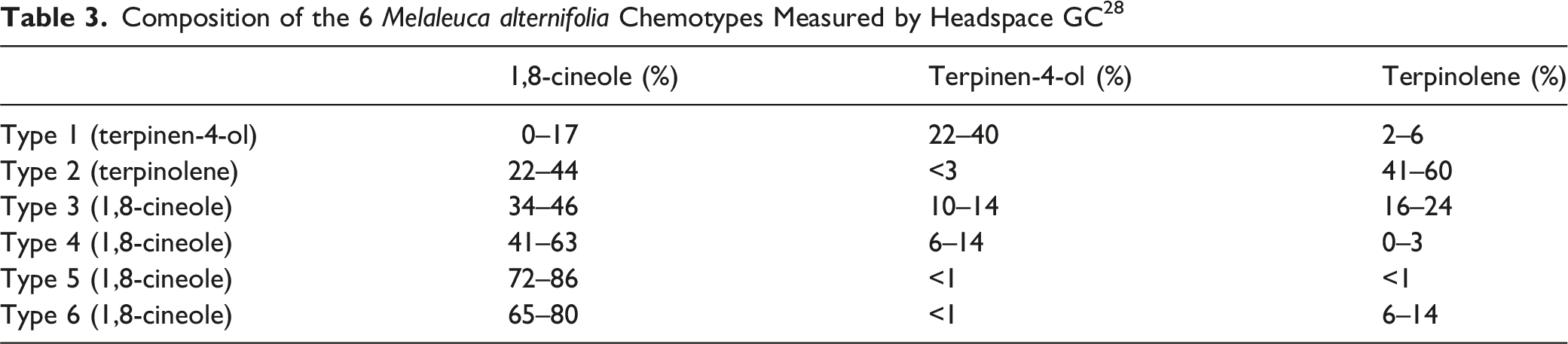
Tea tree oil typically contains approximately 100 constituents 41 ; however, one publication reported that over 220 constituents have been identified in tea tree oil samples, and the concentration of these constituents present in the oil can vary widely depending on the sample. 4 Eight constituents (i.e., terpinen-4-ol, α-terpinene, γ-terpinene, 1,8-cineole, terpinolene, p-cymene, α-pinene, and α-terpineol) typically comprise up to 90% of the oil, 41 and the 3 constituents reported to be present in the greatest amounts are terpinen-4-ol (up to 48%), γ-terpinene, (up to 28%), and 1,8-cineole (up to 15%). 22 Another notable constituent is limonene (up to 4%). The main constituents of tea tree oil have molecular weights ranging from 134 g/mol (p-cymene) to 222 g/mol (globulol and viridiflorol).6,42,43 The log P of the main constituents ranges from 2.73 (α-terpineol) to 6.64 (δ-cadinene).
Tea tree oil is reported to be composed mainly of monoterpene and sesquiterpene hydrocarbons and their associated alcohols. 21 For one sample, GC/MS analysis determined that oxygenated monoterpenes constituted 51% of the oil, monoterpene hydrocarbons constituted 47%, and the remaining 2% of the oil was composed of sesquiterpene hydrocarbons. 44 Another study reported that GC/MS analysis of ethanolic extracts of mature leaf material of Melaleuca alternifolia revealed the presence of 47 compounds, comprising 20 monoterpenes and 27 sesquiterpenes. 45
Standards and Specifications for Tea Tree Oil
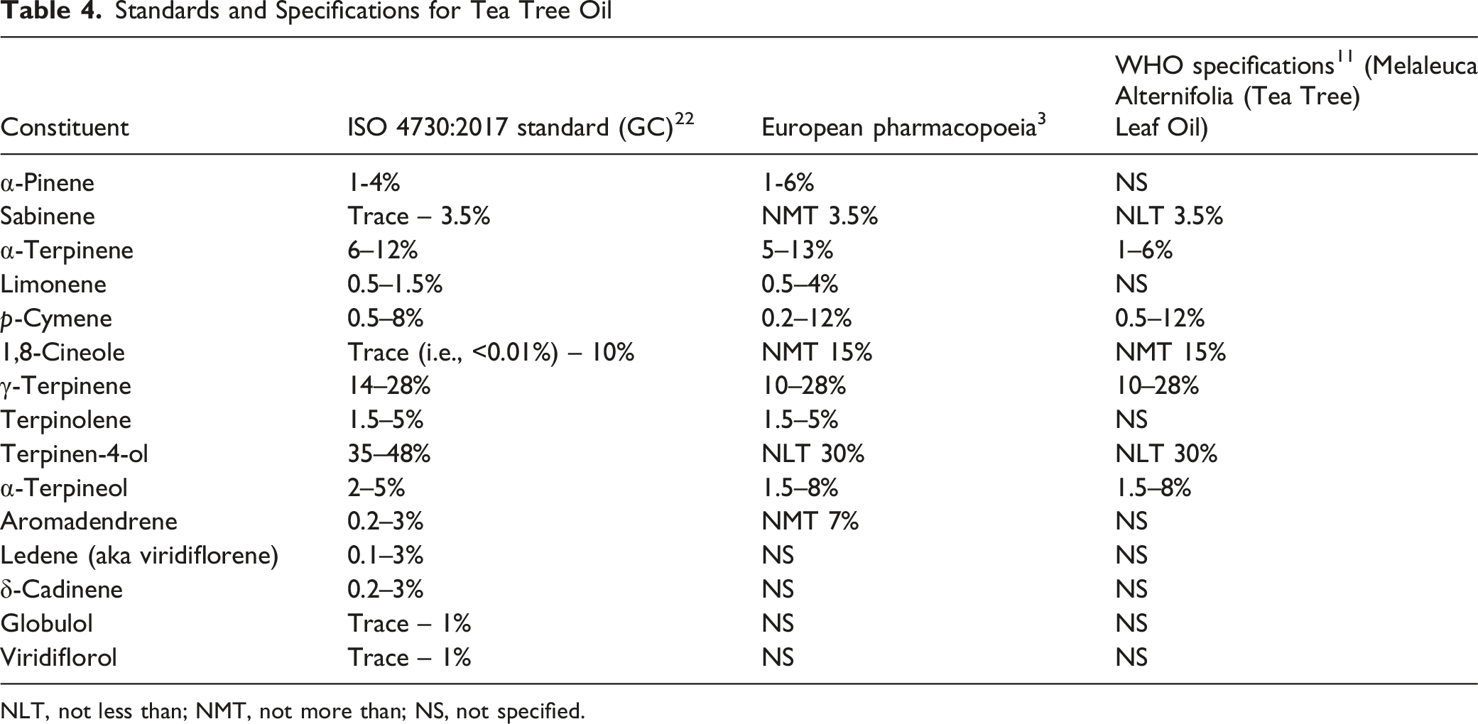
NLT, not less than; NMT, not more than; NS, not specified.
Constituent Profiles of Tea Tree Oil
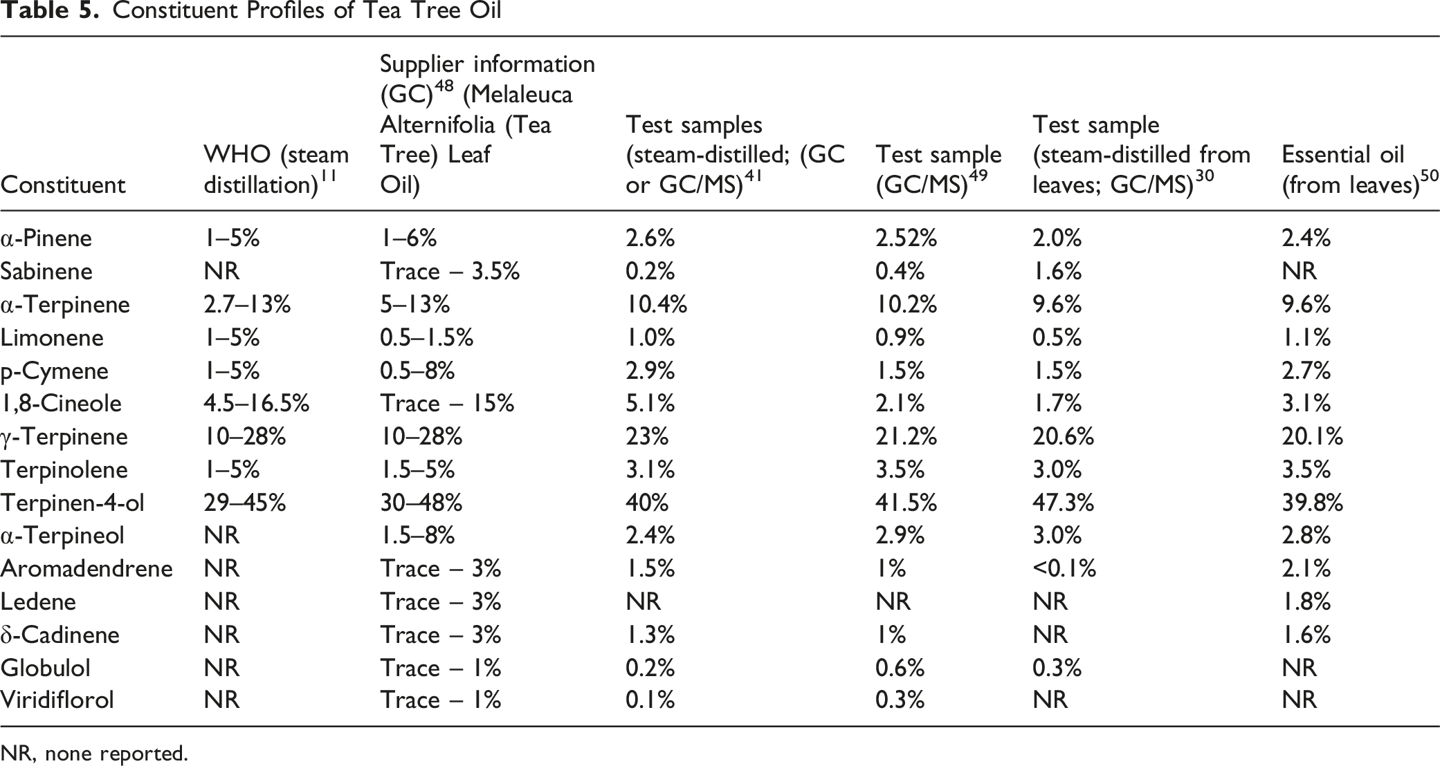
NR, none reported.
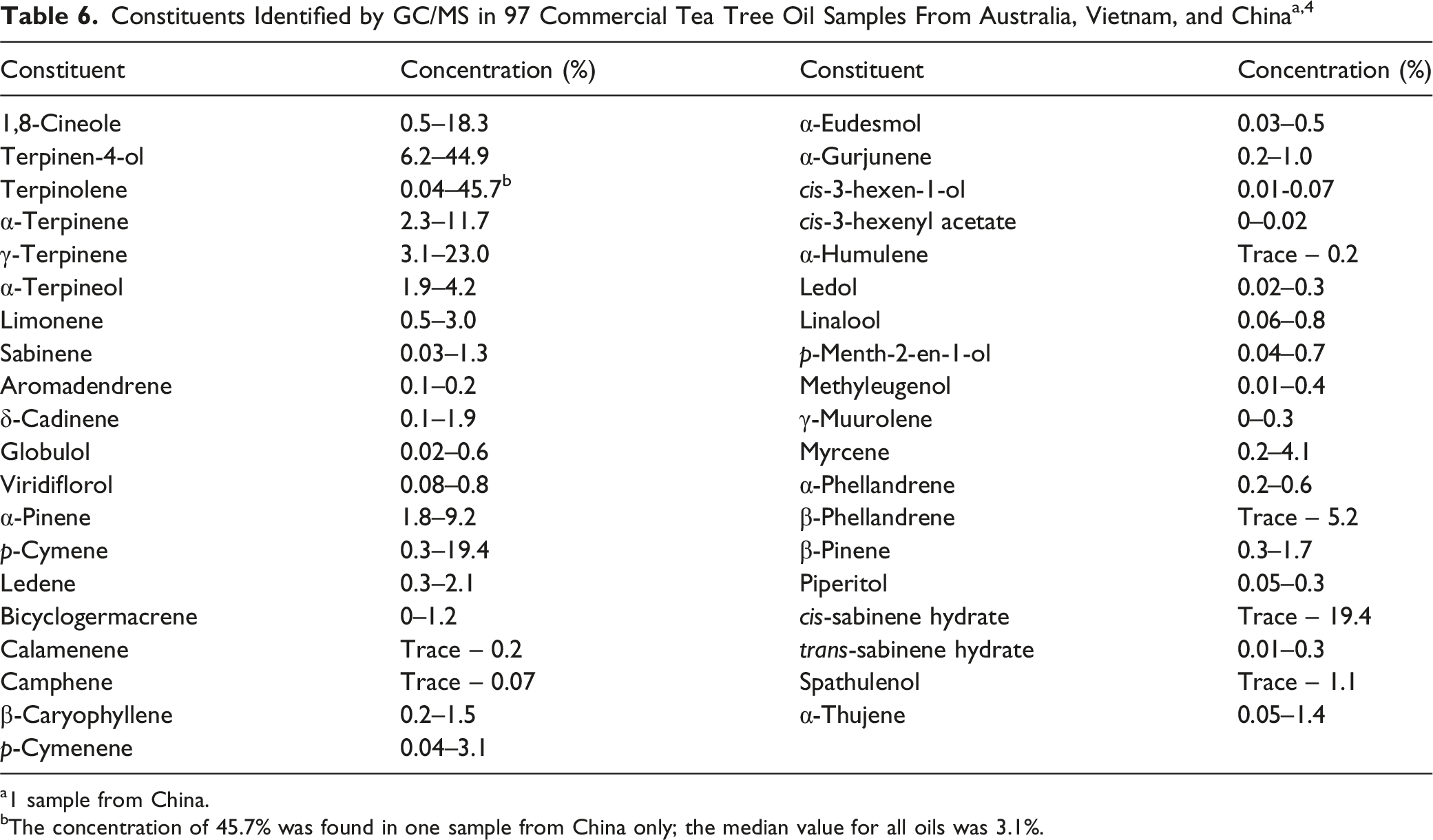
a1 sample from China.
bThe concentration of 45.7% was found in one sample from China only; the median value for all oils was 3.1%.
Composition of Tea Tree Oil at Different Collection Times During Distillation 41
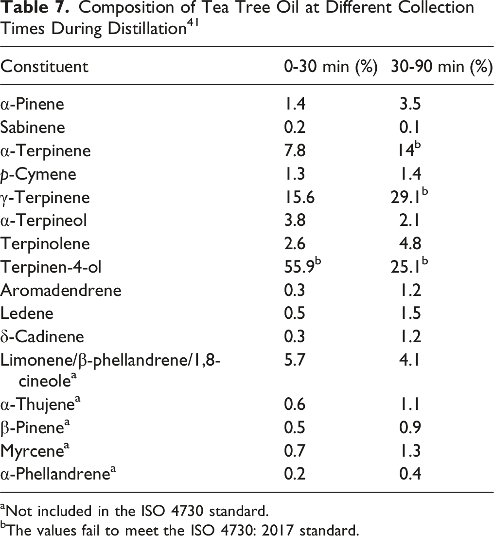
aNot included in the ISO 4730 standard.
bThe values fail to meet the ISO 4730: 2017 standard.
Monoterpenoid Composition Comparison of Aged Oils of Melaleuca alternifolia 41
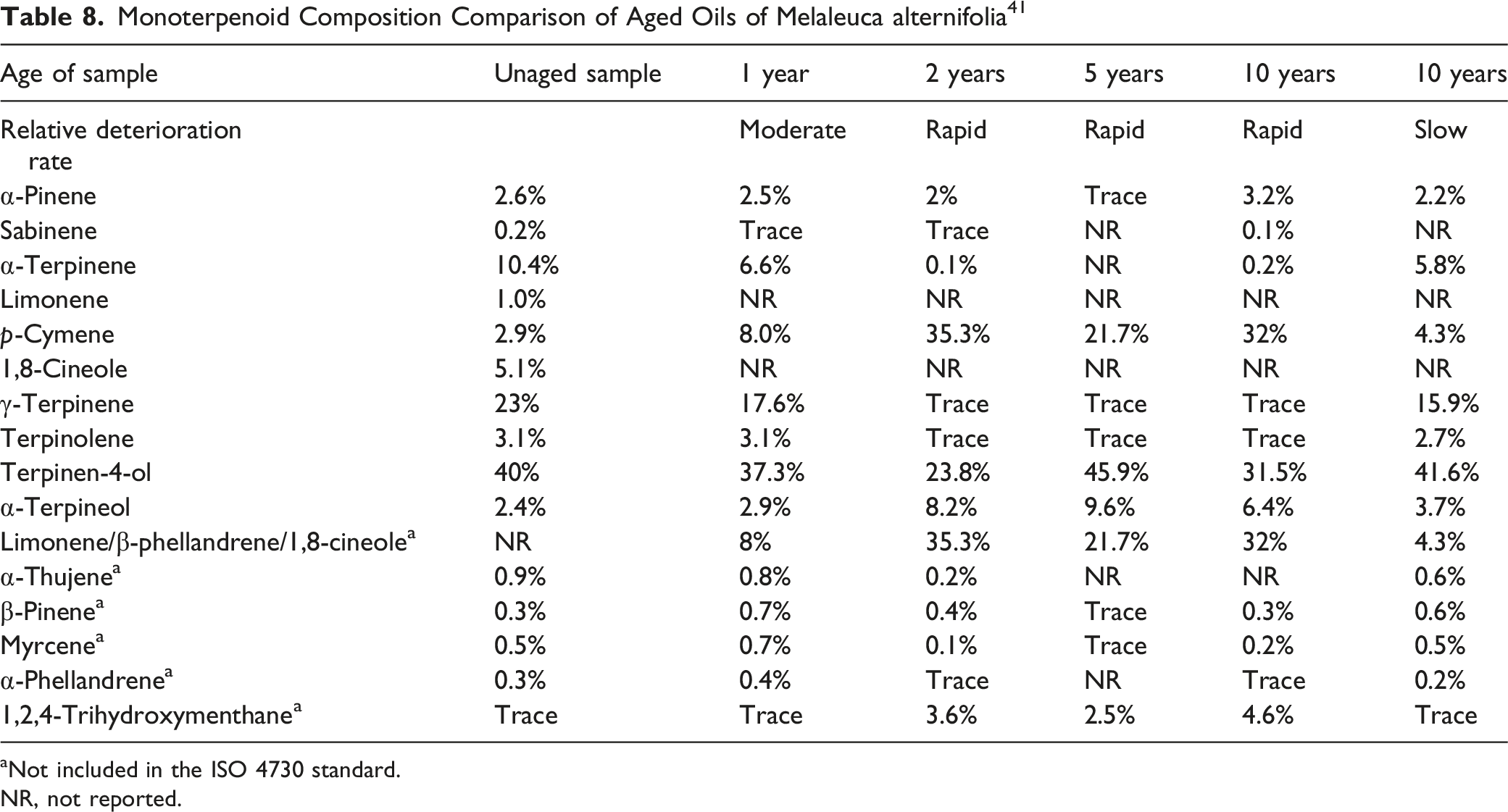
aNot included in the ISO 4730 standard.
NR, not reported.
Composition of Tea Tree Oil at Various Stages of Oxidation 53
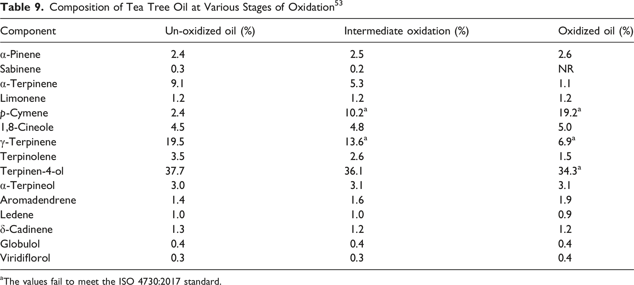
aThe values fail to meet the ISO 4730:2017 standard.
Oxidation processes also lead to the formation of peroxides, endoperoxides, and epoxides.6,41 As tea tree oil undergoes oxidation, peroxide values increase from zero to “unacceptable” levels in the early stages of oxidative degradation. 26 Once the rate of degradation of the peroxides exceeds the rate of their formation, the peroxide values return to zero in highly degraded aged oil. In a study using GC/MS, it was reported that unoxidized, partially oxidized, and oxidized tea tree oil had p-cymene concentrations of 2.5, 10.5, and 19.4%, respectively, and peroxide values of 1.1, 11.7, and 30.5 µeq O2, respectively. 6
According to one supplier, product specifications for tea tree oil stipulate heavy metal limits of ≤3 ppm arsenic, ≤1 ppm cadmium, ≤1 ppm mercury, and ≤10 ppm lead. 54 A certificate of analysis states that the presence of these heavy metals was <1.0 ppm. 23 Heavy metal impurities are expected to be low because steam distillation does not concentrate these impurities. 55
The recommended maximum pesticides residue limits for aldrin and dieldrin in tea tree oil, according to the WHO, are not more than (NMT) 0.05 mg/kg material (ppm). 11 Possible adulterants of tea tree oil include camphor, eucalyptus, cajuput, broadleaf paperbark, Masson pine, maritime pine, and Chir pine. 13 The adulterating materials may not be the essential oil of these species, but materials enriched in terpenes obtained from the waste stream after rectification of camphor, eucalyptus, and pine essential oils.
Melaleuca Alternifolia (Tea Tree) Leaf Powder
Melaleuca Alternifolia (Tea Tree) Powder is reported to contain 3% tea tree oil. 56
Use
Cosmetic
The safety of the cosmetic ingredients addressed in this assessment is evaluated based on data received from the US Food and Drug Administration (FDA) and the cosmetics industry on the expected use of these ingredients in cosmetics. Use frequencies of individual ingredients in cosmetics are collected from manufacturers and reported by cosmetic product category in the Voluntary Cosmetic Registration Program (VCRP) database. Use concentration data are submitted by the cosmetic industry in response to a survey, conducted by the Personal Care Products Council (Council), of maximum reported use concentrations by product category.
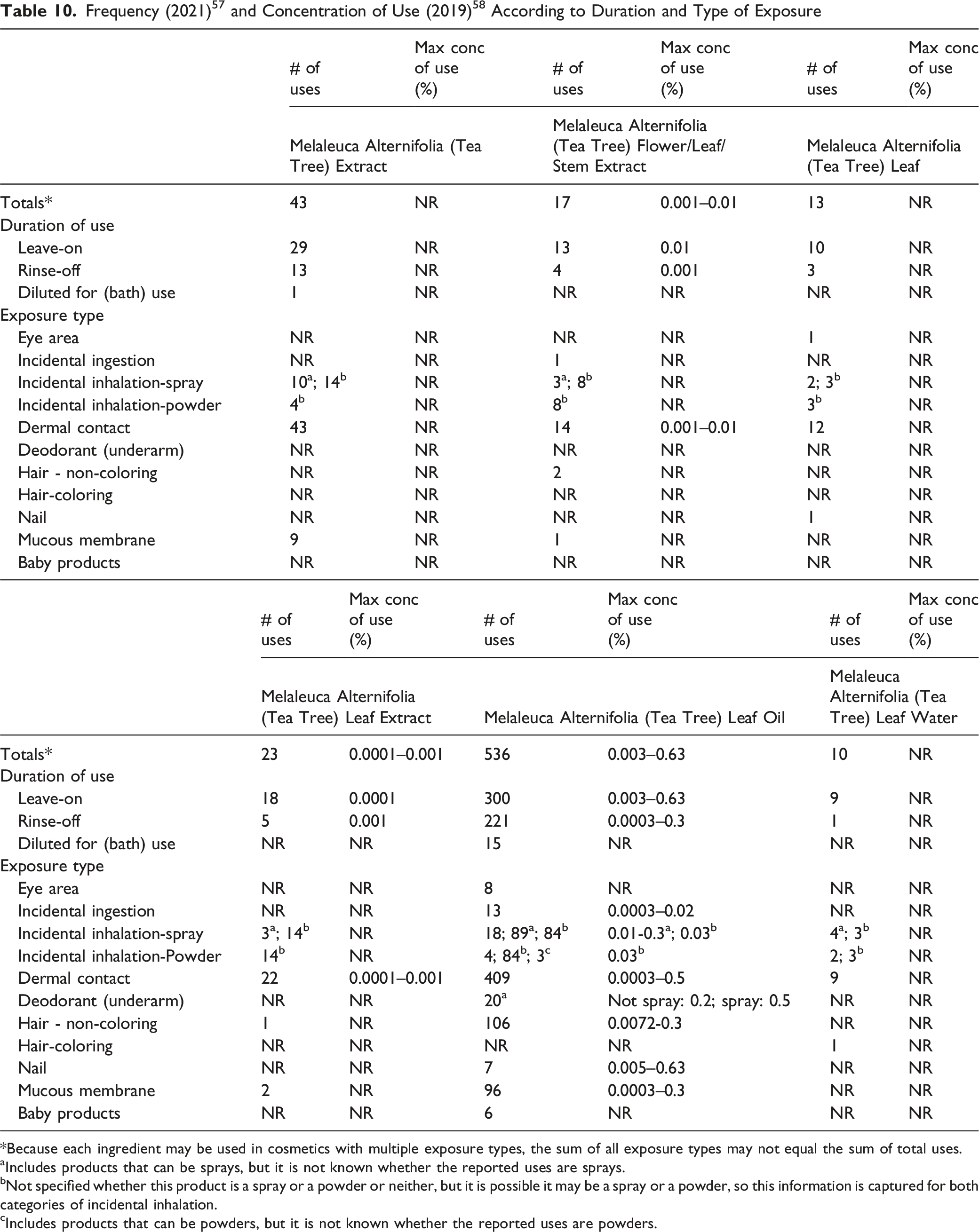
*Because each ingredient may be used in cosmetics with multiple exposure types, the sum of all exposure types may not equal the sum of total uses.
aIncludes products that can be sprays, but it is not known whether the reported uses are sprays.
bNot specified whether this product is a spray or a powder or neither, but it is possible it may be a spray or a powder, so this information is captured for both categories of incidental inhalation.
cIncludes products that can be powders, but it is not known whether the reported uses are powders.
Melaleuca Alternifolia (Tea Tree) Leaf and Melaleuca Alternifolia (Tea Tree) Leaf Oil are reported to be used in products applied near the eye (concentration of use not reported), and Melaleuca Alternifolia (Tea Tree) Flower/Leaf/Stem Extract and Melaleuca Alternifolia (Tea Tree) Leaf Oil in products that can result in incidental ingestion (e.g., at up to 0.02% of the oil in lipstick). Several of the Melaleuca alternifolia (tea tree)-derived ingredients are used in formulations that come into contact with mucous membranes (e.g., 0.3% Melaleuca Alternifolia (Tea Tree) Leaf Oil in bath soaps and detergents). Additionally, Melaleuca Alternifolia (Tea Tree) Leaf Oil is reported to be used in baby products; concentration of use data were not reported for this category.
Additionally, some of the Melaleuca alternifolia (tea tree)-derived ingredients are used in cosmetic sprays and powders and could possibly be inhaled; for example, Melaleuca Alternifolia (Tea Tree) Leaf Oil is reported to be used at up to 0.5% in aerosol deodorant formulations, 58 and according to VCRP data, Melaleuca Alternifolia (Tea Tree) Leaf Oil and Melaleuca Alternifolia (Tea Tree) Leaf Water are reported to be used in face powders. 57 In practice, most droplets/particles released from cosmetic sprays have aerodynamic equivalent diameters >10 µm, with propellant sprays yielding a greater fraction of droplets/particles <10 µm compared with pump sprays.59,60 Therefore, most droplets/particles incidentally inhaled from cosmetic sprays would be deposited in the nasopharyngeal and thoracic regions of the respiratory tract and would not be respirable (i.e., they would not enter the lungs) to any appreciable amount.61,62 There is some evidence indicating that deodorant spray products can release substantially larger fractions of particulates having aerodynamic equivalent diameters in the range considered to be respirable. 61 However, the information is not sufficient to determine whether significantly greater lung exposures result from the use of deodorant sprays, compared to other cosmetic sprays. Conservative estimates of inhalation exposures to respirable particles during the use of loose powder cosmetic products are 400-fold to 1000-fold less than protective regulatory and guidance limits for inert airborne respirable particles in the workplace.63-65
In 2002, the European Cosmetic Toiletry and Perfumery Association (COLIPA) stated “COLIPA recommends that Tea Tree Oil should not be used in cosmetic products in a way that results in a concentration greater than 1% oil being applied to the body. 6 When formulating Tea Tree Oil in a cosmetic product, companies should consider that the sensitization potential increases if certain constituents of the oil become oxidized. To reduce the formation of these oxidation products, manufacturers should consider the use of antioxidants and/or specific packaging to minimize exposure to light.”
In Germany, the Federal Institute for Risk Assessment recommends limiting the concentration of tea tree oil in cosmetics to a maximum of 1%; cosmetic products containing tea tree oil should be protected against light and admixed with antioxidants to avoid oxidation of terpenes. 66 Norway allows Melaleuca Alternifolia (Tea Tree) Leaf Oil to be used at a maximum of 0.5% in mouth care products and 2% in all other cosmetics; it must not be used in products meant for children under 12 years of age. 42 In Australia, typical use concentrations of up to 2% are reported in leave-on (including deodorants and foot sprays) and rinse-off products (including soaps). 12 Use in mouthwash at a typical concentration of 0.2% is also indicated.
Non-Cosmetic
Tea tree oil is listed as a generally recognized as safe (GRAS) flavoring substance by Flavor and Extract Manufacturer’s Association (FEMA).67,68
Tea tree oil is reported to have use as an herbal medicine; it has been used for centuries as a traditional medicine to treat cuts and wounds by the aboriginal people of Australia.31,69 The EMA EU herbal monograph on Melaleuca alternifolia (Maiden and Betch) Cheel, Melaleuca linariifolia Smith, Melaleuca dissitiflora F. Mueller and/or other species of Melaleuca aetheroleum describes traditional cutaneous use (liquid or semi-solid form, up to 100%) in treatment of small superficial wounds and insect bites, small boils, and itching and irritation due to tinea pedis (athlete’s foot), as well as oromucosal use (liquid form, diluted in water) for symptomatic treatment of minor inflammation of the oral mucosa 8 ; the Committee on Herbal Medicinal Products (HMPC) concluded that, on the basis of its long-standing use, tea tree oil preparations can be used for these uses.3,9
According to the WHO, clinical data supports use of tea tree oil in topical applications for symptomatic treatment of common skin disorders (such as acne, tinea pedis, bromidrosis, furunculosis, and onychomycosis), and of vaginitis due to Trichomonas vaginalis or Candida albicans, cystitis, or cervicitis. 11 Tea tree oil is reported to have antimicrobial activity. In traditional medicine, it is used as an antiseptic and disinfectant in the treatment of wounds. Additionally, tea tree oil is reported to have antibacterial, anti-viral, anti-inflammatory activity, analgesic, anti-tumoral, insecticidal, and acaricidal activities.4,12
The US FDA issued a final action in April 2019 (effective April 13, 2020) for tea tree oil, establishing that its use in non-prescription over-the-counter (OTC) consumer antiseptic products intended for use without water (i.e., antiseptic rubs or consumer rubs) is not eligible for evaluation under the OTC Drug Review for use in consumer antiseptic rubs. 70 Drug products containing tea tree oil will require approval under a new drug application or abbreviated new drug application prior to marketing.
Additionally, in a 2016 review, the FDA Pharmacy Compounding Advisory Committee did not recommend Melaleuca Alternifolia (Tea Tree) Leaf Oil for inclusion on the list of bulk drug substances that can be used in pharmacy compounding for topical use in the treatment of nail fungus under Section 503A of the Federal Food, Drug, and Cosmetic Act. 55 The final compounded topical formulations being considered were at strengths of 5%–10%. The Committee considered that although products containing the oil have been commercially available since at least 1982 for use as topical formulations for a wide variety of skin, ocular, oral, and vaginal conditions, the oil may cause local reactions, and a lack of evidence of efficacy in the treatment of onychomycosis and a lack of information on the past use of tea tree oil in pharmacy compounding was cited.
Tea tree oil is reportedly active as an antioxidant. 71 Depending on the testing used, tea tree oil was reported to be a stronger antioxidant than α-lipoic acid, vitamin C, and vitamin E.
Toxicokinetics
Dermal Penetration/Absorption
The EMA monograph on Melaleuca species stated that because tea tree oil is a semi-volatile substance, the majority of an applied dose would be expected to evaporate from the skin surface before it could be absorbed into the skin. 3 In a study in which tea tree oil was applied to filter paper, stored in an oven at 30°C, and then weighed, application of 1.4 mg/cm2 evaporated within 1 h, and 84, 98, and 100% of a 7.4 mg/cm2 application evaporated within 2, 4, and 8 h, respectively. 26
In Vitro
In Vitro Dermal Penetration Studies of Tea Tree Oil Using Skin Samples
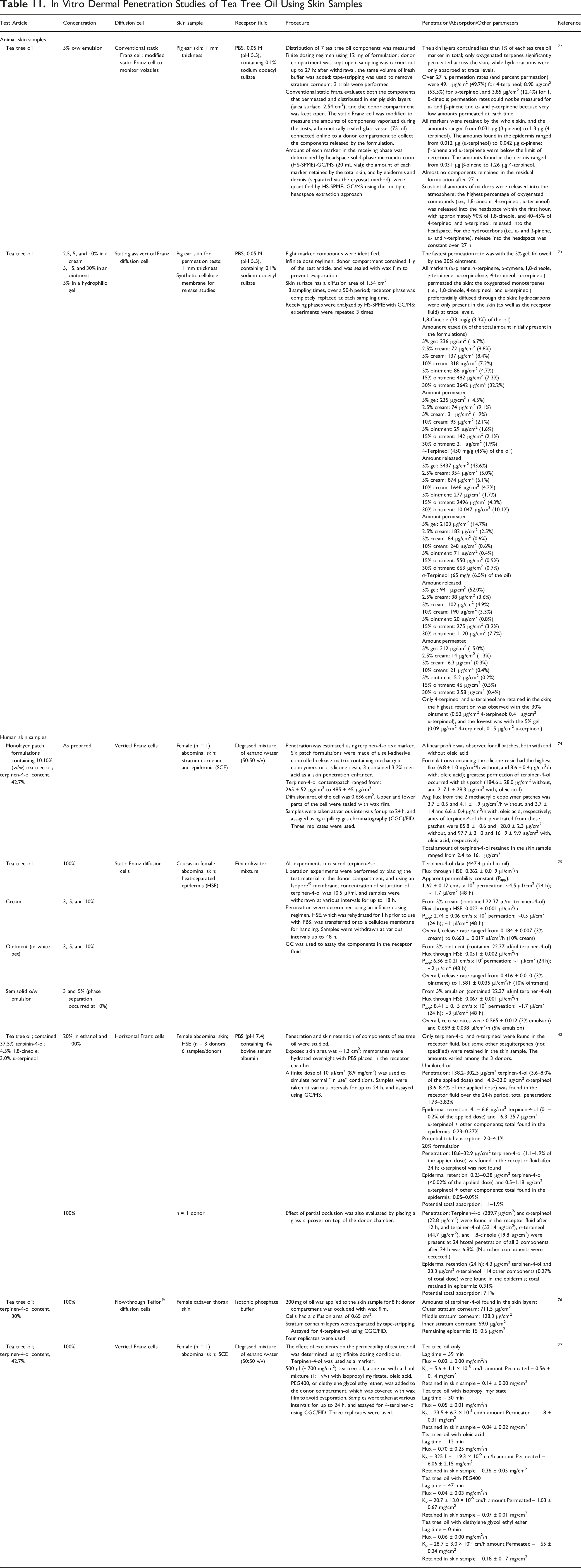
It was also demonstrated that the formulation vehicle affects absorption. 73 Again using pig ear skin, mounted in vertical Franz cell that were sealed to prevent evaporation, and varying amounts of tea tree oil formulated using a cream (2.5–10%), an ointment (5–30%), and a hydrophilic gel (5%), the fastest permeation rate was with the 5% tea tree oil gel, followed by the 30% ointment. Additionally, the effect of excipients used as penetration enhancers on the penetration of pure tea tree oil was investigated. 77 Oleic acid enhanced the penetration of tea tree oil (as determined by using terpinen-4-ol as a marker); the amount permeated increased from 0.56 mg/cm2 pure tea tree oil to 6.06 mg/cm2 with oleic acid used as an excipient, and lag time decreased from 59 min to 12 min, respectively. Other excipients also had an effect, but to a lesser extent.
Volatility of tea tree oil upon application was also investigated. In the study using pig ear skin in which the donor chamber was not covered, substantial amounts of markers were released into the atmosphere; the highest percentage of oxygenated compounds (i.e., 1,8-cineole, 4-terpineol, α-terpineol) was released into the headspace within the first hour, with approximately 90% of 1,8-cineole and 40%–45% of 4-terpineol and α-terpineol released. 72 For the hydrocarbons (i.e., α- and β-pinene and α- and γ-terpinene), release into the headspace was constant over the 27-h test period. The vehicle also affected the amount of each component released; for example, in a study using sealed diffusion cells, 52% of the α-terpineol was released from a 5% gel, but only 0.8% was released from a 5% ointment. 73 In a finite dosing study with human skin samples under open test conditions in horizontal Franz cells, the potential total absorption of undiluted tea tree oil (using terpinen-4-ol, 1,8-cineole, and α-terpineol as markers) was determined to be 2.0–4.1%; at 20% in ethanol, potential total absorption was determined to be 1.1%–1.9%. 43 When the donor chamber was partially occluded, potential total absorption of undiluted tea tree oil was 7.1%.
As demonstrated, a difference in bioavailability of the components exists. Therefore, when using in vitro data related to topical use of tea tree oil, the bioavailability, and more specifically, the absorption profile of the individual constituents of the oil, should be considered for in vitro-to-in vivo extrapolation. 79
Effect on Skin Integrity
Tea Tree Oil
The effect of tea tree oil on skin integrity was determined using full-thickness human breast skin or abdominal skin samples (0.5–1.1 mm; 3–4 donors) mounted in static diffusion cells. 80 The skin samples were exposed for 24 h to solutions of 0, 0.1, 1.0, or 5.0% tea tree oil (50 µl/cm2) in an aqueous solution containing 1% Tween, 0.9% saline, and tritiated water, and to tritiated water, using infinite dosing conditions. The median diffusion area was 2.12 cm2/cell, and donor and receptor cells were covered with wax film to avoid evaporation. Prior to the study, the epidermal site was exposed to ambient laboratory conditions and the dermis exposed to an aqueous solution of 0.9% saline and 1% Tween for 18 h. The maximal flux of tritiated water was significantly reduced with 1.0% tea tree oil, but not at the other 2 concentrations. At 5%, there was some evidence of damage to the barrier integrity, in that the maximal flux the water increased to was 121% of the controls; however, the increase was not statistically significant.
Comparable results were found in a similar study with concentrations of 1 and 5% tea tree oil (48-h exposure) using full-thickness human breast skin or abdominal skin samples (avg thickness, 0.87 mm) mounted in static diffusion cells. 81 Again, 1% tea tree oil (same vehicle as above) did not affect barrier conditions, but there was an increase in the Kp value for tritiated water with 5% tea tree oil. The researchers stated that this demonstrated that the barrier integrity is affected at this concentration of tea tree oil. However, although the effect on the barrier integrity was statistically significant with 5% tea tree oil in the donor phase, the mean permeability coefficient (Kp) value was still considerably below the cut-off level (35 µm/h) used for assessment of barrier function in percutaneous penetration studies.
Penetration Enhancement
Tea Tree Oil
The effect of tea tree oil on permeation of ketoprofen was examined using excised porcine skin mounted in Franz diffusion cells; degassed phosphate-buffered saline (PBS) was placed in the receptor chamber. 82 The skin samples were pre-treated with 500 µl of tea tree oil or deionized water (negative control) for 1 h. After removal of the pre-treatment solution, 500 µl of ketoprofen in polyethylene glycol (PEG)-400 was added to the cell, and the donor chamber was occluded with wax film; the receptor phase was sampled at various intervals for 48 h. The flux of ketoprofen was ∼7.5 times greater with tea tree oil, as compared to the negative control (38.4 vs 5.19 µg/cm2/h, respectively), the Kp of ketoprofen increased from 2.1 × 10⁻⁴ cm/h with deionized water to 15.5 × 10⁻⁴ cm/h with tea tree oil, and the percentage of ketoprofen that was delivered across the skin in 24 h increased from 0.50% to 3.11% with tea tree oil.
Full-thickness samples from human breast or abdominal skin were used to examine the effect of up to 5% tea tree oil on the dermal absorption of methiocarb and benzoic acid (solubilities of 0.03 and 3.0 g/l, respectively). 81 Using static diffusion cells, with a median diffusion area of 2.12 cm2/cell, 50 µl/cm2 of the test substance was applied for 48 h using an infinite dosing regimen. Donor and receptor cells were covered with wax film to limit evaporation. Tea tree oil reduced the maximal flux, thereby reducing the overall amount of benzoic acid and methiocarb entering the receptor chamber.
Absorption, Distribution, Metabolism, and Excretion
Tea Tree Oil
ECHA provided estimates of absorption via various routes 7 Oral, dermal, and inhalation absorption rates were estimated as 70%, 3%, and 100%, respectively. Details were not provided.
Toxicological Studies
Acute Toxicity Studies
Acute Toxicity Studies
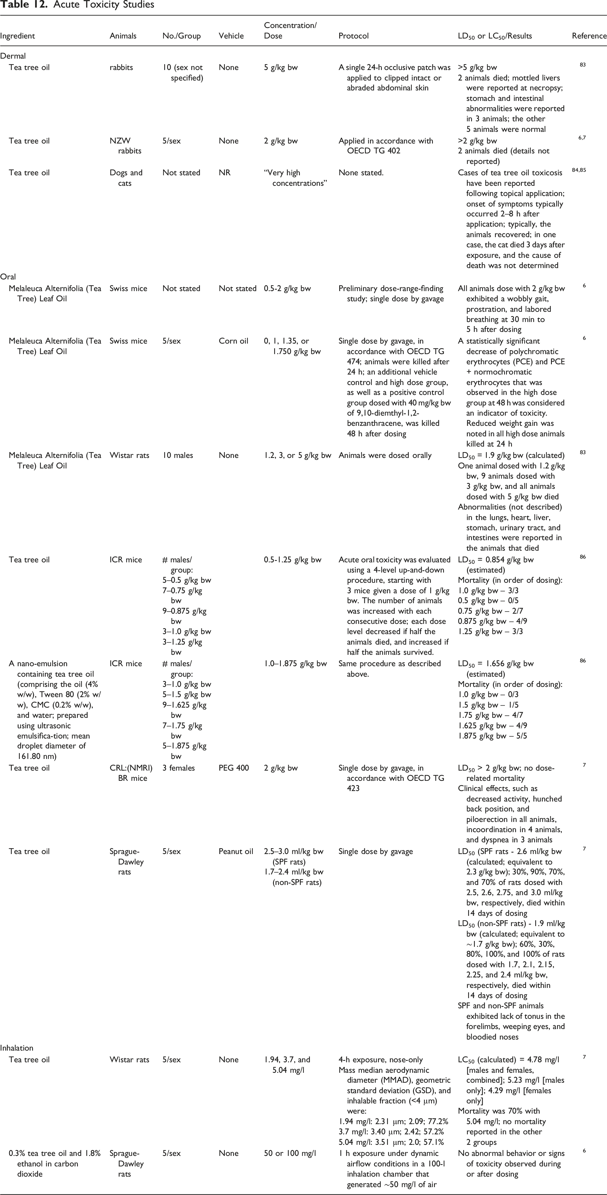
In rabbits, following a single 24-h occlusive patch of tea tree oil that was applied to clipped intact or abraded abdominal skin, the LD50 was >5 g/kg bw; 2 of 10 animals dosed with 5 g/kg bw died, and mottled livers and stomach and intestinal abnormalities were reported in 3 other animals. 83 In another study, tea tree oil had a dermal LD50 > 2 g/kg bw in rabbits.6,7 Dermal applications of “very high concentrations” of tea tree oil have been reported to cause tea tree oil toxicosis in dogs and cats.84,85
In studies in which Swiss mice were given a single dose of up to 2 g/kg bw Melaleuca Alternifolia (Tea Tree) Leaf Oil by gavage, animals dosed with 2 g/kg bw had a wobbly gait, prostration, and labored breathing. 6 In male Wistar rats given a single dose of 1.2–5 g/kg Melaleuca Alternifolia (Tea Tree) Leaf Oil by gavage, the LD50 was calculated to be 1.9 g/kg bw. 83 In one study in ICR (Institute of Cancer Research) mice, the oral LD50s of tea tree oil and a nano-emulsion containing tea tree oil were estimated to be 0.854 g/kg bw and 1.565 g/kg bw, respectively. 86 In another study, the LD50 of tea tree oil was >2 g/kg (in PEG 400) in female mice, 7 and calculated as 2.3 g/kg bw and ∼1.7 g/kg bw (in peanut oil) in specific pathogen-free (SPF) and non-SPF Sprague-Dawley rats, respectively. 7
In an acute inhalation study in which groups of 5 male and 5 female Wistar rats were exposed nose-only to tea tree oil for 4 h, the LC50 was calculated as 4.78 mg/l for males and females combined, as 5.23 mg/l for males only, and as 4.29 mg/l for females only. 7 No abnormal behavior or signs of toxicity were observed during or after dosing when groups of 10 Sprague-Dawley rats were exposed for 1 h to 50 or 100 mg/l of a test substance that contained 0.3% w/w tea tree oil and 1.8% ethanol in carbon dioxide. 6
Short-Term Toxicity Studies
Dermal
Tea Tree Oil
Tea tree oil (2%; 50 µl) was applied to the shaved backs of 3 Wistar rats daily for 28 days. 30 (Additional details, including whether or not collars were used or if the test site was covered, were not provided.) Serum glutamine-oxaloacetic transaminase (SGOT) and serum glutamic-pyruvic transaminase (SGPT) levels were measured on days 0, 14, and 28 using blood samples taken from the tail vein. Repeated dermal applications of tea tree oil did not result in any significant changes in SGOT or SGPT levels.
Oral
Tea Tree Oil
Groups of 10 ICR mice were used in a 28-day oral toxicity study, in accordance with Organisation for Economic Co-operation and Development (OECD) test guideline (TG) 407, to determine the toxicity of a nano-emulsion containing tea tree oil. 86 The test article was prepared using ultrasonic emulsification, and comprised the oil (4% w/w), Tween 80 (2% w/w), carboxymethylcellulose sodium (CMC; 0.2% w/w), and water; the mean droplet diameter was 161.80 nm. The animals were dosed by gavage with 0, 50, 100, or 200 mg/kg bw of the test article, once a day, for 28 days. No effects on food or water consumption, body weights, or mortality were observed. Additionally, there were no physical signs of toxicity during the study, and no gross findings, effects on organs, or microscopic effects observed at necropsy. No differences in hematology parameters were reported. Serum alanine aminotransferase levels showed a dose-related increasing trend, and this value was statistically significantly increased in the high-dose group compared to controls; no other statistically significant differences in serum biochemistry values were noted. The no-observable-adverse-effect-level (NOAEL) of this nano-emulsion containing tea tree oil in mice was >200 mg/kg bw.
Groups of 5 male and 5 female Sprague-Dawley rats were dosed for 28 days with tea tree oil in corn oil by gavage at doses of 0, 5, 15, and 45 mg/kg bw/d, in accordance with OECD TG 407. 7 No mortality was observed, and no test-article related clinical signs of toxicity were reported. Additionally, there were not changes in functional observation battery, motor activity body weight, body weight gain, food consumption, or food efficiency during the study. There were no test-article related gross or microscopic findings reported, and absolute and relative organ weights were similar to controls. The NOAEL was determined to be 45 mg/kg bw/d for both male and female rats.
Subchronic and Chronic Toxicity
Subchronic and chronic toxicity studies on the Melaleuca alternifolia (tea tree)-derived ingredients were not found in the published literature, and unpublished data were not submitted.
Developmental and Reproductive Toxicity
Tea Tree Oil
Groups of 27 mated female Hannover Wistar rats were dosed by gavage with 0, 20, 100, and 250 mg/kg bw/d tea tree oil in PEG 400 on days 5 to 19 of gestation, in a developmental toxicity study performed in accordance with OECD TG 414. 7 The dams were killed on day 20 of gestation. Severe maternal toxicity was observed in dams of the 100 and 250 mg/kg bw/d groups, as evidenced by clinical signs, reduced food consumption, and weight gain reductions of 20% and 45%, respectively, over the gestation period. Seven of the high dose dams died between days 8 and 11 of gestation; there was no mortality in the other test groups. Bilateral enlarged adrenals were observed in all high-dose dams that died during the study and in 6/20 that survived until necropsy; this observation was made in one dam of the mid-dose group. A dose-related decrease in mean fetal weights, related to intrauterine growth retardation, was noted in the mid- and high-dose groups. An increase in the number of late embryonic deaths and post-implantation loss, leading to an overall higher total intrauterine mortality, was observed in the high-dose (but not mid- or low-dose) group; the increase in post-implantation mortality was considered to be secondary to maternal toxicity. There was no statistically significant difference, compared to controls, in the number of visceral malformations in the fetuses of test animals, but there were statistically significant higher numbers of visceral variations reported in the 250 mg/kg bw/d dose group. A statistically significant higher incidence of skeletal malformations unrelated to intrauterine growth retardation was noted in the 250 mg/kg bw/d group, and a statistically significant increase in the number of skeletal variations, secondary to maternal toxicity, was noted in the 100 and 250 mg/kg bw/d groups. The NOAELs for maternal toxicity and for developmental toxicity (secondary to severe maternal toxicity) were 20 mg/kg bw/d tea tree oil.
Effects on Spermatozoa
Animal
The effects of tea tree oil (containing 41.49% terpinen-4-ol, 20.55% γ-terpinene, 9.59% α-terpinene, and 4.42% α-terpineol) on the morpho-functional parameters of porcine spermatozoa were evaluated. 87 Spermatozoa samples (15 × 107 spermatozoa in 5 ml of medium) were exposed to 0.2–2 mg/ml tea tree oil for 3 h. A concentration-dependent decrease in motility was observed with concentrations of 0.4 mg/ml and greater; the decrease was statistically significant at concentrations ≥0.8 mg/ml. Viability of spermatozoa was statistically significantly decreased with ≥1 mg/ml tea tree oil, and sperm acrosome reaction was statistically significantly increased at concentrations of ≥1.4 mg/ml. The effects of terpinen-4-ol alone were also evaluated; a greater concentration of terpinen-4-ol only (relative to the amount in tea tree oil) was needed to have an effect on the morpho-functional parameters.
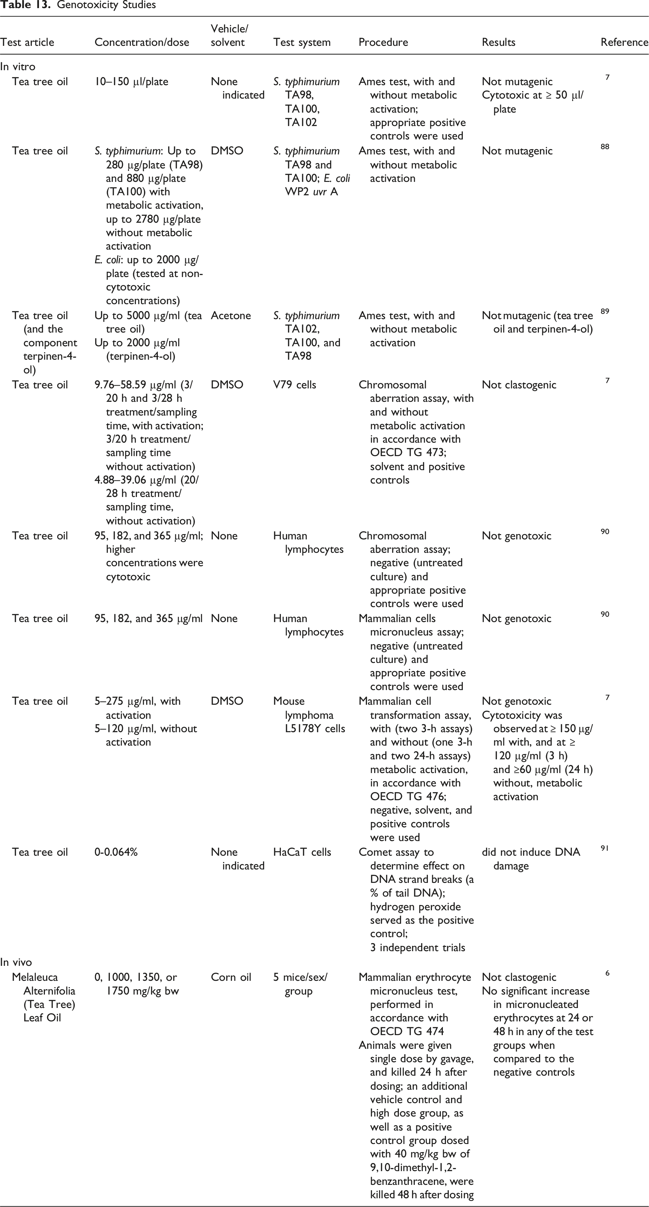
Genotoxicity Studies
Genotoxicity Studies
Carcinogenicity Studies
Carcinogenicity data on the Melaleuca alternifolia (tea tree)-derived ingredients were not found in the published literature, and unpublished data were not submitted.
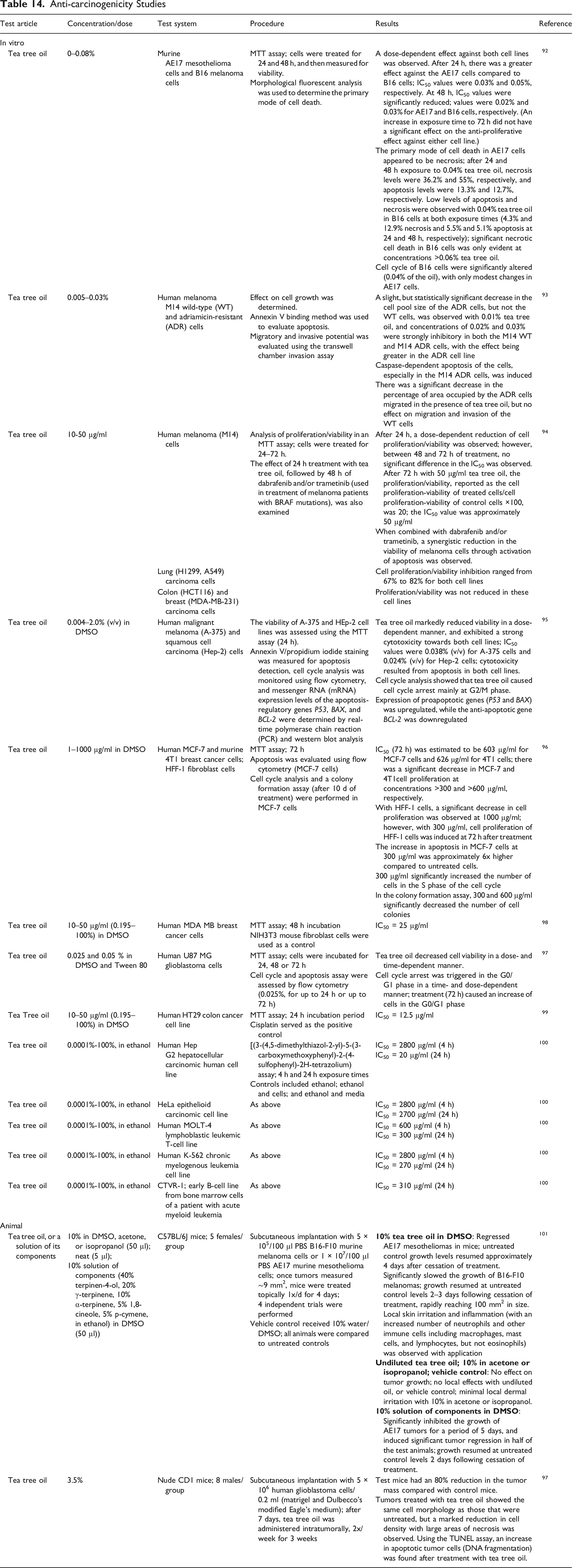
Anti-Carcinogenicity Studies
Anti-carcinogenicity Studies
Other Relevant Studies
Effect on Endocrine Activity
Tea Tree Oil
Effect on Endocrine Activity
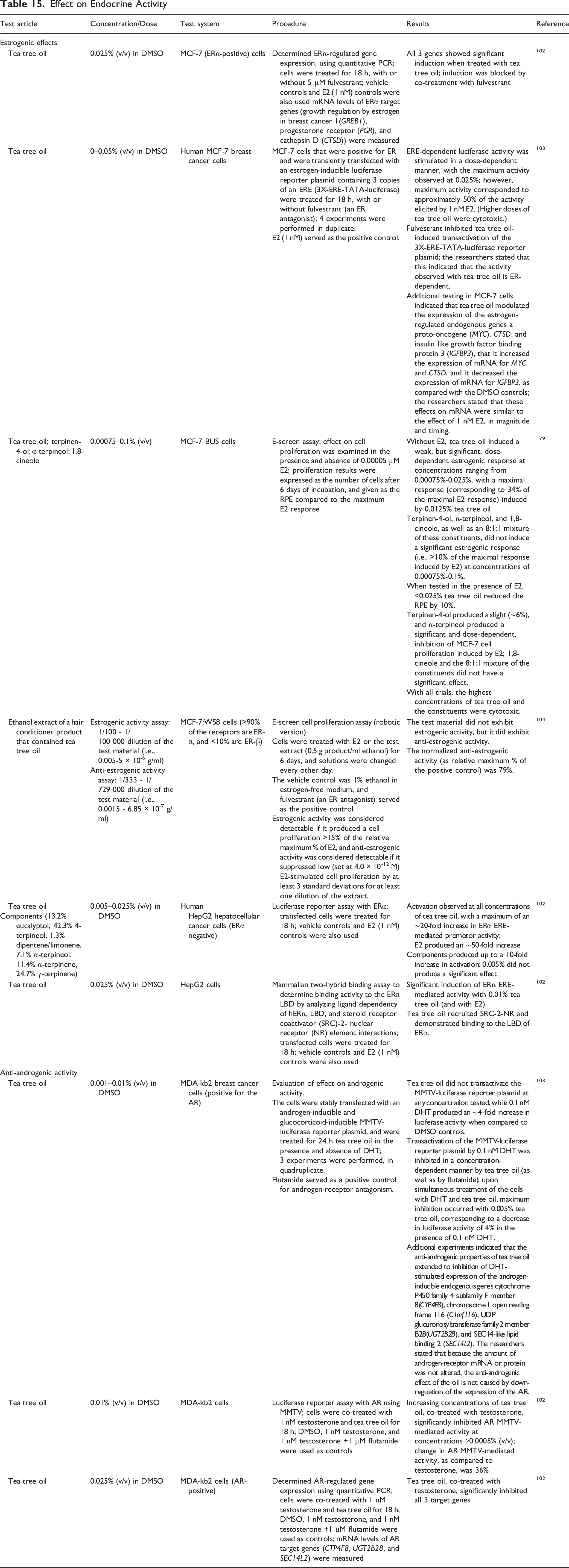
The effect of tea tree oil on estrogen receptor-α (ERα)-regulated gene expression was determined in the human MCF-7 breast cancer cell line; ERα target genes showed significant induction when treated with tea tree oil, and the estrogen response element (ERE)-dependent luciferase activity was stimulated in a dose-dependent manner (maximum activity observed at 0.025%).102,103 Fulvestrant inhibited transactivation of the 3X-ERE-TATA-luciferase reporter, indicating that the activity observed is ER-dependent. In an E-screen assay using MCF-7 BUS cells, tea tree oil (without 17β-estradiol (E2)) induced a weak, but significant, dose-dependent estrogenic response at concentrations ranging from 0.00075% to 0.025%, with a maximal response (corresponding to 34% of the maximal E2 response) induced by a concentration of 0.0125% tea tree oil; when tested in the presence of E2, concentrations of <0.025% tea tree oil reduced the relative proliferative effect (RPE) by 10%. 79 Terpinen-4-ol, α-terpineol, and 1,8-cineole, as well as an 8:1:1 mixture of these constituents, did not induce a significant estrogenic response at concentrations of ≤0.1%. A robotic version of the E-screen cell proliferation assay was performed with MCF-7:WS8 cells to evaluate the estrogenic activity (with ≤5 × 10⁻⁶ g/ml) and the anti-estrogenic activity (with ≤6.85 × 10⁻⁷ g/ml) of an ethanol extract of a hair conditioner product that contained tea tree oil. 104 The formulation did not exhibit estrogenic activity, but it did exhibit anti-estrogenic activity; the normalized anti-estrogenic activity (as relative maximum % of the positive control) was 79%. The effects of tea tree oil were also evaluated with human HepG2 hepatocellular cancer cells (ERα-negative). 102 In a luciferase reporter assay using transfected cells, tea tree oil (≤0.025%) produced a maximum of an ∼20-fold increase in ERα ERE-mediated promotor activity. In a mammalian two-hybrid binding assay to determine binding activity to the ERα ligand-binding domain (LBD), there was a significant induction of ERα ERE-mediated activity with 0.01% tea tree oil, and tea tree oil demonstrated binding to the LBD of ERα.
The effect of tea tree oil (in the presence and absence of dihydrotestosterone (DHT) on androgenic activity was evaluated in MDA-kb2 breast cancer cells transfected with an androgen- and glucocorticoid-inducible mouse mammary-tumor virus (MMTV)-luciferase reporter plasmid. 103 Tea tree oil did not transactivate the reporter plasmid at any concentration tested (≤0.01%), and it inhibited plasmid transactivation by DHT in a concentration-dependent manner; maximum inhibition occurred with 0.005% tea tree oil. Additional experiments in MDA-kb2 cells indicated that the anti-androgenic properties of tea tree oil extended to inhibition of DHT-stimulated expression of androgen-inducible endogenous genes. In another luciferase reporter assay with androgen receptor (AR) MMTV, increasing concentrations of tea tree oil, co-treated with testosterone, significantly inhibited MMTV-mediated activity at concentrations ≥0.0005% (v/v); change in activity, as compared to testosterone, was 36%. 102 The effect of tea tree oil on AR-regulated gene expression was determined in MDA-kb2 cells; tea tree oil, co-treated with testosterone, significantly inhibited the target genes.
In an opinion paper, the SCCP commented that an estrogenic potential of tea tree oil was shown in vitro, but in vivo studies were not available to elucidate the relevance of this finding. 6 The potentially endocrine-active constituents of tea tree oil have not been shown to penetrate the skin; therefore, the (hypothesized) correlation of gynecomastia due to the topical use of tea tree oil, in conjunction with lavender oil, in a 10-year old male, 103 was considered implausible by the SCCP.
Mucosal Toxicity
Tea Tree Oil
The potential for tea tree oil (0.5–500 mg/ml) to induce mucosal damage was examined in porcine uterine mucosa (n = 8) using an Evans Blue permeability assay; the highest concentration of tea tree oil was used as a positive control. 105 Emulsifiers only served as the negative control. Tea tree oil induced a dose-dependent increase in the amount of dye absorbed, and the increase was statistically significant at concentrations of 40 and 500 mg/ml. No damage was observed with 0.2, 0.4, or 20 mg/ml tea tree oil; at 40 mg/ml, moderate damage was induced to the uterine mucosa, with a multifocal detachment of the epithelium.
The same researchers also performed an ex vivo study, filling the uterine horns from 8 female sows with 0.2 or 0.4 mg/ml tea tree oil, and incubating the horns for 1 h. After incubation, each uterine horn was emptied, washed with Dulbecco’s PBS, and 3 cm × 3 cm section was examined. At these test concentrations, tea tree oil did not alter the structure of swine uterine mucosa.
Ototoxicity
Tea Tree Oil
The ototoxicity of tea tree oil was examined in guinea pigs by measuring the thresholds of the compound auditory nerve action potential (CAP) to tone bursts before and after instillation of the oil into the middle ear. 106 After 30 min, undiluted tea tree oil (n = 5) caused a partial CAP threshold elevation at 20 kHz. With 2% tea tree oil in saline (n = 4), no significant lasting threshold change was observed after the same amount of time. Normal saline (n = 4) was used as a negative control.
Immunologic Effects
Tea Tree Oil
In Vitro
The effect of tea tree oil on neutrophil activation was investigated by measuring the tumor necrosis factor-α-induced adherence reaction of human peripheral neutrophils. 107 Tea tree oil was diluted to concentrations of 0.025%–0.2% using dimethyl sulfoxide (DMSO) and Roswell Park Memorial Institute (RPMI) medium (containing 10% fetal calf serum; complete medium). The suppressing activity of tea tree oil was weak; the concentration of tea tree oil providing 50% inhibition (IC50) of neutrophil adherence was 0.033%. Additionally, tea tree oil did not suppress lipopolysaccharide-induced neutrophil-induced adherence.
Animal
Dermal
Five experiments were performed in which BALB/c mice (3/group) were sensitized on shaved abdominal skin with 100 µl of 5% 2,4,6-trinitrochlorobenzene (TNCB) in acetone; after 7 d, a contact hypersensitivity response was elicited (challenge phase) by application of 50 µl of 1% TNCB in acetone to shaved dorsal skin. 108 Undiluted tea tree oil (20 µl) was applied topically to the shaved area 30 min before or 2, 4, or 7 h after challenge, and the change in double skinfold thickness was determined at various time points for up to 120 h. Controls included mice that were treated with tea tree oil alone (sensitized 7 days prior, but not challenged with TNCB) and mice that were not sensitized 7 days previously, but were challenged with TNCB.
For the first 7 h post-challenge, swelling was detected in the skin of both sensitized and non-sensitized mice. The change in double skinfold thickness in the non-sensitized mice (irritant response) subsided significantly in the following 17 h, but remained high in the sensitized mice. Undiluted tea tree oil applied 30 min before TNCB application to the non-sensitized mice did not reduce the increase in double skinfold thickness observed in the first 7 h after TNCB exposure. However, a significant reduction in swelling was observed in sensitized mice that received a single topical application of undiluted tea tree oil before or after challenge.
The researchers then investigated the effect of a single topical application (30 µl) of 5% tea tree oil ointment, 10% gel, or control gel at 7 h after challenge. The 5% tea tree oil ointment and the 10% tea tree oil gel significantly suppressed TNCB-induced swelling by 39 and 35%, respectively. The control gel had little effect, and did not cause a significant suppression when compared with the TNCB control.
The researchers also examined whether tea tree oil alleviated swelling induced by mid-wavelength (UVB) irradiation. Shaved skin of BALB/c mice (3/group) was exposed to 2 kJ/m2 (1 trial) or 8 kJ/m2 (3 trials) UVB (corresponding to a minimal erythema dose of 1 or 4, respectively) using a bank of FS40 sunlamps (250–360 nm; wavelengths <290 nm were screened out). Undiluted tea tree oil (20 µl) was applied topically to the shaved area at either 30 min before or up to 7 h after UVB exposure, and the change in double skinfold thickness was measured at 24, 48, and 120 h. Control mice were treated with tea tree oil, but not exposed to UVB. A single topical application of undiluted tea tree oil after irradiation did not suppress UVB-induced swelling. Furthermore, swelling was significantly increased when tea tree oil was applied before UVB irradiation (8 kJ/m2).
The effect of the cutaneous application of tea tree oil on myeloperoxidase (MPO) activity was examined using groups of 3–4 ICR mice. 109 The mice were injected intradermally with a curdlan suspension (10 mg/ml), followed by application of 0.01 ml tea tree oil to the shaved dorsal skin (immediately, and after 3 h). The animals were killed 6 h after curdlan injection, and skin preparations were obtained. Control mice received applications of 0.1 ml DMSO. Dermal application of tea tree oil decreased MPO activity significantly, from 100% in controls to approximately 55% in the test group.
Inhalation
In mice exposed to tea tree oil via multiple inhalation sessions, there was an increase in the level of circulating blood immunoglobulins and the blood granulocyte number, plus stimulation of the local graft-versus-host reaction of spleen cells. 110 (Details were not available.)
Male C57BI10 x CBA/H (F1) mice (number per group not provided) were exposed to tea tree oil via inhalation, 3x/d (15 min each) for 7 d; the animals were subjected to the vapors by applying 5 drops of the oil to cotton wool, and placing the wool near the cage. 110 A negative control group (no inhalation treatment) and a sham control group (water placed on cotton wool) were used. One day before the termination of dosing, subgroups of mice from each group were injected intraperitoneally with zymosan (to induce peritonitis), PBS, or left untreated. Spleens and peritoneal exudates were collected 24 h after injection. The activity of peritoneal leukocytes in the test group was equivalent to that seen in the negative and sham control groups without inflammation, indicating that tea tree oil had anti-inflammatory action. Additionally, tea tree oil stopped the proliferation of splenocytes in response to T- and B-cell mitogens. The effect of tea tree oil in inflammation was reversed by an opioid receptor antagonist (administered in drinking water). An additional inhalation study reported that the hypothalamic-pituitary-adrenal axis mediated the anti-inflammatory effect of tea tree oil administered to the same strain of mice. 111
Human
Dermal
The effect of tea tree oil on a histamine-induced wheal and flare reaction was examined. 112 Subjects were injected intradermally in each forearm with histamine (50 µl of a 100 µg/ml solution), and after 20 min, undiluted tea tree oil (25 µl) was applied topically at the injection site of one arm (test arm) of 21 subjects. In an additional 6 subjects, paraffin oil (25 µl; oil control) was applied to one arm. The arm not treated with any oil served as a negative control. The flare and wheal responses were measured every 10 min for 1 h; wheal scores were normalized as a percentage of the wheal volume at 20 min due to inter- and intraindividual variability. There was no difference in the mean flare area between the control and test arms in the tea tree oil group. However, the mean wheal volume was statistically significantly decreased as of 10 min after tea tree oil application; at 10 min after application, the mean wheal volume was 92% of that measured prior to application, as opposed to 163% at the same time on the control arm. At 20, 30, and 40 min after oil application, the wheal volume decreased to 83, 62, and 43% of that prior to oil application, respectively, on the test arm; on the control arm, the wheal volumes were 175, 130, and 113%, respectively, at the same times. Liquid paraffin had no effect on wheal or flare response. There was no significant difference in itch (subjective scoring), with or without either oil.
A similar study was conducted in 18 subjects, in which undiluted tea tree oil was applied to the injection site at both 10 and 20 min after histamine injection. 113 In this study, tea tree oil significantly reduced both the flare and the wheal response.
Cytotoxicity
Tea Tree Oil
Emulsions of tea tree oil in culture medium containing 10% fetal calf serum were cytotoxic to adherent peripheral blood mononuclear cells (PBMC); toxicity ranged from 9% (not significant), with 0.004% tea tree oil, to 69% (significant), with 0.016% tea tree oil. 114 In an 3-(4,5-dimethylthiazol-2yl)-2,5-diphenyltetrazolium bromide (MTT) assay evaluating the cytotoxic effects of tea tree oil on HaCaT cells following a 24-h exposure to 0.00%–0.25% w/v, the IC50 was determined to be 0.066%.
Irritation and Sensitization
Dermal Irritation and Sensitization Studies
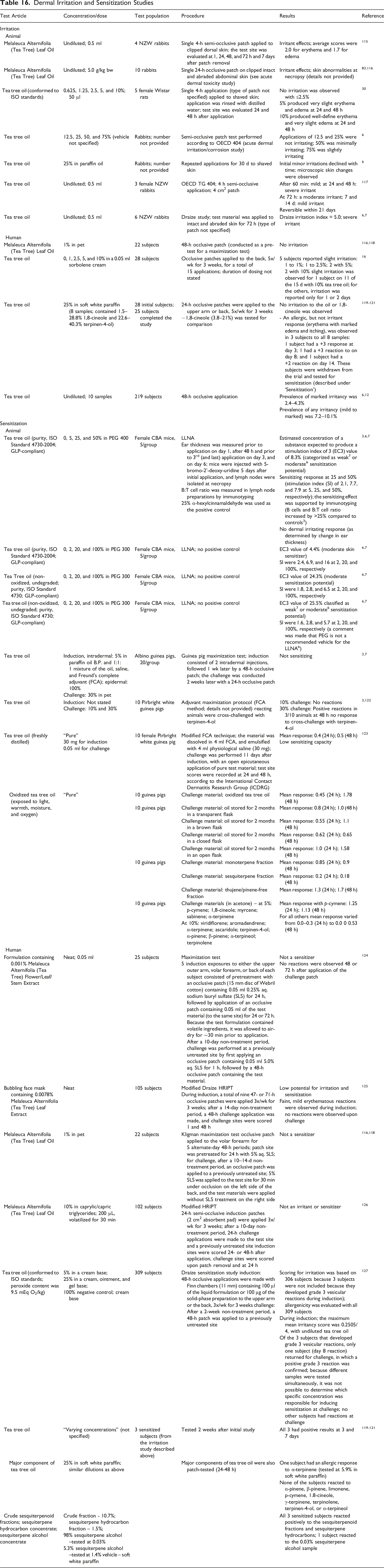
Irritant effects were reported in rabbits after a single 4-h semi-occlusive application, 115 and after a single 24-h occlusive application83,116 of undiluted Melaleuca Alternifolia (Tea Tree) Leaf Oil. Tea tree oil was reported to cause irritation in animals in a concentration-dependent manner; in rats, application of 5% tea tree oil produced very slight erythema, and 10% produced well-define erythema. 30 In rabbits, concentrations of up to 75% were, at most, slightly irritating 6 ; with undiluted tea tree oil, a 4-h semi-occlusive application 117 and application for 72 h to intact and abraded skin produced severe irritation.6,7 In 22 human subjects, a 48-h occlusive patch with 1% Melaleuca Alternifolia (Tea Tree) Leaf Oil in petrolatum (pet) produced no irritation.116,118 In a clinical 3-week occlusive patch test, slight irritation was reported with concentrations of up to 10% tea tree oil in sorbolene cream (5 patches/week, duration not stated; 28 subjects). 16 Two dermal irritation studies were performed with 25% tea tree oil; in one study, no irritation was reported (details were not provided). 16 In the other study, which was a 3-week occlusive patch test in 28 subjects, no irritation was reported with 25% tea tree oil in soft white paraffin; however, an allergic response (erythema with marked edema and itching) was observed in 3 subjects.119-121 In a 48-h patch test with undiluted tea tree oil in 219 subjects, the prevalence of marked irritancy was 2.4%–4.3%, and the prevalence of any irritancy (mild to marked) was 7.2%–10.1%.6,12
In the local lymph node assay (LLNA), tea tree oil was predicted to be a weak or moderate sensitizer at a concentration up to 50%,3,6,7 and a moderate sensitizer when tested undiluted.6,7 In guinea pig studies, tea tree oil was not sensitizing (30% at challenge)3,7 or had a low sensitizing capacity (tested “pure”) 123 ; however, one study indicated that tea tree oil was possibly a weak sensitizer, with 30% tea tree oil producing positive reactions in 3/10 animals at challenge.3,122 In guinea pig studies in which “pure” tea tree oil was used at induction and oxidized tea tree oil was used at challenge, an increase in mean response was observed when compared to challenge with “pure” oil. 123 In clinical studies, a formulation containing 0.001% Melaleuca Alternifolia (Tea Tree) Flower/Leaf/Stem Extract (25 subjects; maximization test), 124 a formulation containing 0.0078% Melaleuca Alternifolia (Tea Tree) Leaf Extract (105 subjects; modified Draize human repeated insult patch test (HRIPT)), 125 and Melaleuca Alternifolia (Tea Tree) Leaf Oil at 1% in pet (22 subjects; maximization test)116,118 and at 10% in caprylic/capric triglycerides (102 subjects; modified HRIPT), 126 were not sensitizers. In a Draize sensitization study with 5%, 25%, or 100% tea tree oil in various excipients, 3 of 309 subjects (0.97%) developed skin reactions suggestive of active sensitization during the induction period; only 1 of the 3 subjects returned for challenge, and the reaction was confirmed in that subject. 127 Because different samples of tea tree oil were tested simultaneously, it was not possible to determine which specific concentration was responsible for inducing sensitization in this subject at challenge; no other subjects had reactions at challenge. The 3 subjects (out of an initial 28 subjects) that developed reactions in the irritation study with 25% tea tree oil in soft white paraffin, described previously, had positive reactions when challenged 2 wk after the initial study; testing was also performed using components of tea tree oil, and all 3 sensitized subjects reacted positively to the sesquiterpenoid fractions and sesquiterpene hydrocarbons.119-121
Phototoxicity
Animal
Tea Tree Oil
A single application of undiluted tea tree oil was applied to the backs (20 µl/5 cm2) of 12 Skh hairless mice.116,128 Thirty minutes after application, the skin was treated with a combination of psoralen and long-wave ultraviolet irradiation or broad light spectrum (UV to infrared), Xenon lamps. The test sites were examined at 4, 24, 48, 72, and 96 h, and tea tree oil was not phototoxic in hairless mice; however, some irritation was observed. (Additional details were not provided).
Cross Allergenicity
Melaleuca alternifolia is contraindicated in cases of known allergy to plants of the Myrtaceae family. 11 Tea tree oil can cross react with colophony. 42
Ocular Irritation
In Vitro
Tea Tree Oil
In a hen’s egg test on the chorioallantoic membrane (HET-CAM) assay, undiluted tea tree oil and water-soluble tea tree oil had mean irritation indices of 16.1 and 14.7, respectively, and both were classified as a severe irritant. 6 In a surfactant, the control (10% surfactant, 0% tea tree oil), 10% tea tree oil in 10% surfactant, and 25% tea tree oil in 5% surfactant were classified as severe irritants, with mean irritation indices of 10.3, 12.1, and 9.8, respectively. However, 5% tea tree oil in 8% surfactant was classified as a slight irritant, with a mean irritation index of 4.5.
A bovine corneal opacity and permeability (BCOP) test was performed in accordance with OECD TG 437 to evaluate the irritation potential of undiluted tea tree oil. 7 Tea tree oil had an in vitro irritancy score of 2.2, and was considered not to be an ocular corrosive or severe irritant. (The negative and positive controls had in vitro irritancy scores of 2.3 and 44.5, respectively).
Tea Tree Powder
Tea tree powder and tea tree ground leaf were classified as non-irritants in the HET-CAM assay. 6 Both test substances had a mean irritation index of 0.0.
Animal
Tea Tree Oil
One-tenth ml of 1% or 5% tea tree oil in liquid paraffin was instilled into the conjunctival sac of Japanese white rabbits (3/group). 6 Conjunctival discharge was observed for up to 6 h following instillation of 1% tea tree oil, and conjunctival redness and discharge were observed for up to 24 h following instillation of 5% tea tree oil. Both test concentrations were classified as minimally irritating to rabbit eyes.
Undiluted tea tree oil (0.1 ml) was instilled into the conjunctival sac of the right eye of 2 New Zealand white (NZW) rabbits. 7 The eyes, which were not rinsed, were examined at 1, 24, 48, and 72 h after instillation. The contralateral eye served as the untreated control. In both animals, conjunctival irritation was moderate at 1 h, minimal at 24 and 48 h, and resolved at 72 h. Tea tree oil produced a maximum group mean score of 9.0, and was classified as a mild ocular irritant.
Clinical Studies
Retrospective and Multicenter Studies
Retrospective, Multicenter, and Cross-Sectional Patch Test Studies With Tea Tree Oil
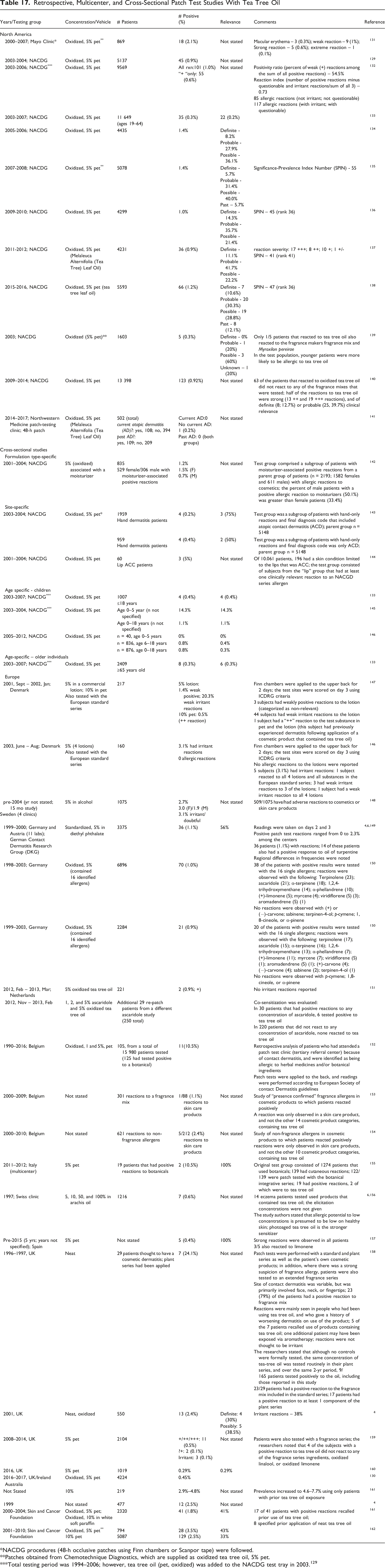
*NACDG procedures (48-h occlusive patches using Finn chambers or Scanpor tape) were followed.
**Patches obtained from Chemotechnique Diagnostics, which are supplied as oxidized tea tree oil, 5% pet.
***Total testing period was 1994–2006; however, tea tree oil (pet, oxidized) was added to the NACDG test tray in 2003. 129
From 2000 to 2007, the Mayo Clinic tested 869 patients with 5% tea tree oil (oxidized); a positive response was found in 18 patients (2.1%). 131 In screening by the NACDG, when tested at 5% (oxidized, in pet) in dermatology patients over 2-year time frames, frequencies of positive reactions ranged from 0.9% (2003–2004; 2011–2012) to 1.4% (2005–2006; 2007–2008).129,134-138 The NACDG measured the positivity ratio (percentage of weak reactions among the sum of all positive reactions) and reaction index (number of positive reactions minus questionable and irritant reactions/sum of all 3) for test results obtained between 2003 and 2006; testing with oxidized tea tree oil had a positivity ratio of 54.5% and a reaction index of 0.73, indicating that 5% tea tree oil (oxidized, in pet) was an “acceptable” patch test preparation. 132 The NACDG also examined the frequency of positive patch test reactions with oxidized tea tree oil as compared to fragrance markers; in 2003, only 1 of the 5/1603 patients that reacted to oxidized tea tree oil also reacted to the fragrance markers fragrance mix and Myroxilon pereirae. 139 During the 2009–2014 time frame, 63 of the 123/13 398 patients that reacted to oxidized tea tree oil did not react to any of the fragrance mixes that were tested. 140 Testing at the Northwestern Medicine patch-testing clinic with 5% Melaleuca Alternifolia (Tea Tree) Leaf Oil (oxidized, in pet) found no difference in positive results between patients with or without atopic dermatitis. 141
Cross-sectional studies were performed by the NACDG. In a subgroup of 835 patients with moisturizer-associated positive reactions (from a parent group of 2193 patients; 2001–2004), 1.2% had positive reactions to oxidized tea tree oil. 142 In subgroups of patients (2003–2004) with hand-only reaction, the percent of positive reactions to oxidized tea tree oil was slightly greater in patients with a final diagnosis code of allergic contact dermatitis only (0.4%), as opposed to those whose diagnosis included allergic contact dermatitis (0.2%). 143 Three of 60 patients (5%) with lip allergic contact cheilitis (ACC) (2001–2004) had positive reactions to oxidized tea tree oil. 144 Cross-sectional NACDG studies also evaluated the sensitization rates in pediatric and older patients. In 2003–2007, 0.4% of pediatric patients (4/1007) that were ≤18 years old had positive reactions to oxidized tea tree oil; during the same time frame, 0.3% of adults (35/11 649) aged 19–64 year old and 0.3% of older patients (8/2409) aged ≥65 years old reacted positively. 133 It was reported that from 2001 to 2004, 14.3% of children aged 0–5 years, and 1.1% of children aged 0–18 years, had a positive reaction to oxidized tea tree oil (total number of patients tested not stated). 145 However, from 2005 to 2012, no pediatric patients (0/40) aged 0–5 years, and 0.3% of patients (n = 876) aged 0–18 years, reacted to the oxidized oil. 146
Testing was also performed in Europe. In Denmark, 44/217 subjects (September 2001 - January 2002) had weak irritant reactions to a commercial lotion that contained 5% tea tree oil, and 1 subject had a ++ reaction to the lotion and 10% tea tree oil in pet 147 ; in June–August 2003, 5/160 subjects had irritant reactions to lotions containing 5% tea tree oil. 147 In Sweden (prior to 2004), 2.7% of 1075 patients tested had a positive reaction to 5% tea tree oil in alcohol. 148 In Germany, testing with 5% tea tree oil (standardized) in diethyl phthalate produced positive results in 1.1% of the 3375 patients tested (1999–2000),4,6,149 and testing at 5% (oxidized) in pet (1998–2003) produced positive results in 0.9%–1.0% of the patients tested. 150 Testing performed in the Netherlands (2012–2013) reported positive results in 0.9% (2/221) of patients patch-tested with 5% tea tree oil (oxidized) in pet. 151 However, when this group and an additional 29 patients from a different study were patch-tested with the 5% oxidized tea tree oil and up to 5% ascaridole (a possible contaminant in aged tea tree oil), 6 of 30 patients that had positive reactions to any concentration of ascaridole also tested positive with tea tree oil; in the 220 patients that did not react to any concentration of ascaridole, none reacted to tea tree oil. In Belgium, 11 of 105 patients (10.5%) had positive reactions to 1 and 5% oxidized tea tree oil in pet; these patients were a sub-group of 15 980 patients that were tested (1990–2016) and identified as being allergic to herbal medicines and/or botanical ingredients. 152 Additional studies performed in Belgium (2000–2010) with fragrance and non-fragrance allergens reported positive reactions to skin care products containing tea tree oil, but not in the other cosmetic product categories.153,154 In testing in Italy with 19 patients that had positive reactions to a botanical integrative series, 2 reacted to 5% tea tree oil in pet. 155 In a Swiss clinic (1997), positive reactions were reported in 0.6% of 1216 patients tested with 5%–100% tea tree oil in arachis oil,6,156 and in Spain (prior to 2015), 0.4% of patients had positive reactions to testing with 5% tea tree oil in pet. 157 In the United Kingdom (UK) (1996–1997), 7 of 29 patients thought to have a cosmetic dermatitis had positive patch test reactions to tea tree oil, applied neat, 158 and in 2001, 2.4% of 550 patients tested with neat, oxidized tea tree oil had positive reactions. 4 Between 2008 and 2016, positive reactions from testing with 5% tea tree oil in pet ranged from 0.1%–0.29% in the UK,159,160 and in 2016–2017, 0.45% of 4224 patients in the UK and Ireland that were patch-tested with 5% tea tree oil (oxidized) in pet had positive reactions. 130
In Australia, positive reaction rates generally appear to be higher than those reported in the US or Europe. The Skin and Cancer Foundation reported a positive reaction rate of 1.8% (41/2320 patients) with 5 and 10% tea tree oil (oxidized) 161 ; however, the same group reported that from 2001 to 2010, the positive reaction rates with 5% (oxidized) and 10% tea tree oil were 3.5% (794 subjects) and 2.5% (5087 subjects), respectively. 162 Additionally, positive reaction rates of up to 4.8% have been reported with 10% tea tree oil. 161
Provocative Testing
Tea Tree Oil
Eight subjects confirmed to previously be sensitized to tea tree oil were tested using occlusive patches to determine their allergic reaction threshold.3,12 Reaction threshold concentrations varied among the subjects, from 0.5% in one subject to a doubtful reaction at 10% in another subject. For the remaining subjects, a 1–3 response was produced in one subject with 1%, in 3 subjects with 2%, and in 2 subjects with 5% tea tree oil. Eleven individual components of tea tree oil were also tested; p-cymene, terpinolene, α-terpinene, and γ-terpinene produced reactions in the sensitized subjects. The study authors commented that they were concerned that the oil samples may have become oxidized during the study.
Forty-three patients with the primary complaint of vulvar pruritus were patch-tested with a battery of allergens, including tea tree oil (undiluted) and common OTC topical vulvar treatments. 163 Of 21 patients that reported using 4 or more topical treatments, 5 of these patients had a positive reaction to tea tree oil. However, tea tree oil was not considered clinically relevant because it was not reported by the patients as being used directly on the vulva to alleviate pruritus.
Cross-Reactivity
Cross-Reactivity With Tea Tree Oil
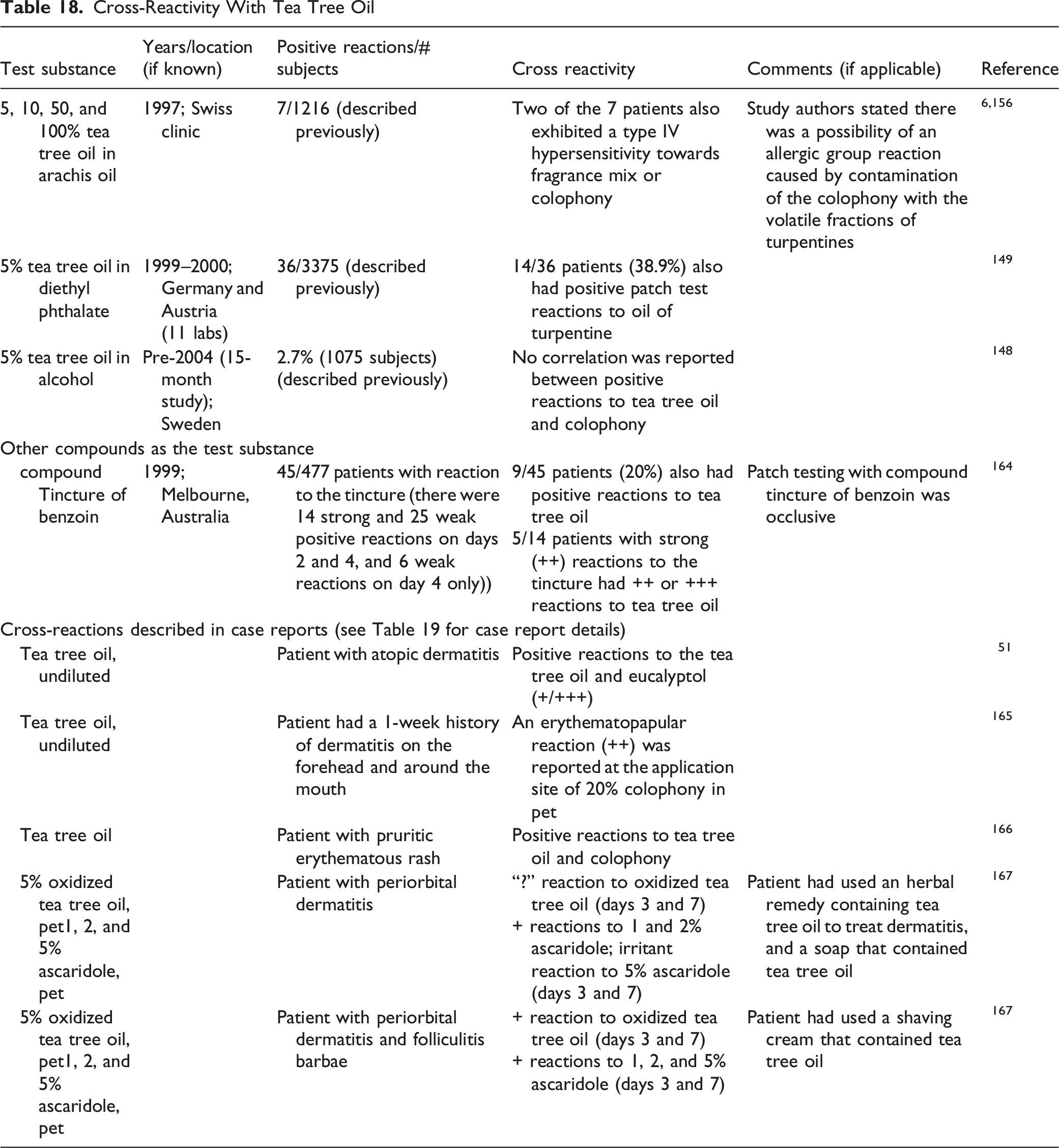
Cross-reactivity with tea tree oil was indicated in some retrospective and multi-center studies. With testing of up to 100% tea tree oil in arachis oil, 2 of the 7 patients that had positive reactions to tea tree oil also exhibited a type IV hypersensitivity towards fragrance mix or colophony; the researchers stated there was a possibility of an allergic group reaction caused by contamination of the colophony with the volatile fractions of turpentine.6,156 In one study in which 36/3375 patients reacted to 5% tea tree oil in diethyl phthalate, 14 of those 36 also had positive patch test reactions to turpentine. 149 However, in another study, no correlation was reported between positive reactions to tea tree oil and to colophony. 148 In 45 patients that had positive patch tests to compound tincture of benzoin, 9 of the 45 also had positive reactions to tea tree oil. 164 In several case reports of reactions to tea tree oil (described later in this report), reactions were also noted with eucalyptol, 51 colophony,165,166 and ascaridole. 167
Case Reports
Tea Tree Oil
Case Reports With Tea Tree Oil

Oral ingestion can be poisonous; serious symptoms, such as confusion and ataxia, can occur. 69 In 2011, the National Capital Poison Center received nearly twice as many calls about tea tree oil than any other named essential oil, including cinnamon oil, clove oil, and eucalyptus oil. 184 In Australia, a retrospective study of essential oil exposure was conducted by analyzing calls to the New South Wales Poisons Information Centre (NSWPIC) during July 2014 to June 2018; NSWPIC takes about half of all calls to poisons information centers in Australia. 185 Tea tree oil was involved in 17% of the reported poisonings.
Risk Assessment
In a 2008 opinion on tea tree oil, the SCCP concluded that a margin of safety (MOS) had not been calculated, and the safety of tea tree oil could not be assessed. 6 The following factors led to this conclusion: tea tree oil is a sensitizer, and sensitization may be enhanced by irritancy; neat tea tree oil and some formulations of 5% or more can induce skin and eye irritation; tea tree oil is prone to oxidation when exposed to air and heat, yielding epoxides and further oxidation products which are considered to contribute to the skin sensitizing potential; and, percutaneous absorption of some constituents of tea tee oil may occur following topical application of the oil and oil-containing products leading to a considerable systemic exposure, but the magnitude of systemic exposure to tea tree oil was uncertain due to a lack of adequate dermal absorption studies.
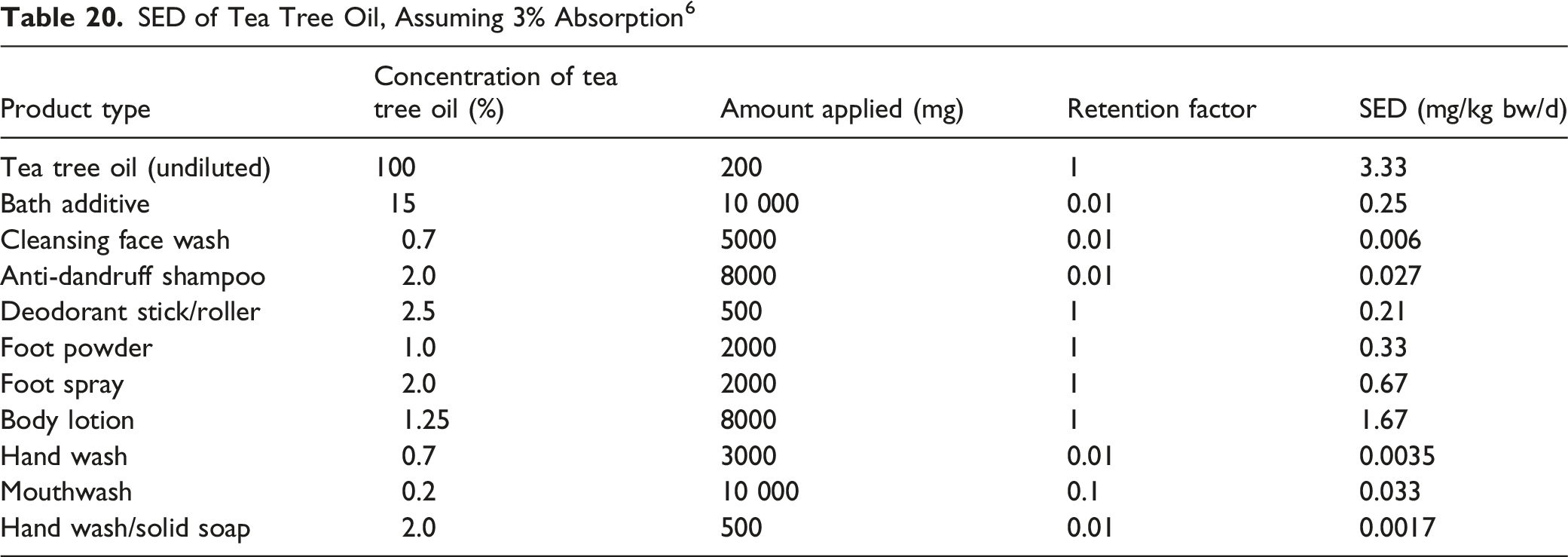
SED of Tea Tree Oil, Assuming 3% Absorption 6
SED and MOS of Tea Tree Oil, Assuming 100% Absorption 42
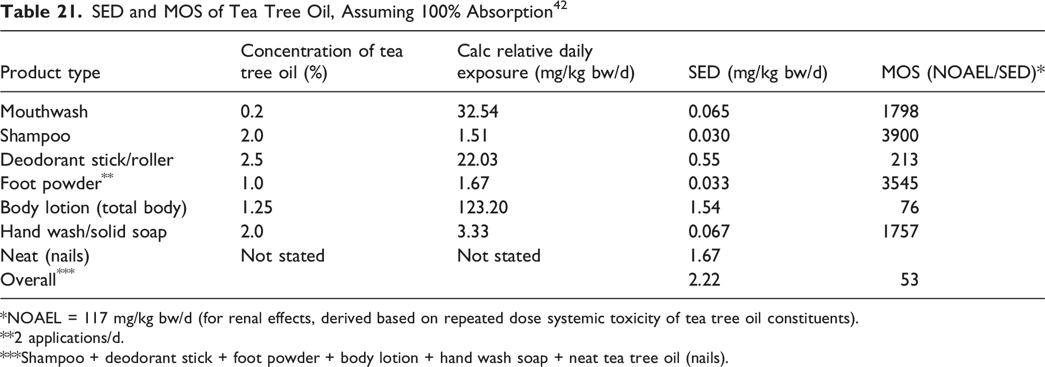
*NOAEL = 117 mg/kg bw/d (for renal effects, derived based on repeated dose systemic toxicity of tea tree oil constituents).
**2 applications/d.
***Shampoo + deodorant stick + foot powder + body lotion + hand wash soap + neat tea tree oil (nails).
Summary
Five of the 8 Melaleuca alternifolia (tea tree)-derived ingredient included in this assessment are reported to function in cosmetics as skin-conditioning agents. Other reported cosmetic functions include abrasive, antioxidant, fragrance ingredient, and flavoring ingredient.
Often, in the published literature, the general name “tea tree” is used, especially, tea tree oil; however, it is not known whether the substance being discussed is equivalent to the cosmetic ingredient. Some constituents of Melaleuca alternifolia have the potential to cause adverse effects. For example, 1,8-cineole (also known as eucalyptol) can be an allergen, and terpinolene, α-terpinene, α-phellandrene, and limonene, ascaridole (a product of tea tree oil oxidation), and 1,2,4-trihydroxymenthane (a product that might be found in aged tea tree oil) are sensitizers. However, the Panel evaluates each ingredient as a whole, complex substance, and not the safety of the individual components.
Melaleuca Alternifolia (Tea Tree) Leaf Water is an aqueous solution of the steam distillates obtained from the leaves of Melaleuca alternifolia. Tea tree oil is the essential oil obtained by steam distillation of the leaves and terminal branchlets of Melaleuca alternifolia (or of Melaleuca linariifolia); it also can be prepared by hydrodistillation, or by solvent extraction.
Six chemotypes have been described for Melaleuca alternifolia; the terpinen-4-ol chemotype is typically used in commercial tea tree oil production. Tea tree oil is reported to contain approximately 100 constituents, with 8 constituents (i.e., terpinen-4-ol, α-terpinene, γ-terpinene, 1,8-cineole, terpinolene, p-cymene, α-pinene, and α-terpineol) typically comprising up to 90% of the oil. Commercial standards for tea tree oil that conform to an ISO specification are indicated. The natural content of the individual constituents of tea tree oil varies considerably depending on the climate, the time of year, the leaf maceration, the biomass used, the age of the leaves, the mode of production, and the duration of distillation. The composition can change as the oil ages, especially when exposed to air, light, and/or high temperatures. Methyleugenol is reported as a minor constituent of Melaleuca Alternifolia (Tea Tree) Leaf Oil.
According to 2021 US FDA VCRP data and Council survey results, 6 of the 8 ingredients included in this safety assessment are currently used in cosmetic formulations. Melaleuca Alternifolia (Tea Tree) Leaf Oil has the greatest frequency and concentration of use; it is reported to be used in 536 cosmetic formulations at a maximum leave-on concentration of 0.63% in cuticle softeners. The highest concentration reported for use in a leave-on product that result in dermal contact is 0.5% Melaleuca Alternifolia (Tea Tree) Leaf Oil, in aerosol deodorants. Collectively, the Melaleuca alternifolia (tea tree)-derived ingredients are reported to be used in products applied near the eye, in products that can result in incidental ingestion, in formulations that come into contact with mucous membranes, and in baby products. Additionally, some of these ingredients are used in spray and powder formulations.
Tea tree oil is listed as a GRAS flavoring substance by FEMA. It is reported to have antimicrobial and antioxidant activity, and has been used as a traditional herbal medicine for centuries. The EMA HMPC concluded that, on the basis of its long-standing use, tea tree oil preparations are approved for a variety of traditional uses. However, the US FDA issued a final action for tea tree oil, establishing that its use in non-prescription OTC consumer antiseptic products intended for use without water is not eligible for evaluation under the OTC Drug Review for use in consumer antiseptic rubs. Additionally, the FDA Pharmacy Compounding Advisory Committee did not recommend Melaleuca Alternifolia (Tea Tree) Leaf Oil for inclusion on the list of bulk drug substances that can be used in pharmacy compounding for topical use in the treatment of nail fungus.
The estimated rates of oral, dermal, and inhalation absorption of tea tree oil were reported to be 70, 3, and 100%, respectively. Because tea tree oil is a semi-volatile substance, the majority of an applied dose would be expected to evaporate from the skin surface before it could be absorbed into the skin. In in vitro studies that used the individual components as markers for penetration, it was demonstrated that the components have distinctly different absorption rates. Additionally, formulation vehicle affects absorption, as does excipients that are used as penetration enhancers.
Tea tree oil increased the percentage of ketoprofen that was delivered across excised porcine skin. However, using human skin samples, it reduced the overall amount of benzoic acid and methiocarb entering the receptor chamber of a static diffusion cell.
In acute dermal toxicity tests in rabbits, the LD50 of tea tree oil was >5 g/kg bw. Dermal applications of “very high concentrations” of tea tree oil have been reported to cause tea tree oil toxicosis in dogs and cats. In an acute oral study, Swiss mice that were given a single dose of 2 g/kg bw Melaleuca Alternifolia (Tea Tree) Leaf Oil by gavage exhibited a wobbly gait, prostration, and labored breathing. In male Wistar rats dosed once with ≤5 g/kg bw Melaleuca Alternifolia (Tea Tree) Leaf Oil by gavage, the LD50 was calculated to be 1.9 g/kg bw. In one study, the oral LD50s of tea tree oil and a nano-emulsion containing tea tree oil were estimated to be 0.854 g/kg bw and 1.565 g/kg bw, respectively. In another study, the LD50 of tea tree oil was >2 g/kg bw (in PEG 400) in female mice, and calculated as 22.3 g/kg bw and ∼1.7 g/kg bw (in peanut oil) in SPF and non-SPF Sprague-Dawley rats, respectively.
In an acute inhalation study in which groups of 5 male and 5 female Wistar rats were exposed nose-only to tea tree oil for 4 h, the LC50 was calculated as 4.78 mg/l for males and females combined, as 5.23 mg/l for males only, and as 4.29 mg/l for females only. No abnormal behavior or signs of toxicity were observed during or after dosing when groups of 10 Sprague-Dawley rats were exposed for 1 h to 50 or 100 mg/l of a test substance that contained 0.3% w/w tea tree oil and 1.8% ethanol in carbon dioxide.
Repeated dermal applications of 2% tea tree oil to the shaved back of rats for 28 days did not result in any significant changes in SGOT or SGPT levels. In a 28-day gavage study (OECD TG 407) in which groups of 10 male ICR mice were dosed with up to 200 mg/kg bw of a nano-emulsion containing tea tree oil (comprising the oil (4% w/w), Tween 80 (2% w/w), CMC (0.2% w/w), and water), the NOAEL was determined to be >200 mg/kg bw. In a 28-day gavage study in which male and female Sprague-Dawley rats were given doses of up to 45 mg/kg bw/d tea tree oil in corn oil, the NOAEL was determined to be 45 mg/kg bw/d for both male and female rats.
A developmental toxicity study was performed in accordance with OECD TG 414, in which gravid female rats were dosed by gavage with up to 250 mg/kg bw/d tea tree oil in PEG 400 on days 5 to 19 of gestation. The NOAELs for maternal toxicity and for developmental toxicity (secondary to severe maternal toxicity) were 20 mg/kg bw/d tea tree oil. An increase in the number of late embryonic deaths and post-implantation loss, leading to an overall higher total intrauterine mortality, was observed in the high-dose group; the increase in post-implantation mortality was considered to be secondary to maternal toxicity. A statistically significant higher incidence of skeletal malformations unrelated to intrauterine growth retardation was noted in the high-dose group, and a statistically significant increase in the number of skeletal variations secondary to maternal toxicity was noted in the 100 and 250 mg/kg bw/d groups.
The effects of tea tree oil on the morpho-functional parameters of porcine spermatozoa were evaluated.by exposing spermatozoa samples to ≤2 mg/ml tea tree oil for 3 h. Viability of spermatozoa was statistically significant decreased with ≥1 mg/ml tea tree oil, and a concentration-dependent decrease in motility was observed with concentrations of 0.4 mg/ml and greater.
Tea tree oil did not demonstrate genotoxic activity. In vitro, tea tree oil was not mutagenic in an Ames test using S. typhimurium and E. coli WP2 uvr A, with or without metabolic activation, in chromosomal assays using V79 cells (≤58.6 µg/ml) or human lymphocytes (≤365 µg/ml), in an in vitro mammalian cell micronucleus assay using human lymphocytes (≤365 µg/ml), in a mammalian cell transformation assay (120 and 275 µg/ml, without and with metabolic activation, respectively), or in a Comet assay using HaCaT cells (≤0.064%). In vivo, Melaleuca Alternifolia (Tea Tree) Leaf Oil was not clastogenic in a mammalian erythrocyte micronucleus test in which mice were dosed orally with up to 1750 mg/kg bw in corn oil.
Carcinogenicity studies were not identified in the published literature. However, numerous studies investigating ant-carcinogenic potential of tea tree oil were found. Tea tree oil exhibited antiproliferative activity against murine AE17 mesothelioma cells and B16 melanoma cells, it impaired the growth of human M14 melanoma cells, and it induced apoptosis in human malignant melanoma (A-375) and squamous cell carcinoma (Hep-2) cells. Tea tree oil also exhibited anti-proliferative activity against human lung carcinoma (H1299, A549) cells; however, in this study, tea tree oil did not have significant effect on the proliferation of breast (MDA-MB-231) or colon carcinoma (HCT116) cell lines. In a different study using human MCF-7 and murine 4T1 breast cancer cells, tea tree oil exhibited an anti-tumor effect by decreasing cell viability and modulating apoptotic pathways. Tea tree oil also inhibited glioblastoma cell growth in vitro (in human U87MG glioblastoma cells) and in vivo (in a subcutaneous model using nude CD1 mice) in a dose- and time-dependent manner, and the mechanisms were associated with cell cycle arrest, triggering DNA damage and inducing apoptosis and necrosis. The IC50 of tea tree oil in human MDA MB breast cancer cells was 25 µg/ml (48 h). The IC50 in several other cancer cell lines ranged from 12.5 µg/ml (24 h) in human HT29 colon cancer cells, to 2800 µg/ml (4 h) in epithelioid carcinomic (HeLa), hepatocellular carcinomic (Hep G2), and human chronic myelogenous leukemia (K-562) cells. In immunocompetent C57BL/6 mice, tea tree oil inhibited the growth of subcutaneous tumors; effectiveness was carrier-dependent.
Human MCF-7 breast cancer cells were used to examine the effect of tea tree oil on ERα-regulated gene expression; ERα target genes showed significant induction when treated with tea tree oil, and the ERE-dependent luciferase activity was stimulated in a dose-dependent manner (maximum activity observed at 0.025%). Fulvestrant inhibited transactivation of the 3X-ERE-TATA-luciferase reporter, indicating that the activity observed is ER-dependent. In an E-screen assay using MCF-7 BUS cells, tea tree oil (≤0.1%; without E2) induced a weak, but significant, dose-dependent estrogenic response at concentrations ranging from 0.00075% to 0.025%, with a maximal response (corresponding to 34% of the maximal E2 response) induced by a concentration of 0.0125% tea tree oil; when tested in the presence of E2, concentrations of <0.025% tea tree oil reduced the RPE effect by 10%. A robotic version of the E-screen cell proliferation assay was performed with MCF-7:WS8 cells to evaluate the estrogenic activity (with ≤5 × 10⁻⁶ g/ml) and the anti-estrogenic activity (with ≤6.85 × 10⁻⁷ g/ml) of an ethanol extract of a hair conditioner product that contained tea tree oil. The formulation did not exhibit estrogenic activity, but it did exhibit anti-estrogenic activity; the normalized anti-estrogenic activity (as relative maximum % of the positive control) was 79%. Human HepG2 hepatocellular cancer cells were also used to examine estrogenic effects. In a luciferase reporter assay using transfected cells, tea tree oil (≤0.025%) produced a maximum of an ∼20-fold increase in ERα ERE-mediated promotor activity, and in a mammalian two-hybrid binding assay to determine binding activity to the ERα LBD, there was a significant induction of ERα ERE-mediated activity with 0.01% tea tree oil, and tea tree oil demonstrated binding to the LBD of ERα.
The androgenic activity of tea tree oil was evaluated in MDA-kb2 breast cancer cells (in the presence and absence of DHT). In cells transfected with an MMTV-luciferase reporter plasmid, tea tree oil did not transactivate the reporter plasmid at any concentration tested (≤0.01%), and it inhibited plasmid transactivation by DHT in a concentration-dependent manner; maximum inhibition occurred with 0.005% tea tree oil. Additional experiments indicated that the anti-androgenic properties of tea tree oil extended to inhibition of DHT-stimulated expression of androgen-inducible endogenous genes. In another luciferase reporter assay AR MMTV, increasing concentrations of tea tree oil, co-treated with testosterone, significantly inhibited MMTV-mediated activity at concentrations ≥0.0005% (v/v); change in activity, as compared to testosterone, was 36%. In a study examining the effect of tea tree oil on AR-regulated gene expression, tea tree oil, co-treated with testosterone, significantly inhibited the target genes.
The potential for tea tree oil to induce mucosal damage was examined in porcine uterine mucosa; no damage was observed with up to 20 mg/ml tea tree oil, but at 40 mg/ml, moderate damage was induced to the uterine mucosa, with a multifocal detachment of the epithelium. In an ex vivo study using uterine horns from female sows, tea tree oil (≤0.4 mg/ml) did not alter the structure of the uterine mucosa.
Immunological effects of tea tree oil were examined in vitro, in mice (via dermal route and inhalation), and in humans (dermal application). In vitro, tea tree oil had a weak effect on suppression of neutrophil activation; the IC50 of neutrophil adherence was 0.033%.
In dermal studies using mice, undiluted tea tree oil (applied before or after challenge) reduced swelling induced by TNCB in sensitized, but not in non-sensitized, mice. In examining whether the oil had an effect on swelling associated with UVB irradiation, a single topical application of undiluted tea tree oil after irradiation did not suppress swelling in mice; additionally, swelling was significantly increased when tea tree oil was applied before UVB irradiation. Cutaneous application of tea tree oil to mice decreased MPO activity, from 100% in controls to approximately 55% in the treated group. In mice exposed to tea tree oil via inhalation, there was an increase in the level of circulating blood immunoglobulins and the blood granulocyte number. Additionally, in mice exposed to tea tree oil vapors, and then induced with peritonitis, peritoneal leukocyte activity in the test group was equivalent to that seen in control groups without inflammation, indicating that tea tree oil had anti-inflammatory action.
In one study using human subjects, undiluted tea tree oil did not have an effect on the mean flare area induced by histamine when the oil was applied 20 min after histamine injection; however, the mean wheal volume was statistically significantly decreased. In another study, in which undiluted tea tree oil was applied to the injection site at both 10 and 20 min after histamine injection, a significant reduction in both the flare and the wheal response was observed.
Emulsions of tea tree oil in culture medium containing 10% fetal calf serum were cytotoxic to adherent PBMCs. Significant toxicity was reported at a concentration of 0.016%.
Irritant effects were reported in rabbits after a single 4-h semi-occlusive application and after a single 24-h occlusive application of undiluted Melaleuca Alternifolia (Tea Tree) Leaf Oil. Tea tree oil was reported to cause irritation in animals, in a concentration-dependent manner; in rats, application of 5% tea tree oil produced very slight erythema, and 10% produced well-define erythema. In rabbits, concentrations of up to 75% were, at most, slightly irritating; with undiluted tea tree oil, a 4-h semi-occlusive application and application for 72 h to intact and abraded skin produced severe irritation. In 22 human subjects, a 48-h occlusive patch with 1% Melaleuca Alternifolia (Tea Tree) Leaf Oil in pet produced no irritation. In a clinical 3-week occlusive patch test, slight irritation was reported with concentrations of up to 10% tea tree oil in sorbolene cream (5 patches/week, duration not stated; 28 subjects). Two dermal irritation studies were performed with 25% tea tree oil; in one study, no irritation was reported. In the other study, which was a 3-week occlusive patch test in 28 subjects, no irritation was reported with 25% tea tree oil in soft white paraffin; however, an allergic response (erythema with marked edema and itching) was observed in 3 subjects. In a 48-h patch test with undiluted tea tree oil in 219 subjects, the prevalence of marked irritancy was 2.4%–4.3%, and the prevalence of any irritancy (mild to marked) was 7.2%–10.1%.
In the LLNA, tea tree oil was predicted to be a weak or moderate sensitizer at a concentration up to 50%, and a moderate sensitizer when tested undiluted. In guinea pig studies, tea tree oil was not sensitizing (30% at challenge) or had a low sensitizing capacity (tested “pure”); however, one study indicated that tea tree oil was possibly a weak sensitizer, with 30% tea tree oil producing positive reactions in 3/10 animals at challenge. In guinea pig studies in which “pure” tea tree oil was used at induction and oxidized tea tree oil was used at challenge, an increase in mean response was observed when compared to challenge with “pure” oil. In clinical studies, a formulation containing 0.001% Melaleuca Alternifolia (Tea Tree) Flower/Leaf/Stem Extract (25 subjects; maximization test), a formulation containing 0.0078% Melaleuca Alternifolia (Tea Tree) Leaf Extract (105 subjects; modified Draize HRIPT), and Melaleuca Alternifolia (Tea Tree) Leaf Oil at 1% in pet (22 subjects; maximization test) and at 10% in caprylic/capric triglycerides (102 subjects; modified HRIPT), were not sensitizers. In a Draize sensitization study with 5, 25, or 100% tea tree oil in various excipients, 3 of 309 subjects (0.97%) developed skin reactions suggestive of active sensitization during the induction period; only 1 of the 3 subjects returned for challenge, and the reaction was confirmed in that subject. Because different samples of tea tree oil were tested simultaneously, it was not possible to determine which specific concentration was responsible for inducing sensitization in this subject at challenge; no other subjects had reactions at challenge. Three of an initial 28 subjects that developed reactions in the irritation study with 25% tea tree oil in soft white paraffin, had positive reactions when challenged 2 weeks after the initial study; testing was also performed using components of tea tree oil, and all 3 sensitized subjects reacted positively to the sesquiterpenoid fractions and sesquiterpene hydrocarbons. Melaleuca alternifolia is contraindicated in cases of known allergy to plants of the Myrtaceae family. Tea tree oil can cross react with colophony.
A single application of undiluted tea tree oil was not phototoxic in hairless mice. However, some irritation was observed.
Tea tree powder and tea tree ground leaf were classified as non-irritants in the HET-CAM assay. Undiluted tea tree oil and water-soluble tea tree oil were both classified as a severe irritant in the HET-CAM assay; however, tea tree oil was classified as not to be an ocular corrosive or severe irritant in a BCOP test. Additionally, using rabbits, tea tree oil was classified as minimally irritating to rabbit eyes when tested at a concentration of up to 5%, and undiluted tea tree oil was considered a mild ocular irritant.
Oxidized tea tree oil (5% in pet) has been part of the NACDG screening series since 2003, and it was added to the British Society for Cutaneous Allergy facial allergy series in 2019. From 2000 to 2007, the Mayo Clinic tested 869 patients with 5% tea tree oil (oxidized); the positive response rate was 2.1%. In screening by the NACDG, when tested at 5% (oxidized) in pet in dermatology patients over 2-year time frames, frequencies of positive reactions ranged from 0.9% to 1.4%. The NACDG also examined the frequency of positive patch test reactions with tea tree oil as compared to fragrance markers; in 2003, only 1 of the 5/1603 patients that reacted to oxidized tea tree oil also reacted to the fragrance makers fragrance mix and Myroxilon pereirae. During the 2009–2014 timeframe, 63 of the 123/13 398 patients (51%) that reacted to oxidized tea tree oil did not react to any of the fragrance mixes that were tested. Testing at the Northwestern Medicine patch-testing clinic with 5% Melaleuca Alternifolia (Tea Tree) Leaf Oil (oxidized, in pet) found no difference in positive results between patients with or without atopic dermatitis.
Cross-sectional studies were also performed by the NACDG examining the effects of oxidized tea tree oil, based on symptoms or age. In patients with moisturizer-associated positive reactions (2001–2004), 1.2% had positive reactions to oxidized tea tree oil. In subgroups of patients (2003–2004) with hand-only reactions, the percent of positive reactions to oxidized tea tree oil was slightly greater in patients with a final diagnosis code of allergic contact dermatitis only (0.4%), as opposed to those whose diagnosis included allergic contact dermatitis (0.2%) among the diagnoses. In 60 patients with lip ACC (2001–2004), 3 (5%) had positive reactions to oxidized tea tree oil. In 2003–2007, 0.4% of pediatric patients that were ≤18 years had positive reactions to oxidized tea tree oil; during the same time frame, 0.3% of adults aged 19–64 years and 0.3% of older patients aged ≥65 years reacted positively. It was reported that from 2001 to 004, 14.3% of children aged 0–5 years, and 1.1% of children aged 0–18 years, had a positive reaction to oxidized tea tree oil; however, from 2005 to 2012, no pediatric patients (0/40) aged 0–5 years, and 0.3% of patients aged 0–18 years, reacted to the oxidized oil.
Testing was also performed in Europe. Frequencies of positive reactions varied greatly, especially when examining reactions in subgroups of patients. In Denmark, 20% of subjects (September 2001 - January 2002) had weak irritant reactions to a commercial lotion that contained 5% tea tree oil, and 1 subject had a ++ reaction to the lotion and 10% tea tree oil in pet; in June to August 2003, 3.1% of subjects had irritant reactions to lotions containing 5% tea tree oil. In Sweden (prior to 2004), 2.7% of patients tested had a positive reaction to 5% tea tree oil in alcohol. 148 In Germany, testing with 5% tea tree oil (standardized) in diethyl phthalate produced positive results in 1.1% of the patients tested (1999–2000), and testing at 5% (oxidized) in pet (1998–2003) produced positive results in 0.9%–1.0% of the patients tested. Testing performed in the Netherlands (2012–2013) reported positive results in 0.9% of patients patch-tested with 5% tea tree oil (oxidized, in pet). However, when this group and an additional 29 patients from a different study were patch-tested with the 5% oxidized tea tree oil and up to 5% ascaridole (a possible contaminant in aged tea tree oil), 6 of 30 patients (20%) that had positive reactions to any concentration of ascaridole also tested positive with tea tree oil; in the 220 patients that did not react to any concentration of ascaridole, none reacted to tea tree oil. In Belgium, 10.5% of patients had positive reactions to 1 and 5% oxidized tea tree oil in pet; these patients were a sub-group of 15 980 patients that were tested (1990–2016) and identified as being allergic to herbal medicines and/or botanical ingredients. Additional studies performed in Belgium (2000–2010) with fragrance and non-fragrance allergens reported positive reactions to skin care products containing tea tree oil, but not in the other cosmetic product categories. In testing in Italy with 19 patients that had positive reactions to a botanical integrative series, 2 (10.5%) reacted to 5% tea tree oil in pet. In a Swiss clinic (1997), positive reactions were reported in 0.6% of patients tested with 5%–100% tea tree oil in arachis oil, and in Spain (prior to 2015), 0.4% of patients had positive reactions to testing with 5% tea tree oil in pet. In the UK (1996–1997), 7 of 29 patients (24%) thought to have a cosmetic dermatitis had positive patch test reactions to tea tree oil, applied neat, and in 2001, 2.4% of 550 patients tested with neat, oxidized tea tree oil had positive reactions. Between 2008 and 2016, positive reactions from testing with 5% tea tree oil in pet ranged from 0.1% to 0.29% in the UK, and in 2016–2017, 0.45% of 4224 patients in the UK and Ireland that were patch-tested with 5% tea tree oil (oxidized) in pet had positive reactions.
In Australia, positive reaction rates generally appear to be higher than those reported in the US or Europe when patch-testing general populations of patients. The Skin and Cancer Foundation reported a positive reaction rate of 1.8% with 5 and 10% tea tree oil (oxidized); however, the same group reported that from 2001 to 2010, the positive reaction rates with 5% and 10% tea tree oil were 3.5% and 2.5%, respectively. Additionally, positive reaction rates of up to 4.8% have been reported with 10% tea tree oil.
Cross-reactivity with tea tree oil was indicated in some retrospective and multi-center studies. With testing of up to 100% tea tree oil in arachis oil, 2 of the 7 patients that had positive reactions to tea tree oil also exhibited a type IV hypersensitivity towards fragrance mix or colophony; the researchers stated study there was a possibility of an allergic group reaction caused by contamination of the colophony with the volatile fractions of turpentine. In one study in which 36/3375 patients reacted to 5% tea tree oil in diethyl phthalate, 14 of those 36 also had positive patch test reactions to turpentine. However, in another study, no correlation was reported between positive reactions to tea tree oil and to colophony. In 45 patients that had positive patch tests to compound tincture of benzoin, 9 of the 45 also had positive reactions to tea tree oil. In several case reports of reactions to tea tree oil, reactions were also noted with eucalyptol, colophony, and ascaridole.
Numerous cases of reaction to tea tree oil have been reported. Adverse reactions were reported with use for treatment of dermatitis and/or psoriasis, other direct skin applications, and from use of hand wash or shampoos. Patients with sensitivity to tea tree oil (dermal and/or oral) were also reported to have reactions to constituents or degradation products of tea tree oil, and positive reactions were reported in a patient with hand eczema following inhalation of tea tree oil vapors. Oral ingestion can be poisonous; serious symptoms, such as confusion and ataxia, can occur.
Daily exposure to tea tree oil was calculated for various product types. Using a rate of percutaneous absorption of 3%, SED estimates between 0.0017 mg/kg bw/d (2% tea tree oil in a hand soap) and 3.33 mg/kg bw/d (undiluted tea tree oil) were obtained. When assuming complete absorption as % of applied dose, SED values for different product types ranged from 0.030 mg/kg bw/d (2.0% tea tree oil in a shampoo) to 1.54 mg/kg bw/d (1.25% tea tree oil in a body lotion). Using 100% absorption and an NOAEL of 117 mg/kg bw/d (for renal effects, derived based on repeated dose systemic toxicity of tea tree oil constituents), and MOS values ranged from 76 (body lotion) to 3900 (shampoo). Based on an aggregate exposure, the SED was calculated as 2.22 mg/kg bw/d, and the overall MOS was 53.
Discussion
This assessment reviews the safety of 8 Melaleuca alternifolia (tea tree)-derived ingredients as used in cosmetic formulations. The Panel concluded that the data included in this review are sufficient for determining the safety of these ingredients as reportedly used in cosmetics.
The majority of the data included in the report is on tea tree oil. Although this name is not an International Nomenclature Cosmetic Ingredient (INCI) name, the Panel considered these data relevant for evaluating the safety all of the cosmetic ingredients named in this report because most constituents of concern are present at the highest levels in oil-derived ingredients, and no signals for additional constituents of concern were noted in the extracts.
The Panel noted that oxidized tea tree oil has the potential to be a sensitizer, and stated that methods should be employed to minimize oxidation of the oil in the final cosmetic formulation. For example, to reduce the formation of oxidation products, manufacturers should consider the use of antioxidants, as well as specific packaging to minimize exposure to light.
Also, because final product formulations may contain multiple botanicals, each possibly containing the same constituents of concern, formulators are advised to be aware of these constituents and to avoid reaching levels that may be hazardous to consumers. For Melaleuca alternifolia (tea tree)-derived ingredients, examples of the constituents the Panel was concerned about include 1,8-cineole (also k as eucalyptol), a possible allergen, and terpinolene, α-terpinene, α-phellandrene, and limonene, which are possible sensitizers. Additionally, the Panel is aware that variances in the composition of tea tree oil, based on geographical or geological differences in growth, have been reported, which could also affect the potential for sensitization. Therefore, when formulating products, manufacturers should avoid reaching levels of plant constituents that may cause sensitization or other adverse health effects.
The Panel expressed concern about pesticide residues, heavy metals, and other plant species that may be present in botanical ingredients. Additionally, the Panel was made aware that some of the Melaleuca alternifolia (tea tree)-derived ingredients could be supplied as adulterated products; the Panel acknowledged this could always be a concern. For these reasons, it was stressed that the cosmetics industry should continue to use current good manufacturing practices (cGMPs) to limit impurities.
Adverse effects that were reported in developmental and reproductive toxicity studies, as well as in studies examining effects on endocrine activity, were noted by the Panel. Because the adverse effects were observed at concentrations that were much higher than those used in cosmetic formulations, concern for these effects with use in cosmetics was mitigated.
The Panel recognized that tea tree oil can enhance the penetration of other ingredients through the skin. The Panel cautioned that care should be taken in formulating cosmetic products that may contain these ingredients in combination with any ingredients whose safety was based on their lack of dermal absorption data, or when dermal absorption was a concern.
Finally, some of the Melaleuca alternifolia (tea tree)-derived ingredients are used in cosmetic sprays or powders, and could possibly be incidentally inhaled during cosmetic use; for example, Melaleuca Alternifolia (Tea Tree) Leaf Oil is reported to be used at up to 0.5% in aerosol deodorant formulations, and Melaleuca Alternifolia (Tea Tree) Leaf Oil and Melaleuca Alternifolia (Tea Tree) Leaf Water are reported to be used in face powders. Little inhalation toxicity data (i.e., only acute studies in rats) were available. In the absence of adequate inhalation data, the Panel noted that in aerosol products, 95%–99% of droplets/particles would not be respirable to any appreciable amount. Furthermore, droplets/particles deposited in the nasopharyngeal or bronchial regions of the respiratory tract present no toxicological concerns based on the chemical and biological properties of these ingredients. Coupled with the small actual exposure in the breathing zone and the concentrations at which the ingredient is used, the available information indicates that incidental inhalation would not be a significant route of exposure that might lead to local respiratory or systemic effects. A detailed discussion and summary of the Panel’s approach to evaluating incidental inhalation exposures to ingredients in cosmetic products is available at https://www.cir-safety.org/cir-findings.
Conclusion
The Expert Panel for Cosmetic Ingredient Safety concluded that the following 8 Melaleuca alternifolia (tea tree)-derived ingredients are safe in cosmetics in the present practices of use and concentration described in this safety assessment when formulated to be non-sensitizing.
Melaleuca Alternifolia (Tea Tree) Extract
Melaleuca Alternifolia (Tea Tree) Flower/Leaf/Stem Extract
Melaleuca Alternifolia (Tea Tree) Flower/Leaf/Stem Oil*
Melaleuca Alternifolia (Tea Tree) Leaf
Melaleuca Alternifolia (Tea Tree) Leaf Extract
Melaleuca Alternifolia (Tea Tree) Leaf Oil
Melaleuca Alternifolia (Tea Tree) Leaf Powder*
Melaleuca Alternifolia (Tea Tree) Leaf Water
* Not reported to be in current use. Were ingredients in this group not in current use to be used in the future, the expectation is that they would be used in product categories and at concentrations comparable to others in this group.
Footnotes
Author’s Note
Unpublished sources cited in this report are available from the Director, Cosmetic Ingredient Review, 555 13th Street, NW, Suite 300W, Washington, DC 20004, USA.
Author Contributions
The articles in this supplement were sponsored by the Cosmetic Ingredient Review.
Funding
The authors disclosed receipt of the following financial support for the research, authorship, and/or publication of this article: The articles in this supplement were sponsored by the Cosmetic Ingredient Review. The Cosmetic Ingredient Review is financially supported by the Personal Care Products Council.
Declaration of Conflicting Interests
The authors declared no potential conflicts of interest with respect to the research, authorship, and/or publication of this article.