
Abstract
Meckel-Gruber syndrome (MKS) is a rare lethal autosomal recessive disorder with typical anomalies including encephalocele, multicystic renal dysplasia, congenital liver fibrosis, and polydactyly. MKS is caused by mutations of genes localized on different chromosomes. Karyotypes of published Meckel–Gruber syndrome cases are without any aberrations. We present a male fetus with meningoencephalocele, multicystic renal dysplasia, congenital liver fibrosis, and other anomalies. Standard cytogenetic examination of cultured fetal skin and muscle fibroblasts showed mosaic trisomy 17. Homozygous deletion in CC2D2A gene was found by Sanger sequencing. This is to our knowledge the first case of genetically confirmed Meckel–Gruber syndrome with incidental cofinding of mosaic trisomy 17. Abnormal karyotype does not exclude diagnosis of MKS with risk of recurrence 25% in next pregnancy. In the case of anomalies typical for Meckel–Gruber syndrome, genetic analysis is indicated.
Keywords
Introduction
Meckel–Gruber syndrome (Meckel syndrome, Gruber syndrome, MKS) belongs to non-motile ciliopathies, genetic disorders with primary ciliary dysfunction.1,2 This syndrome was described for the first time by Meckel in 1822 and later by Gruber in 1934, who named it “dysencephalia splanchnocystica.” 3
The incidence of MKS varies worldwide, from 1:140000 to 1:13250 birth, 4 with high incidence in Finland 1:9000. 4 The highest incidence was observed in the Gujarati Indians (1:1300). 5
MKS is a rare lethal autosomal recessive disease, characterized by central nervous system (CNS) malformations, with the occipital meningoencephalocele occurring in 90% of cases, bilaterally enlarged kidneys with the multicystic dysplasia and fibrotic changes of the liver in probably all cases, and polydactyly in 80% of cases. 3 Other frequent findings are congenital heart defects, lip and palate clefts, microphtalmia and other ocular anomalies, genital, gonadal, and other CNS abnormalities.1,3 Minimal diagnostic criteria of MKS include cystic dysplasia of the kidneys as an obligate feature, fibrotic changes of the liver, and occipital encephalocele or other CNS malformations. 6 Potter-like facial dysmorphology and limb anomalies (club feet, legs flexed beneath the enlarged abdomen) caused by oligo- or anhydramnios are common. 3
MKS is genetically heterogenous disease with 15 known causative genes, MKS1-13, TMEM138, and TMEM237, localized on different chromosomes. 7 Meckel–Gruber case reports described in the literature did not report any chromosomal aberrations. In our presented case report, unusual finding of mosaic trisomy 17 was observed.
Case Report
History
A secundigravida, nullipara was admitted to the hospital in the twenty-third gestational week because of ultrasonographically verified fetal anomalies as described later. No information about the pregnancy course was available. Because of anhydramnios, amnioinfusion was done before cordocentesis. Cordocentesis was carried out because of its higher informative value in comparison to amniocentesis caused by possible contamination of amniotic fluid. An abortion was induced by intraamnial prostaglandins application.
Prenatal Ultrasound Findings (Fetus 1)
During ultrasonographic examination (fetus 1) in the twenty-third ongoing gestational week, fetal anomalies including occipital defect with brain tissue protruding outside the skull and enlarged multicystic kidneys were described (Figure 1(A)–(D)).
A to D, Prenatal ultrasonographic examination (fetus 1) at 22 weeks and 6 days of gestation. Transverse scan of the fetal head (A) and sagittal scan of the cervico-occipital region (B) showing occipital defect with protruding brain tissue outside the skull (arrows). The scull is tightly surrounded by uterine walls due to severe oligohydramnios. Transverse (C) and sagittal (D) scans showing enlarged multicystic kidneys of the fetus (marked by circles on the images). E and F, Prenatal ultrasonographic examination (fetus 2) at 8 weeks and 4 days of gestation. Longitudinal (E) and transverse (F) scan of the embryo with dilated rhombencephalis cavity (5.5 × 4.6 mm). G and H, Prenatal ultrasonographic examination (fetus 2) at 10 weeks and 5 days of gestation. Sagittal (G) and transverse (H) scan of the fetus showing occipital defect with brain tissue protruding outside the scull (arrows).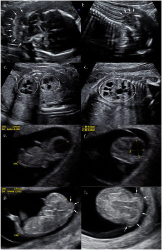
Pathological Examination
A male fetus in the twenty-third gestational week (690 g/34 cm) was submitted to pathological examination. The Potter-like facial deformities, hypertelorism, broad and flat nose, broad cheeks, low set ears, and wide mouth with prominent lips were noted. The most distinctive finding was the defect localized in the occipital region of the head, behind the posterior fontanelle with eventration of meninges and part of the brain tissue measured 20 × 18 mm (Figure 2(A)) containing clear fluid and brain tissue. The abdomen was distended. Leg anomalies included prominent heels bilaterally, club foot on the right, rocker bottom foot, and sandal gap deformity on the left side (Figure 2(B)).
Macroscopical and histological findings (fetus 1) in the twenty-third gestational week. A, Occipital meningoencephalocele. B, Deformities of low extremities. C, Thorax cavity with heart and small lungs, liver in abdominal cavity pushed up and forward. D, Almost the whole abdominal cavity was filled with enlarged kidneys. E and F, Enlarged kidneys with uneven surface and multiple cysts on the cut. G, Fetal lung with decreased amount of interstitial tissue between tubular structures, H&E, 200×. H, Fetal kidney with normal renal parenchyma in the subcapsular part of the kidney and multiple thin walled cysts in renal cortex and medulla lined by one layer of cuboidal epithelium, H&E, 100×. I, Fetal liver with increased amount of fibrous tissue and proliferation of bile ducts in portal areas (arrows), H&E, 100×. J, Immunohistochemistry with antibody against CK19 demonstrating peripheral proliferating bile ducts, 100×, DAB.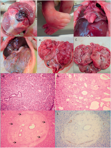
Internal examination
Lungs were smaller bilaterally and weighted 8.4 g with the lung weight/body weight (LW/BW) ratio 0.012 (1.2%) (Figure 2(C)). Almost the whole abdominal cavity was filled with enlarged kidneys, with other abdominal organs pushed up and forward (Figure 2(D)). The size of the kidney on the right side was 55 × 35 × 25 mm and on the left side was 60 × 35 × 30 mm, with uneven rough surface (Figure 2(E)). Multiple cysts up to 10 mm, filled with clear fluid were present on the cut, with thin rim of renal parenchyma preserved under the capsule (Figure 2(F)).
Histological examination
The lung parenchyma was collapsed bilaterally, with decreased amount of interstitial tissue between tubular epithelial structures, findings typical for lung hypoplasia. Developmentally, lung parenchyma represented glandular to tubular stage (Figure 2(G)). The renal parenchyma with glomeruli and normal-sized tubules was preserved in the subcapsular part of the kidney. The finding of numerous thin-walled cysts in renal cortex and medulla lined by one layer of cuboidal epithelium and separated by loose connective tissue was histologically consistent with the multicystic renal dysplasia (Figure 2(H)). The increase of fibrous tissue in the liver, with proliferation and peripheral arrangement of biliary ducts in portal areas was suggestive of the congenital liver fibrosis (Figure 2(I) and (J)).
Genetic Testing
The standard cytogenetic examination of amniocytes was unsuccessful because of contamination of amniotic fluid. Array-comparative genomic hybridization (CGH) examination of umbilical blood did not show any genomic imbalances.
Standard cytogenetic examination of the long-term cultured fetal skin and muscle fibroblasts showed mosaic trisomy 17 with karyotype 46, XY/47, XY + 17 with the lines ratio of 20:2. The presence of C-metaphase with the pathological findings was also evidenced in the cultured skin and muscle fibroblasts, so the pseudomosaicism was excluded. However, the cultivation of placental sample (chorionic villus) was unsuccessful. Negative results of array CGH examination of umbilical blood was likely due to the inability of this method to detect genomic imbalance in such low percentage mosaicism.
The DNA of both parents was extracted from peripheral blood using QIAamp DNA Blood Mini Kit (Qiagen, Hilden, Germany). The DNA was processed using TruSight One (Illumina, San Diego, CA) gene panel target enrichment and massively parallel sequencing was carried out on Illumina MiSeq system. In both parents, the same single-nucleotide deletion in CC2D2A gene was revealed. Both parents were heterozygous for the deletion of c.3829-1delG (p.Val1097Phefs) in the gene CC2D2A, described earlier as pathogenic in homozygous form. 8 This mutation was subsequently confirmed by Sanger sequencing in the fetus in homozygous state. First, the DNA was extracted from formalin-fixed and paraffin-embedded (FFPE) tissue sample using GeneRead DNA FFPE Kit (Qiagen, Hilden, Germany). Following that, the extracted DNA was amplified in polymerase chain reaction (PCR) with primers designed specifically to detect the particular deletion in CC2D2A gene using Primer3 software 9 with following sequences—CC2D2A-F: CATTGGGAACTCAGAATTTGC and CC2D2A-R primer: TCATTCCAGCTAGGGTTTGG. Subsequently, the PCR product was analyzed by Sanger sequencing using BigDye Terminator 3.1 cycle sequencing chemistry (ThermoFischer Scientific, Waltham, MA) and ABI 3500 genetic analyzer. We consider the detected variant (deletion in CC2D2A gene) to be causative for the observed phenotype of Meckel–Gruber syndrome type 6 in the fetus.
Gene panel analysis using massively parallel sequencing was repeatedly attempted on fetal tissue sample with no success. Therefore, the panel sequencing was carried out instead in samples from both parents. The same mutation was revealed in both parents in heterozygous state by panel sequencing which was later confirmed also in the fetus by Sanger sequencing to be in homozygous state.
Family History
Both parents denied consanguinity, with an otherwise noncontributory family history without known birth defects, genetic conditions, recurrent miscarriages, or sudden unexplained deaths. Parents ancestors came from the neighboring villages in Slovak Republic.
Nearly one and half year after previously described induced abortion, the patient visited the same ultrasonographer in the ninth and subsequently in the eleventh gestational week of the next pregnancy. Fetal anomalies very suspicious of Meckel–Gruber syndrome were present again (Figure 1(E)–(H)). Unfortunately, the track of the patient was lost and no further information is available for this pregnancy.
Prenatal Ultrasound Findings (Fetus 2)
Prenatal ultrasonographic examination (fetus 2) showed dilated rhombencephalis cavity (5.5 × 4.6 mm) at ongoing ninth week of gestation (Figure 1(E) and (F)) and occipital defect with brain tissue protruding outside the scull at eleventh ongoing gestational week (Figure 1(G) and (H)).
Discussion
Meckel–Gruber syndrome is a rare lethal disorder with autosomal recessive inheritance. Although suggestive, the chromosomal aberration has apparently never been observed in connection with MKS.2,10
The presented case is to our knowledge the first reported case of genetically confirmed Meckel–Gruber syndrome with additional and probably incidental mosaic trisomy 17.
Mosaic trisomy 17 belongs to the rarest aneuploidies identified. Only 29 cases with this aneuploidy were reported to date. 11 Most of them were diagnosed prenatally, usually with no anomalies after birth and with normal karyotype in peripheral blood lymphocytes. Just in few cases, the mosaicism was confirmed in skin fibroblasts postnatally. Ultrasonographic findings typical for these fetuses were intrauterine growth retardation, nuchal thickening, cerebellar hypoplasia, cardiac abnormalities, asymmetric anomalies, foot position abnormalities, pleural effusions, and single umbilical artery. None of these clinical features overlap with clinical features of MKS cases. 11
Differential diagnosis of MKS includes trisomy 13, trisomy 18, autosomal dominant polycystic kidney disease, Smith-Lemli-Opitz syndrome, Joubert syndrome, and Bardet-Biedl syndrome.5,12
HNF1B (hepatocyte nuclear factor-1 beta) is a gene involved in development of different organs including the kidneys. It is localized on the long arm (q) of chromosome 17 at position 12 (17q12). Mutations of this gene result in different renal anomalies, especially renal cysts. 13 The most frequent mutation is deletion; however, association of 17q12 duplication with multicystic dysplastic kidney was described.13,14
17q12 duplication as possible causative factor of multicystic renal dysplasia could lead to misinterpretation of etiology of renal cystic dysplasia in our presented case report. Panel sequencing is expensive and not generally available method in comparison to standard karyotype examination. The fetus in our case was phenotypically typical for Meckel–Gruber syndrome. However, cases of MKS without classical symptomatology, eg, Meckel syndrome with Dandy–Walker malformation without encephalocele 15 associated with incidental mosaic trisomy 17 karyotype demonstrated in our case could explain fetal renal anomaly. Incorrect diagnosis could lead to inappropriate management of subsequent pregnancies. Trisomy 17 is without increased risk for the next pregnancy due to postzygotic mosaic formation.
We faced a challenge in identification of the causative mutation in the fetus. We originally planned to carry out comprehensive gene panel analysis using massively parallel sequencing but the formalin fixed or formalin-fixed paraffin embedded samples of the fetus were incompatible with the Trusight One kit by Illumina. We therefore changed strategy and analyzed samples from both parents. Meckel Gruber candidate genes were searched for disease causing or probably disease-causing mutations in both parents and single mutation was found in both in heterozygous state. This was later confirmed also in the fetus by Sanger sequencing in homozygous state.
Considering autosomal recessive inheritance of Meckel–Gruber syndrome, the risk of recurrence in next pregnancy is 25%. Prenatal diagnosis of MKS includes ultrasonography and amniotic fluid or chorionic villi genetic analysis. Mothers with positive history of pregnancy with Meckel–Gruber syndrome may benefit from earlier ultrasonographic examination. Signs of the occipital defect are apparent before 12 weeks of gestation as we demonstrate in Figure 1(E)–(H). This fact allows earlier and thus more safe decision for termination of such pregnancy. However, the diagnosis in early pregnancy does not require the presence of all signs, and classical triad encephalocele, polydactyly, and polycystic kidneys do not manifest on imaging until much later, which may cause diagnostic problems. 16
Prenatal genetic counseling with diagnostic sequencing may be necessary in similar cases with unusual phenotypes as the genetic proof is crucial in diagnosing the disease. Options to prevent recurrence of MKS in next pregnancy are in preimplantation genetic diagnosis and in vitro fertilization.
In conclusion, the Meckel–Gruber syndrome is lethal disorder because of pulmonary hypoplasia and other developmental abnormalities with a recurrence risk of 25%. Abnormal karyotype does not exclude diagnosis of Meckel–Gruber syndrome, therefore in suspicious cases, genetic testing should be performed.
Footnotes
Declaration of Conflicting Interests
The author(s) declared no potential conflicts of interest with respect to the research, authorship, and/or publication of this article.
Funding
The author(s) disclosed receipt of the following financial support for the research, authorship, and/or publication of this article: This contribution is the result of implementation of the project “REVOGENE – Research centre for molecular genetics” (ITMS 26240220067) supported by the Research & Developmental Operational Programme funded by the ERDF.