
Abstract
Background:
Fetal hemorrhage (FH) is an important and difficult cause of death to recognize at perinatal autopsy. Existing literature is restricted to case reports and small case series, and none comprehensively describe both placental and autopsy findings. We sought to characterize fetal and placenta findings where the ultimate cause of death was determined to be FH to aid in the identification of these cases.
Materials and Methods:
This is an autopsy series of perinatal deaths with fetomaternal hemorrhage (FMH) and FH into the amnionic sac. We included singleton pregnancies with Kleihauer Betke (KB) testing, and divided cases into 3 groups: FMH with ≥40% of fetal blood volume, FMH ≤39%, and suspected FH into the amnionic sac with negative KB test.
Results:
We identified 20 cases from 691 perinatal autopsies, 10 with FMH ≥40%, 5 with FMH ≤39%, and 5 suspected FH into the amnionic sac. Cases with FMH≥40% were likely to have a normal placental weight, villous edema, and fetal hydrops. While FH into the amnionic sac was more likely to have a small placenta with disruption/defect of fetal vessels, and less likely to have fetal hydrops.
Conclusion:
This study describes the differences and similarities between cases with FMH and FH in to the amnionic sac, and highlights the need for careful placenta and fetal autopsy examinations.
Introduction
Perinatal autopsy serves as an important part of the investigation of death in the fetal and neonatal period.1,2 While there is no universally accepted and utilized classification of perinatal death, the existing systems generally divide causes of death into categories such as maternal medical conditions during pregnancy, obstetric complications, maternal or fetal hematologic conditions, fetal genetic/karyotypic and structural karyotypic abnormalities, placental and/or fetal infection, and pathologic placental conditions.3-5 Acute in utero events can lead to brain injury in the neonate, but may also contribute to perinatal death. They can be difficult to detect at autopsy and are sometimes referred to as “sentinel events.” 6 These events include placental abruption or uterine rupture, acute umbilical cord occlusion, maternal hypotension, and fetal hemorrhage. 6 Fetal hemorrhage occurs in utero, by definition, and includes hemorrhage into a body cavity or organ, into the placenta/intervillous space, or into the amnionic sac. Detecting fetal hemorrhage into the amnionic sac or placenta/intervillous space (fetomaternal hemorrhage (FMH)) can be difficult at the autopsy bench, and definitive diagnosis usually requires correlation with clinical and laboratory findings such as the appearance of the amnionic fluid and Kleihauer-Betke (KB) test.7,8
Literature review reveals several case reports and small series of fetal hemorrhage, variably addressing the diagnoses of FMH or fetal hemorrhage into the amnionic sac, and variably addressing pathologic features of either the placenta or fetus or both.9-15 Despite these case examples, the diagnosis of fetal hemorrhage at autopsy continues to be a diagnostic challenge to the pathologist. Our aim was to describe the placental and perinatal autopsy findings in a relatively large cohort of patients diagnosed with fetal hemorrhage as a cause of death and bring attention to the most relevant findings in these cases with the goal of improving pathologists' recognition and diagnosis.
Methods
This was a retrospective cohort of perinatal autopsy cases at Endeavor Health (formerly NorthShore University HealthSystem) located in Evanston, Illinois. The study was reviewed and approved by the Endeavor Health Institutional Review Board (IRB2026-0007). A search of the Pathology laboratory PowerPath system was conducted to find perinatal autopsy cases between 1/1/2017 and 12/31/2025. Search terms included “fetomaternal hemorrhage” and “fetal hemorrhage.” We included singleton perinatal autopsies and excluded autopsies of twins due to the difficulty in interpreting the source of the hemorrhage. We also excluded any autopsy case in which a KB test was not performed and/or the results of KB test were not identified in the available medical records since the KB test is the most definitive test for FMH. Based on the KB result (mL of fetal blood in the maternal circulation), the % of the fetal blood volume was calculated using the estimation of 100 mL of fetal blood per kg fetal weight. To establish cause of fetal death, we used the Initial Causes of Fetal Death (INCODE) system requirements for fetomaternal hemorrhage, 3 and applied these same quantitative standards to liveborn infants. Cases with FMH were divided into those with ≥40% (FMH ≥ 40) of the fetal blood volume, defined in INCODE as a probable cause of fetal death, or those with ≤39% (FMH ≤ 39) of the fetal blood volume, defined in INCODE as a possible cause of fetal death. Cases with suspected fetal hemorrhage into the amnionic sac and a KB test which was resulted as no evidence of FMH were considered a third group: fetal hemorrhage into the amnionic sac (FH-AS). Maternal demographic data, medical history, and delivery information were manually extracted from the autopsy reports and available medical records.
Placental pathology information was collected from either the autopsy or surgical pathology report. Placentas were grossed within 24-72 hours after fixation in 10% neutral-buffered formalin. Standard microscopic sections were submitted of the membranes, umbilical cord, full-thickness parenchyma x2, maternal surface, and any parenchymal lesions. Placental data included trimmed placental weight and umbilical cord abnormalities defined as furcate cord insertion, velamentous cord insertion, hypercoiling (>3 coils/10 cm), true knot, marginal cord insertion (within 2 cm of the placental margin), and nuchal cord or body cord, if documented in the medical record. Microscopic findings included stage/grade of acute inflammation (AI), chronic inflammation, fetal vascular malperfusion (FVM), and maternal vascular malperfusion (MVM).16,17 We also recorded the presence or absence of increased nucleated erythrocytes in the fetal vasculature (>10 per 10 high-power fields) and villous edema.
Performance of the autopsy cases was completed by the staff of the autopsy department, including a resident, pathologists’ assistant, and an autopsy technician, all under the direct supervision of a perinatal autopsy pathologist. All examined perinatal autopsy cases underwent the institution's routine autopsy procedure, which includes external examination, in situ and internal examination, organ dissection, and histological sampling of all visceral organs. Autopsy data collected from the autopsy reports included cause of death, body weight, results of the KB test, presence or absence of fetal hydrops (defined by presence of excessive fluid in at least 3 of the following: subcutaneous, pleural cavity, peritoneal cavity or pericardial sac), increased hepatic extramedullary hematopoiesis for gestational age, and bone marrow erythroid hyperplasia (defined as myeloid to erythroid ratio 1:1-2 or greater).
Characteristics of only the FMH ≥ 40 group and FH-AS group were compared using Chi-square tests for categorical variables and Mann-Whitney U test for continuous variables. Analyses were performed using IBM SPSS Statistics V28.0.0.0.
Results
Case Selection
691 perinatal autopsies were performed over the 8-year study period, and 57 (8%) were identified by the initial search. Of these, 30 of the autopsy reports included the search terms, but indicated that fetomaternal or fetal hemorrhage was not found, and therefore, these cases were excluded. There were 27 (4%) cases where the cause of death was identified to be related to suspected fetomaternal hemorrhage or fetal hemorrhage; however, cases without KB results (N = 5) and cases of twins (N = 2) were excluded. Therefore, 20 cases were identified in total (2.9% of all perinatal autopsies). Ten cases were identified as FMH ≥ 40, 5 cases as FMH ≤ 39, and 5 cases as FH-AS.
Demographics
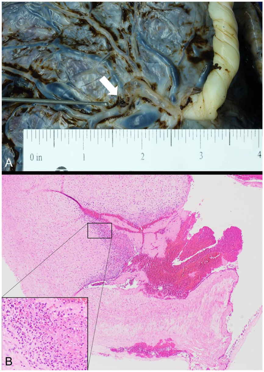
In general, the 3 study groups showed similar demographic characteristics (Table 1). Patients in the FMH ≥ 40 had higher parity compared to those in the FH-AS group. Interestingly, most of the patients in all of the groups had no prior medical history at all, with only 2/20 (10%) having maternal diabetes mellitus and 2/20 (10%) cases having maternal hypertensive disease. No cases examined showed maternal drug use, trauma, clinical chorioamnionitis, or preterm rupture of membranes. Of note, the mean gestational ages trended closer to term in the group FH-AS, whereas the mean gestational ages of FMH ≥ 40 and FMH ≤ 39 were between 29 and 31 weeks. All cases of FH-AS had bloody amnionic fluid upon delivery, except 1 case which did not indicate the appearance of the amnionic fluid. No cases in the FMH ≥ 40 or FMH ≤ 39 groups had documented bloody-appearing amnionic fluid.
Maternal Demographic Data, Clinical Information, and Placental Pathology.
Bold text in the table data represents statistically signficant differences between FMH ≥ 40% and FH-AS.
Abbreviations: FMH, fetomaternal hemorrhage; FH-AS, fetal hemorrhage into amnionic sac; GA, gestational age; NRBC, nucleated red blood cells.
Values represent either N (%) or mean (standard deviation).
Placental Pathology
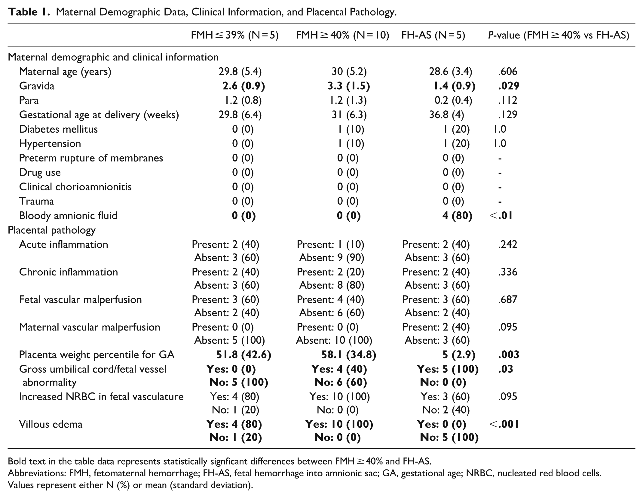
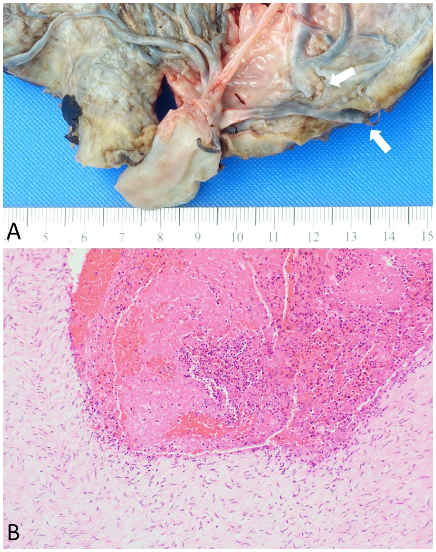
There was no statistically significant difference in the prevalence of AI, CI, FVM, or MVM between the groups; however, MVM was completely absent from the 2 FMH groups, and was present as high-grade MVM in 40% (2/5) of FH-AS cases (Table 1). Mean placental weight percentile was between 50th and 60th percentile for FMH ≥ 40 and FMH ≤ 39, but all placentas from FH-AS group were below the 10th percentile for gestational age. Villous edema was present in nearly all of the FMH cases (14/15, 93.3%) and absent from those with FH-AS (P < .001) while a majority of cases in all 3 groups showed increased nucleated erythrocytes in the fetal vasculature (Table 1). All of the FH-AS cases had umbilical cord or large fetal vessel abnormalities (ruptured velamentous vessels in the setting of vasa previa, furcate insertion with vessel rupture (Figure 1), chorionic vessel defect (Figure 2), ruptured umbilical cord hematoma/intrafunicular hemorrhage, and umbilical cord ulceration in the setting of intestinal atresia). In the 3 cases examined with ruptured velamentous, furcate, or chorionic vessels, all showed inflammation in the wall of the ruptured vessel, and 2 also demonstrated thrombosis. The 2 cases with umbilical cord pathology showed acute hemorrhage in Wharton's jelly and smooth muscle disruption/injury in the umbilical vessels.
(A) Furcate umbilical cord insertion with disrupted furcate vessel (arrows). (B) Histology showing thrombosis and acute inflammation in the disrupted vessel wall. Hematoxylin and Eosin, 10×.
(A) Chorionic vessel defect (arrow). (B) Eosinophilic T-cell vasculitis in disrupted vessel wall. Hematoxylin and Eosin, 4×.
Autopsy Results
The majority of the autopsies were performed on stillborn fetuses (n = 17) while only 3 of the perinates died in the neonatal period. Mean body weight percentile for gestational age was not statistically significantly different between the groups (Table 2).
Autopsy Findings.
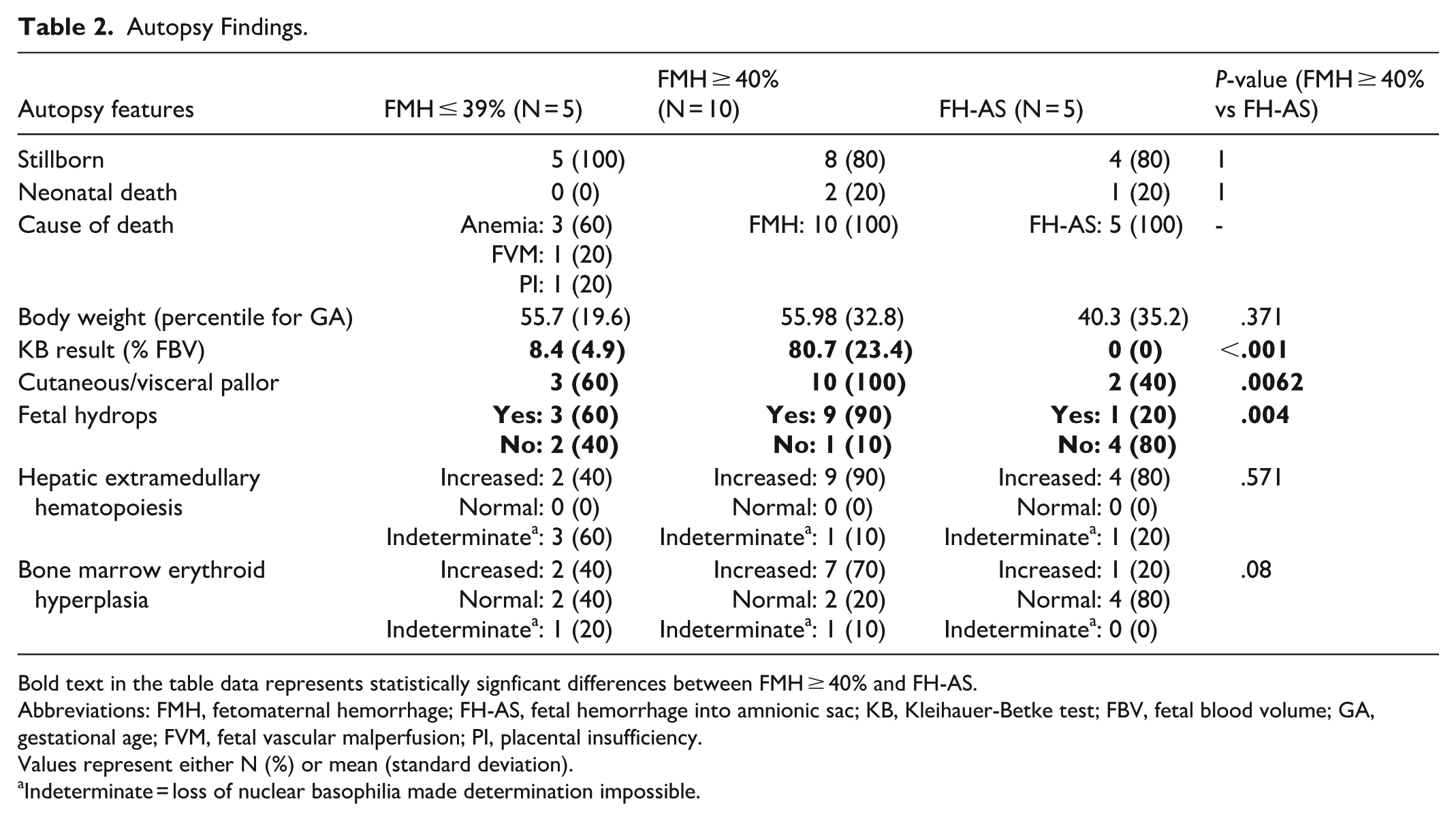
Bold text in the table data represents statistically signficant differences between FMH ≥ 40% and FH-AS.
Abbreviations: FMH, fetomaternal hemorrhage; FH-AS, fetal hemorrhage into amnionic sac; KB, Kleihauer-Betke test; FBV, fetal blood volume; GA, gestational age; FVM, fetal vascular malperfusion; PI, placental insufficiency.
Values represent either N (%) or mean (standard deviation).
Indeterminate = loss of nuclear basophilia made determination impossible.
For cases from FMH ≥ 40, the cause of death was fetomaternal hemorrhage in all cases (Figure 3). From this cohort, 5/10 (50%) of the cases showed a KB result of 100% of the fetal blood volume in the maternal circulation. Of the remainder of the cases, 3/10 (30%) showed the loss of between 60% and 90% of the fetal blood volume and 2/10 (20%) showed between 40% and 50% of fetal blood volume. Visceral findings consistent with fetal anemia were seen in the majority of cases including 90% with fetal hydrops, 100% with cutaneous pallor, 70% with bone marrow erythroid hyperplasia (Figure 4(A)), and 90% with increased hepatic extramedullary hematopoiesis (Figure 4(B)). All cases showed signs of fetal stress in the viscera, including thymic involutional changes (100%), aspirated squamous cells in the pulmonary airspaces (100%), and hypoxic ischemic changes in the brain (70%) which ranged from acute (40%) to subacute/chronic (30%) (Figure 4(C)).
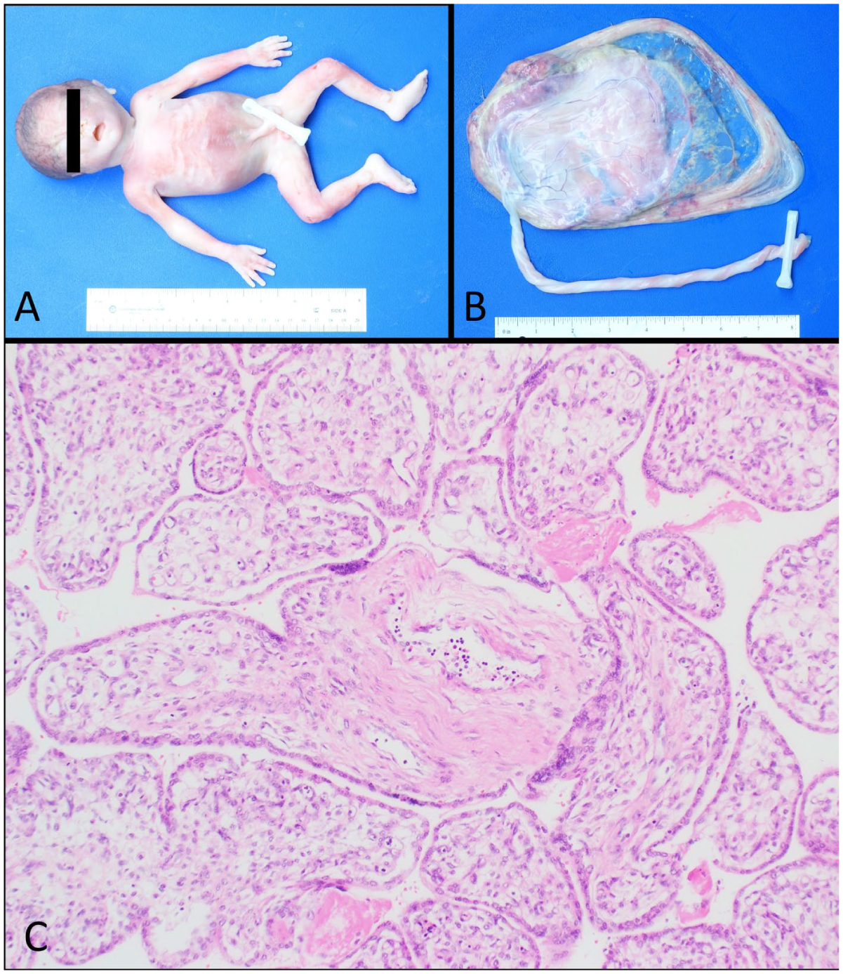
(A) Fetus and (B) placenta exhibiting the characteristic pallor seen in fetomaternal hemorrhage. (C) Placenta demonstrating villous edema and increased nucleated erythrocytes in the fetal vasculature. Hematoxylin and Eosin, 4×.
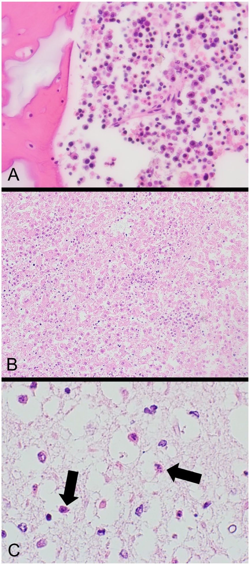
Fetal hypoxic changes. (A) Bone marrow erythroid hyperplasia. Hematoxylin and Eosin, 60×. (B) Liver of term stillborn fetus with increased extramedullary hematopoiesis. Hematoxylin and Eosin, 20×. (C) Brain showing shrunken, eosinophilic neurons with karyorrhexis (arrows) in the subiculum. Hematoxylin and Eosin, 60×.
Causes of death for the FMH ≤ 39 group included anemia (possible FMH), placental insufficiency, and umbilical cord blood flow obstruction/fetal vascular malperfusion. The 3 cases with fetal anemia as a cause of death had signs of possible blood loss, including cutaneous pallor, lack of intravascular blood, villous edema, nucleated erythrocytes in the fetal vasculature, and erythroid hyperplasia in the bone marrow and/or increased extramedullary hematopoiesis in the liver. However, in these cases, the KB results were insufficient to assign fetomaternal hemorrhage (7%-16% of fetal blood volume) as a probable cause of death. In another case, the cause of death was determined to be obstruction of umbilical cord blood flow, with thrombosis in the fetal vasculature and regionally distributed avascular villi. The KB results did show a small FMH of 6% of the fetal blood volume, indicating FMH as possible contributing factor to the fetal demise. The last case had a cause of death of placental insufficiency secondary to probable congenital infection associated with chronic villitis in the placenta and congenital pneumonia in the fetus. The KB result for this case also indicated a small FMH representing 3% of the fetal blood volume.
Autopsies from the FH-AS cohort were significantly less likely to show fetal hydrops than those with FMH as a probable cause of death (Table 2). Cutaneous and visceral pallor were also seen in only 40% of the cases.
Discussion
This autopsy case series of fetal hemorrhage identified FMH 2 times more often than fetal hemorrhage into the amnionic sac. While there is no pathognomonic finding in FMH, our data show that the most distinctive autopsy and placental findings associated with large volume FMH are a normal placental weight with villous edema and a hydropic fetus. In contrast, fetal hemorrhage into the amnionic sac is associated with disruption or defect of the umbilical cord or fetal vessels, bloody amnionic fluid, a small placenta, and the fetus is less likely to show signs of hydrops. These findings suggest that some cases of FMH may develop over a longer period of time as opposed to the more acute development of FH-AS.
It is hypothesized that some fetal blood (approximately 1% of fetal blood volume) crosses the placenta into maternal circulation during pregnancy without apparent clinical significance in many pregnancies.18,19 FMH, which accounts for 4% of all stillbirths, does not have a known cause. 20 This entity can be acute or chronic, and can lead to severe anemia, disseminated intravascular coagulopathy, cardiac failure, and death. Chronic cases can cause hypoxia and thus lead to neurologic pathology, such as hypoxic ischemic encephalopathy.6,18,21 INCODE (Initial Causes of Fetal Death), created by the Stillbirth Collaborate Research Network, set the percentage of fetal blood in maternal circulation sufficient to be a probable cause fetal death at 40% of the fetal blood volume. 3 Cases in which a KB test or flow cytometry for fetal hemoglobin are ≥40% are considered to be “probable” causes of death, whereas cases between 5% and 39% are only considered to be “possible” causes of death. Though FMH is uncommonly detected antenatally, it can be treated by cordocentesis with intrauterine transfusion to correct the fetal anemia. 18
Umbilical cord rupture can be seen in cases with cord abnormalities, including velamentous cord insertion, or nuchal/body cord wrapping which significantly shortens the cord length. 22 The most frequently reported type of cord rupture is a Benckiser's hemorrhage, where one or several vessels are ruptured in the setting of a velamentous umbilical cord insertion. 12 Some studies report fetal exsanguination in approximately 75% to 100% of cases, while others report closer to 50% mortality.10,23 Chorionic or umbilical cord vessel rupture can be secondary to mechanical or toxin-induced injury, vascular dilatation, vessel wall injury, or structural abnormalities of the umbilical cord vessels. 11 Studies have shown that the most common site for a rupture of the cord is near the cord insertion site on the placental disc. 23 This demands careful examination of the placenta at the surgical bench or during autopsy to identify potential small defects in a vessel of the cord or chorionic plate, if there is suspicion of fetal hemorrhage.
Laceration of fetal vessels can occur on unprotected fetal vessels, such as in cases of vasa previa, velamentous cord insertion, furcate cord insertion, or accessory lobes with membranous linking vessels.8,24 Other causes of injury to fetal vessels can include umbilical cord ulceration, iatrogenic injury form diagnostic procedures, or extensive tension on the umbilical cord during descent of the fetus.14,25 These diagnoses are often not made until autopsy, and can be easily overlooked even during postmortem examination. 8 A high index of suspicion and familiarity with the array of autopsy findings in fetal hemorrhage can potentially improve diagnostic recognition. If disrupted vessels are seen during a postmortem examination, documentation of artifactual tearing after delivery versus antemortem vessel injury is paramount. Microscopic sections of the disrupted vessels may show evidence of inflammation or thrombosis. Of the 5 cases of FH-AS examined in this study, 3 showed inflammation and/or thrombosis, while 2 showed intrafunicular hemorrhage with umbilical vessel wall injury.
Fetal findings of anemia are variable in cases of fetal hemorrhage and can typically include cutaneous, visceral, conjunctival, and oral mucosal pallor, as well as a lack of intravascular blood or flattening of the inferior vena cava or innominate vein.9,15,26-28 Postmortem hematocrits can also be performed to confirm anemia if it is suspected at the time of autopsy, 29 but may be difficult to obtain. In our series, postmortem hemocrit was obtained in only 1 case. Findings of fetal anemia are typically insufficient alone to make the definitive diagnosis of fetal hemorrhage from umbilical cord rupture, chorionic vessel rupture, or FMH. In addition to the KB test, careful examination of the placenta should be considered required in these cases. This study demonstrates the significance of evaluating placental pathology in cases of suspected fetal hemorrhage, as the placental and umbilical cord findings determined the source of hemorrhage in all 5 cases with fetal hemorrhage into the amnionic sac. See Figure 5 for suggested approach of cases in which fetal hemorrhage is suspected.
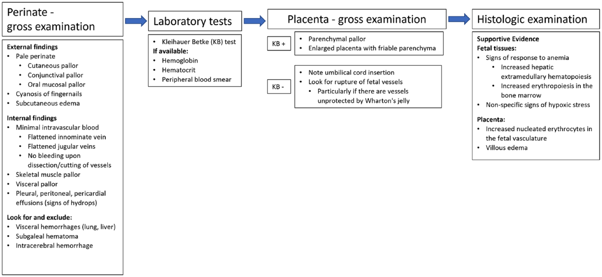
Suggested approach for perinatal autopsy and placenta examination in cases of fetal hemorrhage.
When genetic testing is desired from placental villous tissue, oftentimes the specimen is taken in the delivery room from the fetal side near the umbilical cord insertion per local protocol. This practice can disrupt the insertion site and potentially limit the pathologist’s ability to assess cord insertion abnormalities or large fetal vessel pathology. This study demonstrates the importance of examining this area on the fetal surface in establishing a cause of death in FH-AS. Therefore, this practice can be harmful, potentially obscuring the cause of death, particularly in cases of FH-AS and also potentially in cases of cord accident when thrombi are disrupted in this area. Pathologists can educate their clinical colleagues about the potential impact of the specimen sampling protocol on their placental pathology examination, and suggest sampling away from the cord insertion site.
To our knowledge, this case series is the largest descriptive study of fetal hemorrhage in the literature and is the first to compare the placental and fetal findings of FMH with fetal hemorrhage into the amnionic sac. Although this is a retrospective study, a major strength is that the perinatal autopsy cases were performed systematically with a careful gross and histologic examination along with comprehensive placental examination in all cases. An additional strength is that we only included cases with a documented KB test to be absolutely sure of the source of the fetal hemorrhage. However, the largest limitation of this study was the lack of KB testing performed in many perinatal mortalities, which led to the relatively small sample size despite this being the largest case series. In summary, our study highlights the differences and similarities between cases with large FMH and fetal hemorrhage into the amnionic sac. Our results show that careful inspection of fetal and placental tissues as well as a high index of suspicion for the diagnosis of fetal hemorrhage is important, particularly when faced with a pale perinate on the autopsy table lacking intravascular blood suggestive of blood loss anemia.
Footnotes
Funding
The authors received no financial support for the research, authorship, and/or publication of this article.
Declaration of Conflicting Interests
The authors declared no potential conflicts of interest with respect to the research, authorship, and/or publication of this article.