
Abstract
To the Editor:
Paragonimiasis is a parasitic disease caused by paragnonimus species; the primary site of infection is the lung. 1 Herein, we report a case of paragonimiasis presenting as an abdominal mass.
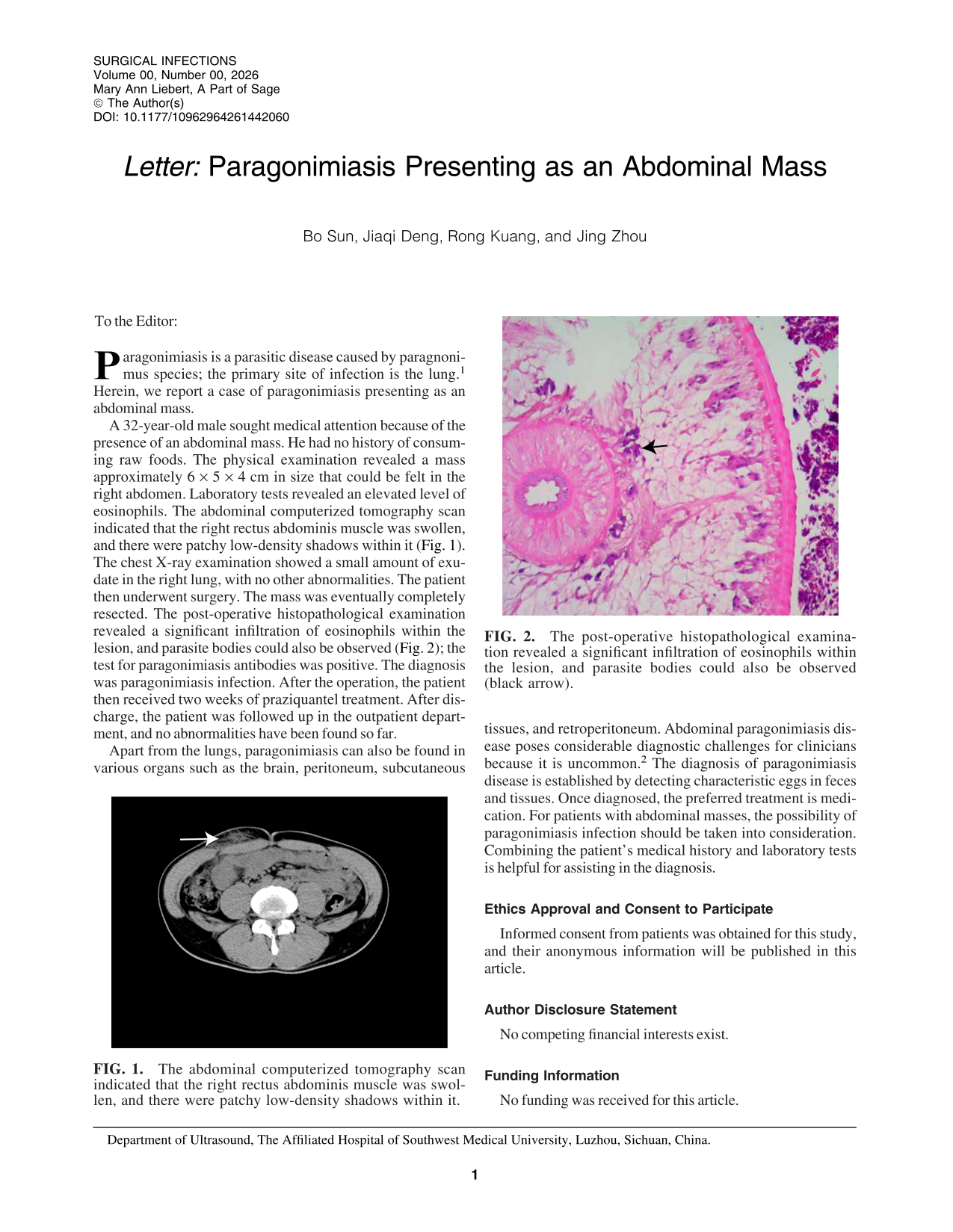
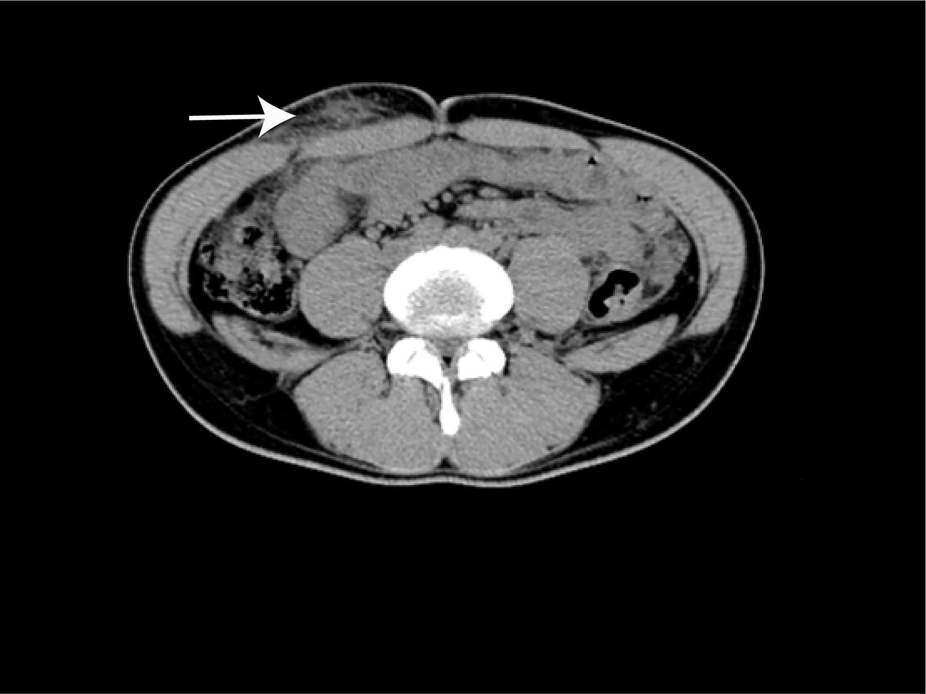
A 32-year-old male sought medical attention because of the presence of an abdominal mass. He had no history of consuming raw foods. The physical examination revealed a mass approximately 6 × 5 × 4 cm in size that could be felt in the right abdomen. Laboratory tests revealed an elevated level of eosinophils. The abdominal computerized tomography scan indicated that the right rectus abdominis muscle was swollen, and there were patchy low-density shadows within it (Fig. 1). The chest X-ray examination showed a small amount of exudate in the right lung, with no other abnormalities. The patient then underwent surgery. The mass was eventually completely resected. The post-operative histopathological examination revealed a significant infiltration of eosinophils within the lesion, and parasite bodies could also be observed (Fig. 2); the test for paragonimiasis antibodies was positive. The diagnosis was paragonimiasis infection. After the operation, the patient then received two weeks of praziquantel treatment. After discharge, the patient was followed up in the outpatient department, and no abnormalities have been found so far.
The abdominal computerized tomography scan indicated that the right rectus abdominis muscle was swollen, and there were patchy low-density shadows within it.
The post-operative histopathological examination revealed a significant infiltration of eosinophils within the lesion, and parasite bodies could also be observed (black arrow).
Apart from the lungs, paragonimiasis can also be found in various organs such as the brain, peritoneum, subcutaneous tissues, and retroperitoneum. Abdominal paragonimiasis disease poses considerable diagnostic challenges for clinicians because it is uncommon. 2 The diagnosis of paragonimiasis disease is established by detecting characteristic eggs in feces and tissues. Once diagnosed, the preferred treatment is medication. For patients with abdominal masses, the possibility of paragonimiasis infection should be taken into consideration. Combining the patient’s medical history and laboratory tests is helpful for assisting in the diagnosis.
Ethics Approval and Consent to Participate
Informed consent from patients was obtained for this study, and their anonymous information will be published in this article.
Footnotes
Author Disclosure Statement
No competing financial interests exist.
Funding Information
No funding was received for this article.