
Abstract
The antibacterial effect of chloramphenicol, hibiscus acid, and a mixture of hibiscus acid and chloramphenicol against antibiotic-resistant enterohemorrhagic Escherichia coli (EHEC) and Salmonella Typhimurium (ST) was determined in Caesarean-Derived (CD)-1 mice. Hibiscus acid was isolated from Hibiscus sabdariffa calyces. The minimum inhibitory concentration (MIC) and the minimum bactericidal concentration (MBC) of chloramphenicol (CH), hibiscus acid (HA), and mixtures of HA/CH were determined for EHEC and ST. 11 groups of six mice each were formed. Four groups were inoculated orally with 4 log10 Colony forming units (CFU) of ST, four groups were inoculated with 4 log10 CFU of EHEC, and the remaining three groups were not inoculated. Six hours post inoculation, the mice in some groups received, via the oral route, solutions of hibiscus acid (7 mg/mL), chloramphenicol (82 µg/mL), a mixture of HA/CH (5.7 mg/mL HA and 4 µg/mL CH), or isotonic saline solution. The MIC and MBC values were between 7 and 5 mg/mL for hibiscus acid, between 17.6 and 82 µg/mL for chloramphenicol, and between 4.2 mg/mL/0.3 µg/mL and 5.7 mg/mL/0.4 µg/mL (concentration of hibiscus acid/concentration of chloramphenicol) or HA/CH. EHEC and ST were not detected in the feces of mice that were administered hibiscus acid alone or in mixture with chloramphenicol. By contrast, pathogens were isolated from the feces of untreated mice and those treated with chloramphenicol alone throughout the study.
INTRODUCTION
Antimicrobial resistance is a major public-health challenge: resistant pathogens erode the effectiveness of existing therapies and threaten both human and animal health. 1 Secondary metabolites from medicinal plants, including alkaloids, flavonoids, tannins, and terpenes, serve defensive functions in planta and exhibit antimicrobial activity. These bioactive molecules position plants as a reservoir of chemical scaffolds and potential adjuvants to counter drug-resistant pathogens.2–4
Foodborne pathogens such as enterohemorrhagic Escherichia coli (EHEC) and Salmonella enterica serovar Typhimurium remain global concerns, being implicated in outbreaks of diarrhea, hemorrhagic colitis, and other forms of foodborne disease.5–8
It is known that chloramphenicol at the concentrations used for the treatment of bacterial infections in humans can be toxic and cause serious conditions such as aplastic or hypoplastic anemia, thrombocytopenia, granulocytopenia, and pancytopenia. 9 Other antibiotics currently used in the therapy against bacterial infections can also be toxic, so reducing the concentrations in which they are administered could reduce the probability of a possible toxic or harmful effect in humans and animals.
Hibiscus sabdariffa is widely cultivated in tropical and subtropical regions. 10 Prior studies show that hibiscus acid (HA), isolated from its calyces, contributes to the plant’s antimicrobial activity and displays a favorable safety profile. In vivo antibacterial effects have been reported for H. sabdariffa extracts and for HA in animal models.2,11
Here, we evaluate purified HA alone and in combination with chloramphenicol (CH) in a standardized murine infection model. The objective of this study was to assess the antibacterial effect of HA alone or with CH in CD-1 mice infected with multidrug-resistant EHEC and S. Typhimurium.
METHODS, RESULTS, AND DISCUSSION
Dried calyces of H. sabdariffa L. (Oaxaca variety), harvested in Oaxaca, Mexico, were used in a total amount of 1 kg. Hibiscus acid was isolated from acetone extracts of H. sabdariffa calyces using the procedure previously reported by Portillo-Torres et al.
2
To determine the antibacterial effects in the culture medium, solutions of hibiscus acid (HA, final concentration of 100 mg/mL), chloramphenicol (CH, final concentration of 1.5 mg/mL), and mixtures of hibiscus acid/chloramphenicol (HA/CH; 80%/20%, 60%/40%, 40%/60%, and 20%/80%) were prepared. Two multidrug-resistant strains were employed: S. Typhimurium C65 (originally isolated from coriander)
14
and enterohemorrhagic E. coli (EHEC) (isolated from raw beef in our laboratory). Both strains had previously been characterized as resistant to multiple antibiotics
12
as determined using Clinical and Laboratory Standards Institute (CLSI) criteria.
15
The antimicrobial activity on solid media was assessed using a disc diffusion assay adapted from our previous protocol.
2
Each sterile 6-mm paper disc was loaded with 20 µL of the test solutions, resulting in final contents of 2 mg HA or 30 µg CH, and the HA/CH mixtures were prepared at 80/20, 60/40, 40/60, and 20/80 ratios. All experiments were performed in triplicate, and inhibition zones were interpreted following CLSI guidelines.
15
The minimum inhibitory concentration (MIC) and minimum bactericidal concentration (MBC) were determined by the broth macrodilution method, following our previously reported procedure with minor adjustments. Inocula of approximately 1 × 10⁵ CFU/mL of S. Typhimurium or EHEC were prepared in Trypticasein Soy Broth (TSB). Tubes containing different concentrations of HA, CH, or their mixtures (HA/CH) were incubated with the bacterial suspensions, and all assays were carried out in triplicate.
2
The fractional inhibitory concentration index (FICI) was determined using the MIC values of each compound (HA or CH) when tested individually and in combination (HA/CH). The FICI was calculated as:
16

Values of FICI ≤0.5 were considered synergistic, those between >0.5 and 1.0 indicated an additive effect, values from >1.0 to 2.0 were interpreted as indifference, and values ≥2.0 indicated antagonism. 17
The evaluation of the antimicrobial effect of HA, CH, and HA/CH mixtures in CD-1 mice infected with R+ EHEC or S. Typhimurium was conducted as previously reported. 12 Rifampicin-resistant (R+) mutants of S. Typhimurium (Sigma-Aldrich, Mexico) and EHEC were obtained and characterized according to earlier descriptions. 18 Bacterial suspensions were adjusted to 1 × 10⁵ CFU/mL in TSB. Animal experiments were performed with 66 male CD-1 mice (8 weeks old, 30–35 g), under conditions approved by the Ethics Committee for the Care and Use of Laboratory Animals at Autonomous University of the State of Hidalgo (UAEH), as previously described. 12
The MBC values obtained for the R+ pathogens were used to define the treatment doses: 7 mg/mL (HA), 82 µg/mL (CHL), and 5.7 mg/mL/4 µg/mL (HA/CH). The animals were distributed into 11 groups (I–XI, n = 6 each). All groups underwent a 7-day acclimatization period with a standard diet and free access to water. After this period, the animals were orally challenged with suspensions of R+ EHEC and R+ S. Typhimurium. Mice were inoculated orally with bacterial suspensions or treatment solutions using an esophageal cannula, following the procedure described previously. 12 Group I served as an uninfected/untreated control and received only oral isotonic saline solution (ISS). Groups II and III were also uninfected but were given CH or HA, respectively, as treated controls. Groups IV and V were orally challenged with 1 × 10⁴ CFU of R+ S. Typhimurium or R+ EHEC, respectively; six hours later, these animals received 0.5 mL of ISS instead of treatment, serving as infected/untreated controls. Groups VI, VIII, and X were orally infected with 1 × 10⁴ CFU of R+ S. Typhimurium (0.1 mL suspension) and, 6 h later, treated by esophageal gavage with 0.5 mL of CHL, HA, or HA/CH, respectively. Groups VII, IX, and XI received 1 × 10⁴ CFU of R+ EHEC (0.1 mL suspension) and were subsequently treated in the same manner. All treatments and ISS administrations were given every 12 h over a 7-day period. Pathogen excretion in feces, together with animal mortality and clinical alterations, was monitored throughout the experiment according to previously published methods. 12 Table 1 summarizes the inhibition zones obtained with the different treatments. HA produced inhibition diameters of 14.5 ± 0.1 mm against R+ S. Typhimurium and 10.0 ± 0.3 mm against R+ EHEC. As previously reported, 12 CH produced inhibition zones of 14.3 ± 0.6 mm and 10.7 ± 0.5 mm, respectively; in the present study these values were again confirmed and used as reference points for evaluating HA and HA/CH combinations. As previously reported, 12 CH also inhibited both pathogens, and according to CLSI guidelines for susceptibility testing, 15 those results had been interpreted in the earlier study as indicating resistance of R+ EHEC to CH and an intermediate response for R+ S. Typhimurium. These published values were considered here only as reference points for comparison. When HA was combined with CH, inhibition was generally enhanced against R+ EHEC, with the most pronounced effect observed for the 80/20 mixture, which produced the largest inhibition zone (14.9 mm). In contrast, no improvement was detected for R+ S. Typhimurium; most mixtures yielded smaller zones than either compound alone, and only the 20/80 combination produced a diameter (14.4 mm) comparable with CH (Table 1). An additional experiment was conducted to determine the MIC and MBC values of HA, CH, and their combinations against R+ S. Typhimurium and R+ EHEC, with the results summarized in Table 2. For HA, the MIC was 7 mg/mL for R+ S. Typhimurium and 5 mg/mL for R+ EHEC. The inhibitory parameters for CH against both pathogens (MIC 17.6 µg/mL; MBC 82 µg/mL) have already been documented in a previous study and are included here only as reference. 12 When HA and CH were tested in combination (HA/CH), the required concentrations of both compounds were reduced, with MIC values of 4.2 mg/mL (HA) and 3 µg/mL (CH) for both strains. A similar situation was observed for the MBC values of HA and CH, alone and in a mixture (Table 2). These outcomes suggest that HA may enhance the activity of CH by promoting its entry into bacterial cells, in agreement with earlier observations on membrane permeability. 2 The HA/CH mixture produced FICI values of 0.77 for R+ S. Typhimurium and 1.0 for R+ EHEC, indicating an additive interaction in both cases (Table 2). Similar additive or synergistic effects between antibacterial compounds have been reported to enhance efficacy compared with single treatments.13,19 However, not all combinations yield positive interactions; for example, Farooqui et al. 19 found that CH combined with methanolic extract of Camellia sinensis did not improve the activity against S. Typhi compared with the compounds tested individually.
Diameters of Inhibition Halos of Hibiscus Acid, Chloramphenicol, and Hibiscus Acid/Chloramphenicol (HA/CH) against Salmonella and Escherichia coli
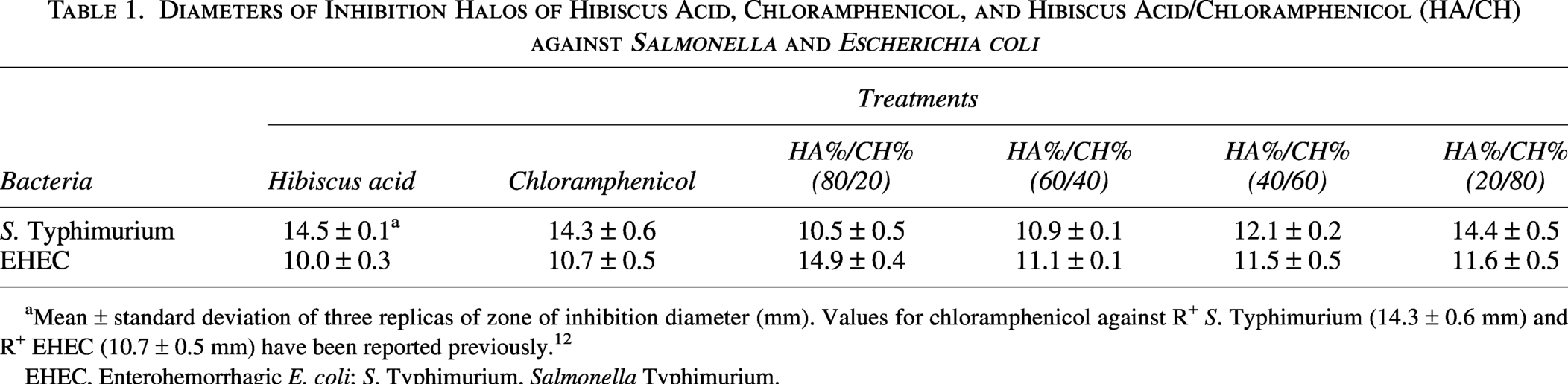
Mean ± standard deviation of three replicas of zone of inhibition diameter (mm). Values for chloramphenicol against R+ S. Typhimurium (14.3 ± 0.6 mm) and R+ EHEC (10.7 ± 0.5 mm) have been reported previously. 12
EHEC, Enterohemorrhagic E. coli; S. Typhimurium, Salmonella Typhimurium.
Minimum Inhibitory Concentration (MIC), Minimum Bactericidal Concentration (MBC), and Fractional Inhibitory Concentration Index (FICI) Values of Hibiscus Acid, Chloramphenicol, and Hibiscus Acid/Chloramphenicol (HA/CH)
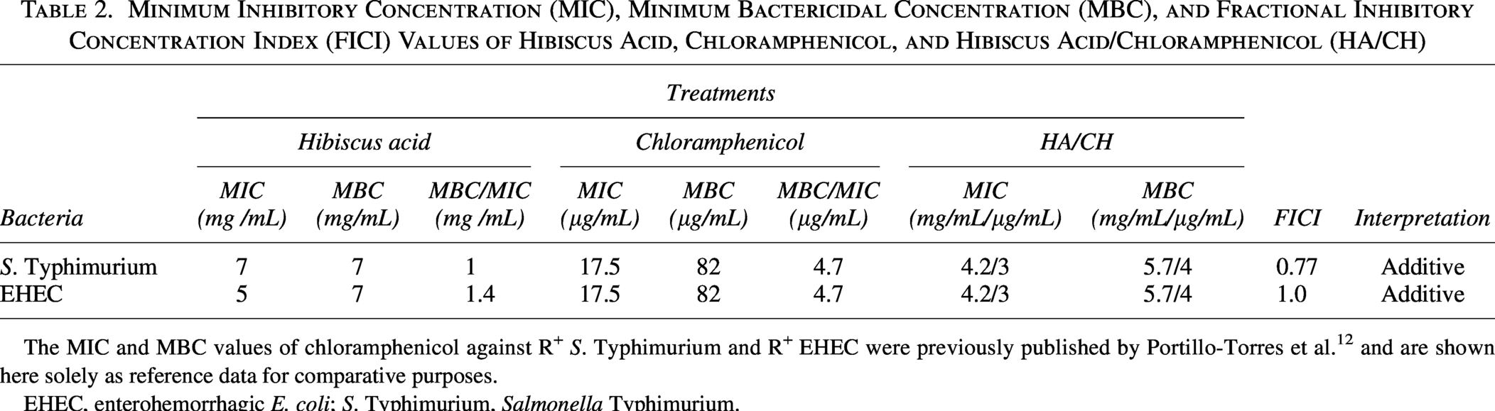
The MIC and MBC values of chloramphenicol against R+ S. Typhimurium and R+ EHEC were previously published by Portillo-Torres et al. 12 and are shown here solely as reference data for comparative purposes.
EHEC, enterohemorrhagic E. coli; S. Typhimurium, Salmonella Typhimurium.
Combination therapy, where two or more agents are administered together, offers advantages such as improved efficacy, broader antibacterial coverage, reduced resistance, and lower host toxicity. 20 In our study, the combination of HA with CH made it possible to significantly reduce the concentration of CH required for effective treatment (Table 2). Since CH can be toxic at the concentrations commonly used against bacterial infections, lowering its dosage when administered with HA may decrease the likelihood of harmful effects in humans and animals while maintaining therapeutic efficacy. The continuous rise of antibiotic resistance underscores the need for alternative compounds that can be used alone or in combination with conventional drugs to control resistant pathogens. The outcomes of the in vivo experiments are summarized in Table 3. Both R+ S. Typhimurium and R+ EHEC successfully colonized CD-1 mice and increased in number in animals that received only ISS or CH treatment. In contrast, no viable bacteria were detected in the feces of mice treated with HA alone or with the HA/CH mixture, indicating a strong protective effect of HA either independently or in combination with CH (Table 3). The present findings are in agreement with our previous study using H. sabdariffa calyx extract. In that report, a higher extract concentration was required, and the effect of combining it with CH was not evaluated. 12 In the current study, the results summarized in Table 4 show that mice infected with R+ EHEC or R+ S. Typhimurium and treated with ISS or CH after infection developed clinical signs, and some animals died. In contrast, in groups infected with the same pathogens but treated with HA alone or the HA/CH mixture, only one mouse in each group exhibited transient symptoms during the first two days after infection; these animals subsequently recovered, and no deaths occurred. These findings demonstrate that HA, at the minimum bactericidal dose tested, exerted a clear antimicrobial effect in animals infected with both pathogens. Moreover, the HA/CH mixture was also effective, even though the amount of HA in the combination was lower than when administered alone. This suggests that HA enhances the action of CH, allowing the use of very low CH concentrations to inactivate both R+ S. Typhimurium and R+ EHEC, even when these pathogens are resistant to CH individually. Considering that CH at therapeutic doses is associated with toxicity in humans, its use at reduced, nontoxic levels in combination with plant-derived antimicrobials such as HA may represent a safer alternative. Overall, the present study provides evidence that HA from H. sabdariffa calyces, either alone or in combination with CH, holds promise as an antibacterial agent for infections caused by multidrug-resistant and nonresistant bacteria. Further investigations are warranted to confirm and extend these observations.
Effect of Treatments in CD-1 Mice on the Fecal Excretion of EHEC R+ and Salmonella Typhimurium R+ (CFU/g)
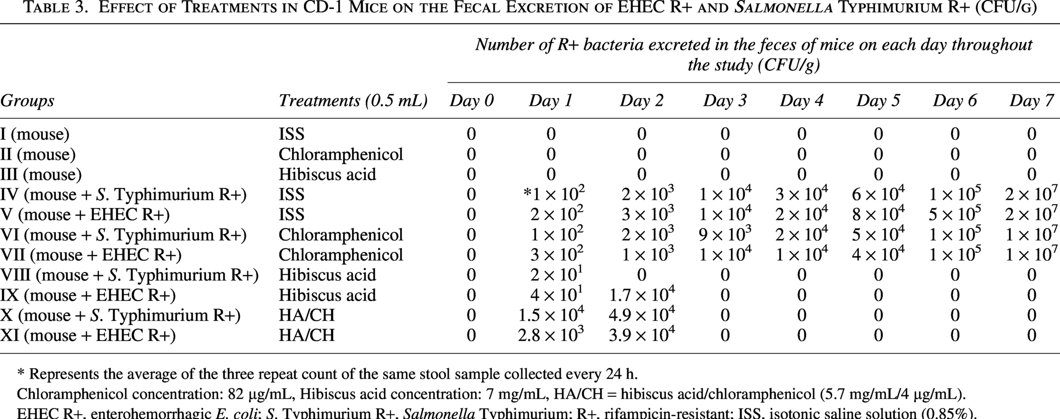
* Represents the average of the three repeat count of the same stool sample collected every 24 h.
Chloramphenicol concentration: 82 µg/mL, Hibiscus acid concentration: 7 mg/mL, HA/CH = hibiscus acid/chloramphenicol (5.7 mg/mL/4 µg/mL).
EHEC R+, enterohemorrhagic E. coli; S. Typhimurium R+, Salmonella Typhimurium; R+, rifampicin-resistant; ISS, isotonic saline solution (0.85%).
Clinical Signs and Mortality Observed in Groups of Mice Infected and Not Infected with S. Typhimurium R+ and EHEC R+ During the Course of the Experiment
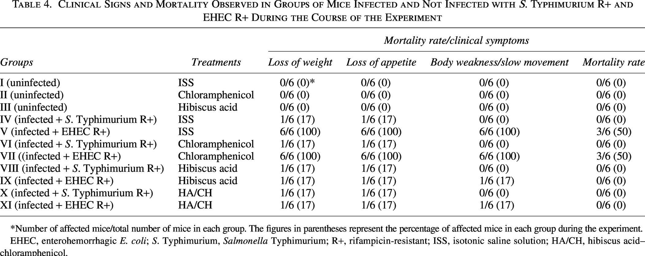
*Number of affected mice/total number of mice in each group. The figures in parentheses represent the percentage of affected mice in each group during the experiment.
EHEC, enterohemorrhagic E. coli; S. Typhimurium, Salmonella Typhimurium; R+, rifampicin-resistant; ISS, isotonic saline solution; HA/CH, hibiscus acid–chloramphenicol.
Footnotes
ACKNOWLEDGMENTS
The work was supported by Secretaría de Ciencia, Humanidades, Tecnología e Innovación (SECIHTI) for financial support to the project number A1-S-8288 “Antimicrobials from Jamaica flower calyxes alone and in combination with antibiotics: determination of the mechanisms of action on resistant and nonresistant pathogenic bacteria to antibiotics, the antimicrobial effect in vivo and adverse reactions in animals.”
AUTHOR DISCLOSURE STATEMENT
The authors declare no conflict of interest. The funders had no role in the design of the study; in the collection, analyses, or interpretation of data; in the writing of the article; or in the decision to publish the results.
FUNDING INFORMATION
The work was supported by National Council for Science and Technology (CONACyT) for financial support to the project number A1-S-8288 “Antimicrobials from Jamaica flower calyxes alone and in combination with antibiotics: determination of the mechanisms of action on resistant and non-resistant pathogenic bacteria to antibiotics, the antimicrobial effect in vivo and adverse reactions in animals.”
ETHICAL CONSIDERATIONS
The study was approved by the Laboratory Animal Research Ethics Committee of the Autonomous University of the Hidalgo State (UAEH2019-A1-S-8288).