
Abstract
The article’s aim was thus to evaluate the antiproliferative activity of gold nanoparticles associated with the methanolic extract of Chrysophyllum cainito in human breast cancer cell line (MCF-7), cervical cancer tissue (HeLa), and colorectal cancer cell line (HCT-116) cancer cell lines. The methanolic extract showed the presence of polyphenols and organic acids related to anticancer properties. In relation to the gold nanoparticles functionalized with the methanolic extract of C. cainito peels, these nanoparticles showed significant cytotoxic activity (p = .0001) in the HCT-116 cell line at 72 h after exposure to these. We consider this the first contribution to the use of the methanolic extract of C. cainito for the green synthesis of gold nanoparticles. In conclusion, the green synthesis of gold nanoparticles associated with C. cainito methanolic peel extract may be an interesting option for the search for new cancer treatments.
INTRODUCTION
Cancer is known as a group of diseases characterized by uncontrolled cell proliferation, giving rise to tumors that can invade and colonize new organs. 1 The cancers with the highest mortality rate include lung cancer, followed by colorectal cancer, liver cancer, stomach cancer, and breast cancer. 2 In this regard, several therapies are available nowadays to fight these diseases, such as chemotherapy, radiotherapy, surgery, and combinations of the above. However, there are still several limitations, such as drug resistance and a lack of drug specificity, which make research into new treatments crucial.
At present, there are several interesting approaches to the study and discovery of novel compounds that might aid in the development of new treatments to fight cancer, mainly in the field of basic and preclinical research. In this context, tropical plants like Chrysophyllum cainito L. (C. cainito) that have traditionally been associated with anticancer properties promise to be an interesting reservoir of bioactive compounds. 3
In the Yucatan Peninsula, in southeastern Mexico, fruits such as C. cainito have been traditionally used as plants that can combat cancer and other conditions; They grow in backyards and are easy to access since they are sold locally.3,4 In fact, there are some reports that have been conducted on C. cainito have shown anti-inflammatory, 5 anti-hypertensive, 6 antidiabetic properties7–9 and anticancer properties.10,11 In addition, phytochemical studies of different parts of the plant like stem bark, leaf, fruits, seeds, or fruit peel, have revealed the presence of chemical compounds, including alkaloids, sugars, tannins, saponins, terpenoids, flavonoids, and phenolic compounds, some of which have even been identified in the peels of both fruits and are deemed responsible for the cytotoxic and antiproliferative capacity of this specie. 4 Likewise, it is worth mentioning that methanolic peel extracts from C. cainito (CE) have been reported to exhibit pharmacological properties such as antioxidant, cytotoxic, anti-inflammatory, immunosuppressive and antihypertensive activity.5,12 Additionally, it has been proven experimentally that gold nanoparticles synthesized with plant extracts have pharmacological effects such as the cytotoxic effect of Mimosa tenuiflora, 13 the antioxidant effect of Sambucus nigra L., 14 tumor therapy of Mangiferin 15 and anticancer activity of Scutellaria barbata, 16 Panax notoginsen 17 and Annona muricata. 18 However, there are still no reports on the use of C. cainito in the synthesis of nanoparticles and its evaluation in cancer models.
The aim of the present research was thus to evaluate the antiproliferative activity of associated gold nanoparticles with methanolic extracts of C. cainito on MCF-7, HeLa, and HCT-116 cell lines, thereby contributing to the search for new matrices for use in cancer treatment.
MATERIALS AND METHODS
Fruit procurement, processing, and extract preparation
Samples of C. cainito L. (10 kg approximately) were purchased at commercial maturity from local markets in Yucatan, Mexico (city of Merida) in 2014 and authenticated by Susana Peralta Gómez (Biology academy, UACM, Mexico). Then the samples were washed, disinfected, dried thoroughly with a paper towel, and manually peeled. Subsequently, the peels were freeze-dried on a Labconco Model 6 (Labconco, MO, USA) at 0.04 mBar and −50°C for 48 h. Then the peels were processed and turned into dust and stored at −20°C until use. 4 The extract was obtained through alcoholic maceration. About 10 g of freeze-dried peel were homogenized in 50 mL of methanol at room temperature for 24 h. After the methanolic extracts were filtered, dried at room temperature, and subsequently stored at −20°C until use. 4
Phytochemical analysis
Phenolic and flavonoid compounds quantification
Total phenolic compounds were assessed by means of the Folin–Ciocalteu reagent according to the method described by Singleton and Rossi 19 2.5 mg of methanolic extract were diluted in 5 mL of H2Od, and 200 µL of diluted extract were transferred to test tubes. After that, 1 mL of Folin-Ciocalteu 10% (v/v) was added and incubated for 1 min at room temperature. Subsequently, 0.8 mL of Na2CO3 7.5% (w/v) was added and incubated for 60 min. Finally, the phenolic compounds were measured by spectrophotometry at 765 nm (JENWAY 6705 UV/vis). A gallic acid curve was used as a standard curve and performed in triplicate. Flavonoid quantification was performed according to the method described by Chang et al. 20 About 500 µL of diluted methanolic extract were transferred to test tubes, and immediately 1500 µL of methanol, 100 µL of AlCl3 10% (w/v), 100 µL of potassium acetate 1 M, and 2800 µL of H2Od were added. Immediately, the test tubes were mixed and incubated for 30 min at room temperature. The determination was performed by spectrophotometry at 450 nm. A quercetin curve was used as a standard curve and performed in triplicate.
Caffeic, gallic, and chlorogenic acid determination by High-performance liquid chromatography (HPLC)
Caffeic, gallic, and chlorogenic acid determination were performed according to the method described by Pellati et al. 21 A sample of methanolic extract (5 mg/mL) were diluted 1:10 (v/v) and 20 µL of the extracts were injected into an Agilent 1200 Series HPLC system and a Hi-Plex Ca column (Agilent Technologies. Serial No:0006236070-136) was used for the determination. A C18 µBondapack™ column (3.9 × 300 mm, SN. W00671T018) was used for determination. The mobile phase consisted of (A) acetic acid 0.1% (v/v) and (B) acetonitrile. An elution gradient was used according to the following: 0–10 min 85% (A)/15% (B); 10–20 min 705 (A)/30% (B) and 20–35 min 85% (A)/15% (B). The flow rate was 1 mL/min and column temperature of 30°C. Data were recorded at 320 nm, and concentrations were determined by interpolation on calibration curves of caffeic, gallic, and chlorogenic acids.
Sugar and organic acids determination by HPLC
Sugar determination (sucrose, glucose, and fructose) was performed by HPLC according to Aarland et al. 0.5 g of freeze-dried peel of C. cainito L. was macerated in 15 mL of methanol and maceration elapsed for 24 h at room temperature. Subsequently, 2 mL of extract were filtered through a 0.2 µm nylon filter (Millex, Millipore, Bedford, MA). 22 Then 20 µL of filtered extract were injected into the HPLC previously described. A Hi-plex Ca (8% crosslinked, 7.7 × 300 mm, 8 µm, Agilent Tecnologies©, SN. 0006236070–136) column at 80°C. The run was performed according to the following flow rate: 0.6 mL/min, 30 min of run, and the mobile phase consisted of H2O HPLC grade. Sucrose, glucose, and fructose dilutions were used as standard curves. The determination of organic acids was performed as follows. A solution of each methanolic extract was prepared (1 mg/mL w/v) in H2O HPLC grade. Subsequently, the extracts were filtered with 0.2 µm filters (Millex, Millipore, Bedford, VA), and 20 µL of filtered extracts were injected into the HPLC previously described. A C18 column (X-Terra MS, 5 µm; 4.6 × 250 mm) was used, and the mobile phase consisted of Phosphate Buffer Solution (50 mM, pH 2.8) in isocratic mode. The flow was adjusted to 0.7 mL/min. The data were recorded at 210 nm, and they were interpolated with calibration curves of citric, malic, oxalic, and tartaric acid. The results were expressed in grams of compound per 100 g of freeze-dried peel (g/100 g lyophilized peel [LP]). In all cases, sugars and organic acids were measured in triplicate under three different experimental conditions.
Pharmacological assays
Cell lines
Cervical carcinoma (HeLa), mammary carcinoma (MCF-7), and colon cancer (HCT-116) cell lines were obtained from the American Type Culture Collection of the Department of Pharmacy, Faculty of Chemistry, National Autonomous University of Mexico. All cell lines were maintained in RPMI 1640 medium supplemented with 10% (v/v) fetal bovine serum and cultured at 37°C with 5% CO2 and 100% humidity. 4
Cell proliferation assay
Cell numbers were addressed by the Cell Counting Kit-8 (CCK-8, Dojindo Lab, Kumamoto, Japan). A curve was seeded with HeLa, MCF-7, and HCT-116 cell lines, respectively, in the following order: 0, 2500, 5000, 10,000, 20,000, 40,000 cells/well, and were incubated normally for 12 h. After that the supplemented medium was replaced by unsupplemented medium, and 10 µL of CCK-8 reagent was added to each well, and the cells were reincubated for 1 h more. Data from each cell curve was recorded in a 96-well plate at 450 nm, and the cell number was determined by a linear regression.
Synthesis of gold nanoparticles and gold nanoparticles functionalized using the methanolic extract of C. cainito
The synthesis of naked gold nanoparticles (Au-NPs) was performed using the AuCl4Na 2H2O (Sigma-Aldrich®, catalogue number 298174) reduction according to Frens. 23 The synthesis of gold nanoparticles associated with the methanolic extract of C. cainito (Au-CE-NPs) was performed by reducing AuCl4Na·2H2O in the presence of the extract of C. cainito,24,25 with the following modifications. AuCl4Na 2H2O solution (0.01% w/v; 25 mL) was placed in an Erlenmeyer flask, under constant stirring at 90°C in the dark. Na3C6H5O7 solution (1% w/v; 0.375 mL) was immediately added for the synthesis of non-associated Au-NPs, and the synthesis was allowed to proceed for 30 min. Then, for the synthesis of functionalized Au-NPs, a reduction solution was prepared by taking C. cainito dry methanolic extract (4 mg) and then dissolving it in 1 mL of double-distilled water. After that, the solutions were filtered through a 0.45 µm nylon filter and stored in amber vials at 5°C until use. For the synthesis of functionalized nanoparticles, 25 mL of an AuCl4Na 2H2O (0.01% w/v) solution was added to an Erlenmeyer flask, which was placed under agitation at 90°C in the dark. 0.35 mL of C. cainito extract (4 mg/mL) was immediately added for the formation of Au-CE-NPs. Reaction was maintained under the initial conditions for 30 min, and then 0.05 g of gum arabic was added as a stabilizer. Finally, the two resulting reactions were cooled to room temperature, filtered through a 0.45 µm nylon filter, and stored in amber vials at 5°C until further use.
Chemical and physicochemical characterization of the gold nanoparticles
Spectrophotometric characterization of synthesized gold nanoparticles
For the characterization, 2 mL aliquot was taken from each colloidal solution of naked Au-NPs and Au-CE-NPs. The two colloidal solutions were subjected to a spectrophotometric scan from 450 to 800 nm to determine their maximum absorbance range using a spectrophotometer, Thermo Fisher Scientific (GENESYS™ 10S UV-Vis).
Size determination of gold nanoparticles synthesized by high-resolution transmission electron microscopy (HRTEM)
The HRTEM images were obtained by drying a drop of Au-NPs and Au-CE-NPs samples on a Ted Pella formvar-coated copper grid. Images were obtained on a JSM7600-F(JEOL) Instrument operated at 200 kV.
Determination of conjugation of synthesized gold nanoparticles by Fourier-transform infrared spectroscopy (FTIR)
To determine the functionalization of the extracts with the synthesized Au-NPs, 1 mL of the Au-CE-NPs was transferred to microtubes and centrifuged at 20,000 g for 10 min (IEC MicroCL 21 Centrifuge, Thermo Electron Corporation). Subsequently, the supernatant was removed, and the procedure was repeated two additional times. Once the 20 µL pellets of each solution were obtained, they were placed on a slide and dried in an oven at 50°C for 30 min. Once dry, the nanoparticle samples were subjected to analysis by FTIR (Carry 630 FTIR, Agilent Technologies) to determine the functional groups present in the colloidal solution of Au-NPs.
Determination of the antiproliferative capacity of synthesized nanoparticles
The antiproliferative capacity of the synthesized nanoparticles was assessed using the CCK-8 technique. HeLa, MCF-7, and HCT-116 cell lines were seeded at a density of 5000 cells per well in a 96-well plate, identified, and incubated for 12 h. Following this period, the culture medium was replaced by supplemented medium plus 10 µL of Au-NPs in the control group and 10 µL of Au-CE-NPs in the treatment group. Plates were incubated for 24, 48, and 72 h, and finally, 10 µL of CCK-8 reagent was added at the end of each interval (Sigma-Aldrich, Cat. 96992) and incubated for an additional hour. Once this time had elapsed, the absorbance at 450 nm was determined using a plate reader (Thermo Scientific Multiskan FC, 51119000 Model). The number of cells was determined as described previously.
Statistical analysis
Results are presented as mean ± standard deviation. Analysis of Variance (ANOVA) followed by Tukey’s test was used for multiple comparisons of parametric data, and a Kruskal–Wallis test followed by Dunn’s test was used for multiple comparisons of non-parametric data, considering a p < .05 as statistically significant. GraphPad Prism version 8.0.1 software was used (GraphPad Software, Inc., La Jolla, CA, USA).
RESULTS AND DISCUSSION
Phytochemical analysis
Quantitative analysis
Methanolic extract of C. cainito (CE) peels contained 533.66 ± 12.11 mg GAE/g DME of phenolic compounds (Table 1). These results differ from those reported in 2018 by Chel Guerrero et al., 4 where it was also mentioned that methanolic extracts contained a greater amount of total phenolic compounds. Phenolic compounds are known to be sensitive to various factors, such as temperature. There are reports linking storage temperature with the degradation of phenolic compounds, such as green tea myricetin, which, starting from 0.35 ± 0.04 mg/g at the beginning of the evaluation, contains 0.77 ± 0.04 mg/g after 5 years of storage at 4°C. 26 Although it is known that at low temperatures phenolic compounds have less degradation, this is also proportional to the storage time.
Quantitative Phytochemical Analysis of C. caimito Fruit Peel
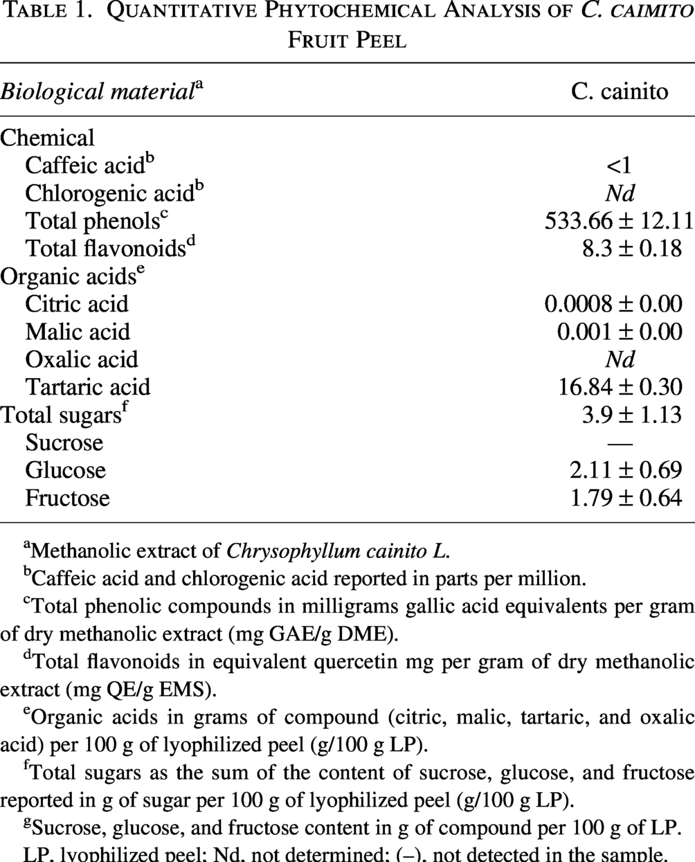
Methanolic extract of Chrysophyllum cainito L.
Caffeic acid and chlorogenic acid reported in parts per million.
Total phenolic compounds in milligrams gallic acid equivalents per gram of dry methanolic extract (mg GAE/g DME).
Total flavonoids in equivalent quercetin mg per gram of dry methanolic extract (mg QE/g EMS).
Organic acids in grams of compound (citric, malic, tartaric, and oxalic acid) per 100 g of lyophilized peel (g/100 g LP).
Total sugars as the sum of the content of sucrose, glucose, and fructose reported in g of sugar per 100 g of lyophilized peel (g/100 g LP).
Sucrose, glucose, and fructose content in g of compound per 100 g of LP.
LP, lyophilized peel; Nd, not determined; (–), not detected in the sample.
Moreover, in another study, the authors refer to a smaller concentration of phenolic compounds (109.5 ± 1.4 TAE), however, the authors used a much higher concentration of extract (50 mg/mL) than the one we used (2.5 mg/mL). These findings are also analogous to those reported by Silva et al., 19 who determined that different tropical fruits, including C. cainito, contained large amounts of phenolic compounds in pulp and peel. 19
The polarity of the extraction solvent has an important role in the selective extraction due to the different polyphenol composition.27,28 In this context, the difference of electronegativity between -OH groups and carbon and hydrogen atoms makes a negative and positive region, respectively. Besides, methanol has a lower evaporation point than water. It also has a low boiling point, which allows it to evaporate at a lower temperature than water, which allows compounds to be extracted without risk of temperature degradation.29,30 This reinforces the idea of methanol being the most efficient solvent for the extraction of many biologically active compounds from fruit peels.31,32 Similarly, when applied to leukemia cell lines, phenolic compounds such as gallic acid have been shown to inhibit the activity of the enzyme ribonucleotide reductase (which catalyzes the synthesis of deoxyribonucleotides from ribonucleotides), resulting in growth arrest of these cells. 32 In addition, polyphenols have a potential use in cancer treatment through cell cycle arrest33–35 or the apoptosis pathway. 36 These contributions support the idea that the methanolic extracts of C. cainito could have pharmacological effects, particularly those related to antiproliferative activity.
The extracts of C. cainito showed a total sugar amount of 3.9 ± 1.13 g per 100 g of LP. Likewise, 2.11 ± 0.69 g/100 g of glucose and 1.79 g/100 g of fructose were obtained for C. cainito peel extracts. The absence of sucrose indicated that the fruits were in a state of commercial maturity (Table 1). These results differ from those found by Chel-Guerrero et al., 4 who reported a total sugar amount of 0.15 ± 0.00 g/100 g in the LP for C. cainito. The same authors also reported levels of 0.07 ± 0.00 g/100 g and 0.08 ± 0.00 g/100 g of glucose and fructose for C. cainito, respectively.
In agreement with the findings of Chel-Guerrero et al., 4 the presence of sucrose could not be determined in either of the fruit peels because fruits were used at commercial maturity. Organic acid determination (citric, malic, oxalic, and tartaric acids) showed that the LP of C. cainito showed an amount of citric (0.0008 ± 0.00 g/100 g LP), malic (0.001 ± 0.00 g/100 g LP), and tartaric acids (16.84 ± 0.30 g/100 g LP). However, oxalic acid was not detected in the C. cainito (Table 1).
Some reports indicate that C. cainito has an anticancer effect against U-2 osteosarcoma cells and human hepatocarcinoma HepG2 cells through the generation of reactive oxygen species that lead to the induction of apoptosis through an intrinsic pathway.10,37 It is important to note that phenolic compounds such as gallic acid have been implicated in various metabolic pathways, such as the generation of reactive oxygen species, activation of apoptosis through intrinsic and extrinsic pathways, and also in cell cycle arrest.38–40 The results obtained in this work help to establish a comprehensive phytochemical profile of the peels of this fruit since their phytochemical properties have not yet been described in the literature.
Pharmacological assays
Synthesis of gold nanoparticles associated and not associated with the extract of C. cainito
It has been previously described that the synthesis of Au-NPs depends on several factors, such as temperature, reaction time, and the concentration of reducing compounds used.23,24 It was observed that the concentration of C. cainito had a direct impact on the synthesis outcome. A high concentration of 50 mg/mL led to the immediate precipitation of Au-NPs (data not shown). At concentrations of 10 and 5 mg/mL, nanoparticle precipitation was observed at 4 and 7 min after the reaction began, respectively (data not shown). Conversely, a concentration of 1 mg/mL appeared insufficient to promote nanoparticle formation, likely due to the inadequate presence of reducing agents (data not shown).
The color of the resulting colloidal solution, which correlates with nanoparticle size due to surface plasmon resonance (SPR), served as a visual indicator of successful synthesis.23,41 Generally, the color shifts from pale red (∼5 nm) to ruby-red (∼20 nm), red-violet (∼40 nm), violet (∼60 nm), and purple (∼90 nm), reflecting changes in particle size. Based on these observations, the optimal synthesis of Au-NPs using C. cainito was achieved at a concentration of 4 mg/mL, under conditions of 90°C for 30 min. The formation of Au-NPs was confirmed visually by the characteristic color change attributed to SPR.
Nanoparticle synthesis under two different reducing conditions (Na3C6H5O7 and C. cainito extract) produced nanoparticles of different colors (Fig. 1). While the Au-NPs exhibit a pale-red color (Fig. 1A) and the Au-CE-NPs exhibit a violet color (Fig. 1B). The change in color can be explained by the size of the Au-NP 42 and the differential content of organic acids in the extracts, and particularly some polyphenolic compounds in C. cainito peel.
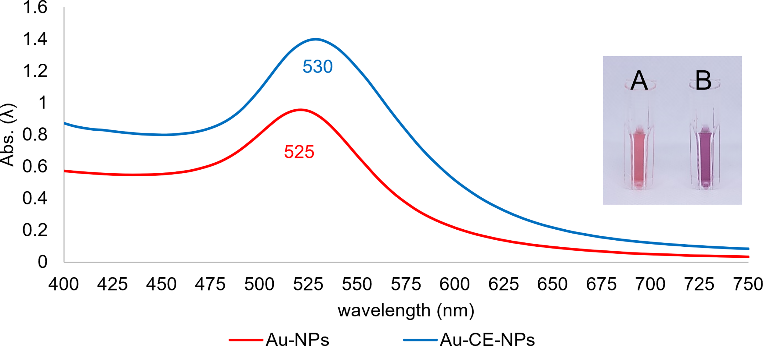
Gold nanoparticles (Au-NPs) color and absorbance spectrum. Absorbance spectrum and color of unassociated nanoparticles Au-NPs (red line and
Characterization of the synthesized gold nanoparticles
The characterization of the Au-NPs synthesized was performed using three different techniques. Spectrophotometry, FTIR, HRTEM, and Dynamic Light Scattering (DLS). The colors of the synthesized nanoparticles provide a preliminary and approximate indication of their sizes (Fig. 1A and B).
Furthermore, the maximum absorbance peaks for Au-NPs, Au-CE-NPs, were at 525 nm and 530 nm, respectively (Fig. 1). The spectra of the nanoparticles indicate that the synthesis produced nanoparticles of 20 nm approximately, and regarding those associated with C. cainito extract, the absorbance spectra indicate an approximate increase in size of 50 and 80 nm. SPR allows partial confirmation of the shape of the synthesized nanoparticles, as the absorbance spectra only show a single peak of maximum absorption corresponding to approximately spherical nanoparticles. The above is consistent with previous findings in the literature, where maximum absorption peaks between 520 and 550 nm have been reported. Nanoparticles were approximately spherical in all cases,18–20 differing from other types of nanoparticles, such as nanorods, that show two peaks of maximum absorbance corresponding to their length and diameter.21–24
Through TEM characterization of the synthesized Au-NPs (Fig. 2), it was possible to confirm that the Au-NPs are approximately spherical (Fig. 2A) with a diameter of 17.32 ± 2.1 nm (Fig. 2A1). These results confirm the formation of Au-NPs by the Frens method.
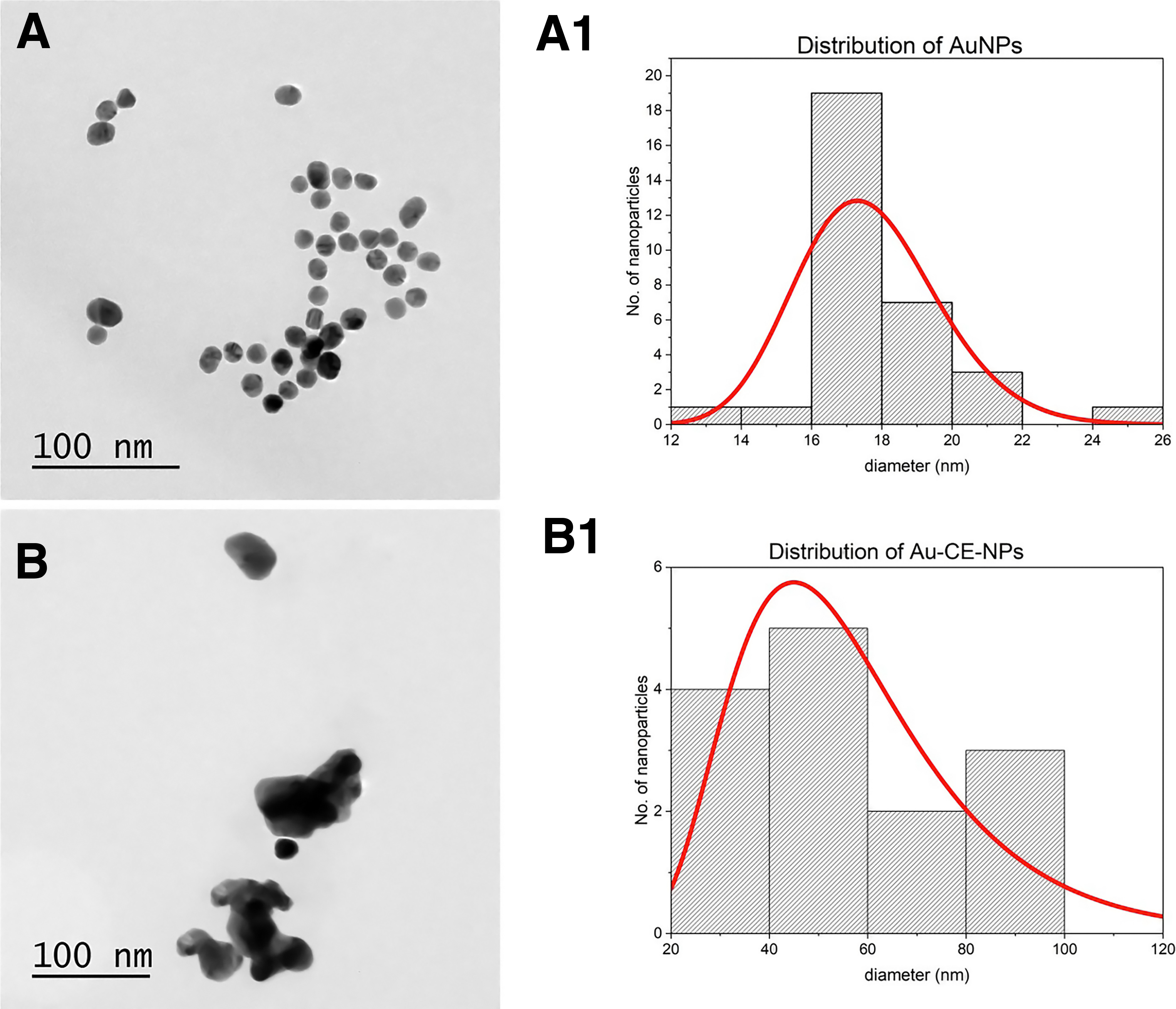
TEM of gold nanoparticles synthesized and size distribution histograms of synthesized gold nanoparticles.
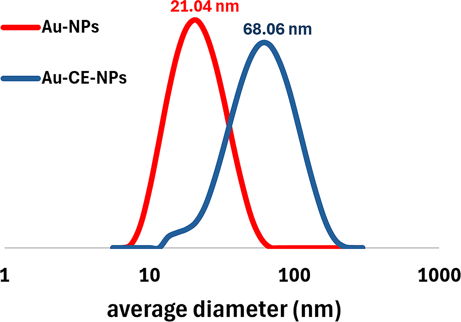
Regarding the associated Au-NPs, the Au-CE-NPs (Fig. 2B) showed a heterogeneous cluster shape and spherical-shaped particles of diameter 59.08 ± 20.9 nm (Fig. 2B1). These results confirm the presence of synthesized Au-NPs and the increment in diameter by the C. cainito methanolic extract reduction. The DLS analysis shows that the synthesized Au-NPs have an average diameter of 21. 04 nm for Au-NPs (Fig. 3, red line), and 68.06 nm for Au-CE-NPs (Fig. 3, blue line). This result reinforces what was found by HRTEM analysis.
FTIR analysis
The transmittance spectrum of the methanolic extract and the Au-NPs were compared based on the changes before and after the bioreduction. FTIR spectra of C. cainito, and synthesized Au-NPs (Fig. 4) showed absorbance bands between 3600–3100 cm−1 and 1602 cm−1 corresponding to the stretching of the hydrogen bond (−OH); 2920 and 2847 cm−1 corresponding to the C–H asymmetrical stretching vibration of the C–H2 groups; 2456 cm−1 corresponding to groups C≡C or C≡N; between 1000 and 1100 cm−1 corresponded to C = O stretching, and at 660 cm−1 it was due to O–H stretching of aromatic compounds. 43
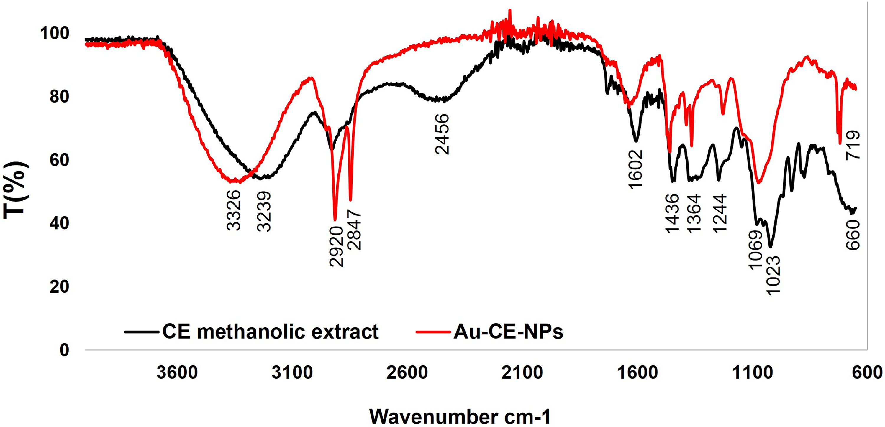
Fourier-transform infrared spectroscopy (FTIR) spectra of CE methanolic extracts and conjugated gold nanoparticles. CE methanolic extract spectrum (black line) and Au-CE-NPs spectrum (red line).
The FTIR analysis of C. cainito versus Au-CE-NP indicated changes in the absorption bands in 2920 and 2847 cm−1 due to the stretching vibration of the C–H groups of polyphenol compounds; 1436, 1364, 1244, and 719 cm−1 in the fingerprint region. The presence of bioactive compounds like polyphenols in the C. cainito methanolic extract determined the Au-NP size. Through the reduction of hydroxyl groups into carbonyl groups, the nanoparticles begin to aggregate, generating an increase in the size of the nanoparticle. 44
In conclusion, FTIR analysis indicated that Au-CE-NPs were stabilized due to the direct interaction of carbonyl groups in the polyphenolic compounds. 45 That is why we propose a stabilization model for this reaction (Fig. 5).
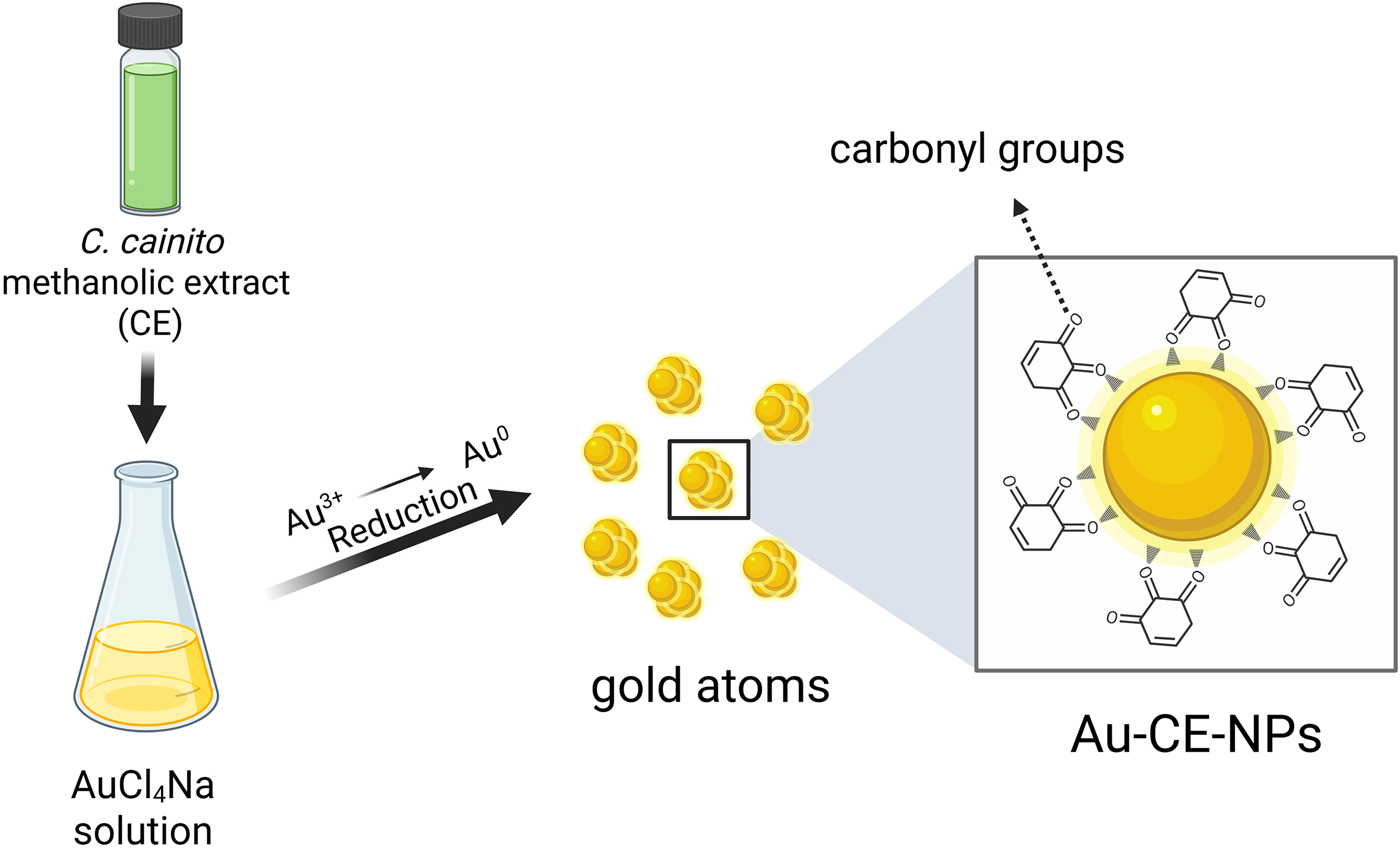
Proposed schematic representation of reduction, stabilization, and association of gold nanoparticles with C. cainito methanolic extract. (created with BioRender.com).
Determination of the antiproliferative capacity of synthesized nanoparticles
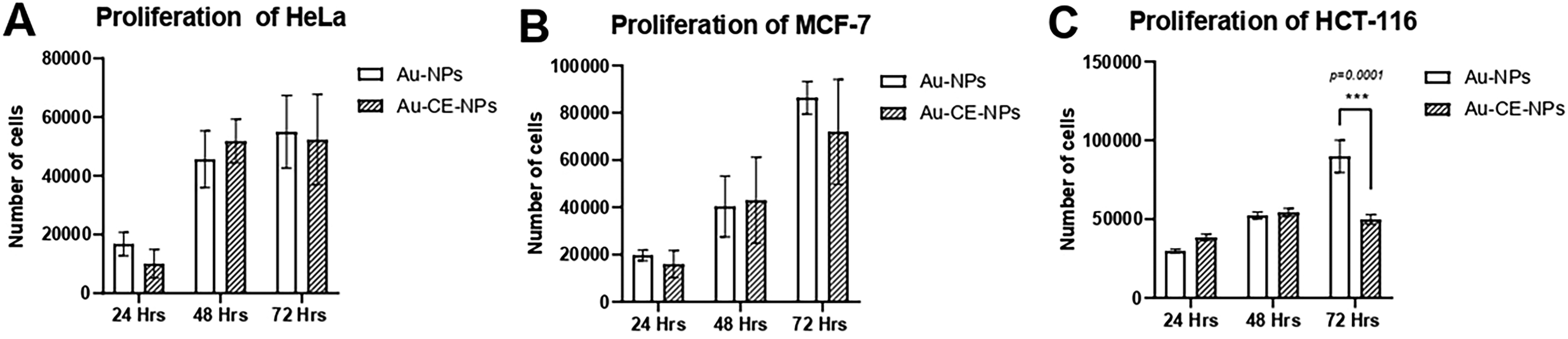
To determine whether the binding of the extract to nanoparticles could show proliferative activity through nanoparticle synthesis, the synthesized nanoparticles were subjected to the antiproliferative capacity assay using CCK-8 with HeLa, MCF-7, and HCT-116 cell lines (Fig. 6A–C, Table 2). CCK-8 solution utilizes a highly water-soluble WST-8 salt that produces a water-soluble formazan dye upon reduction. The quantity of formazan dye generated by intracellular dehydrogenase activity is directly proportional to the number of living cells; CCK-8 does not stain viable cells, see Cell Counting Kit-8, Dojindo Lab, Kumamoto, Japan.
Determination of the antiproliferative capacity of unassociated Au-NPs and CE methanolic extract (Au-CE-NPs).
Percentage Viability of Exposed Cell Lines
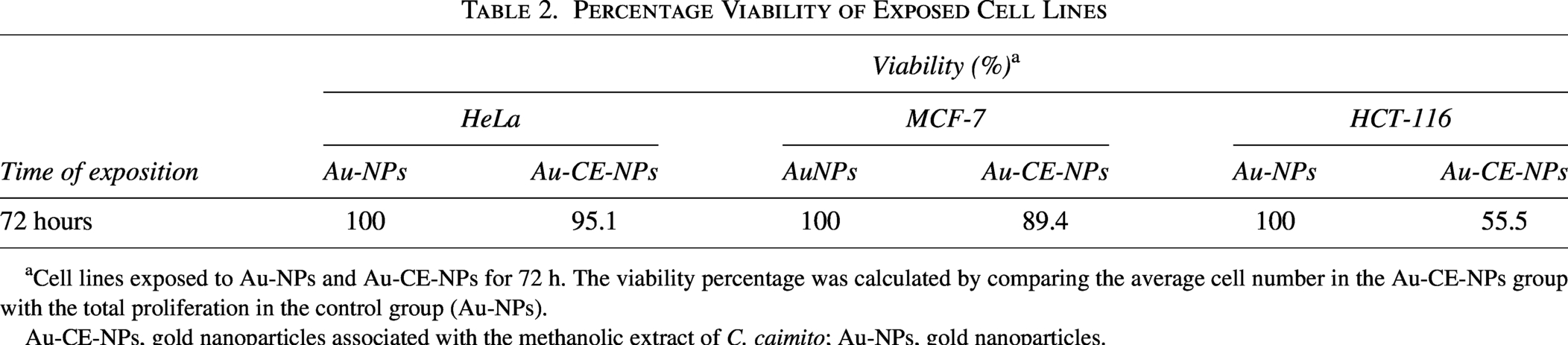
Cell lines exposed to Au-NPs and Au-CE-NPs for 72 h. The viability percentage was calculated by comparing the average cell number in the Au-CE-NPs group with the total proliferation in the control group (Au-NPs).
Au-CE-NPs, gold nanoparticles associated with the methanolic extract of C. caimito; Au-NPs, gold nanoparticles.
Likewise, it was determined that Au-CE-NPs showed only significant antiproliferative activity (p = 0.0001) in the HCT-116 cell line at 72 h post-treatment, resulting in 44.5% inhibition of proliferation with respect to Au-NPs exposure (Fig. 6C). Some reports establish that Au-NPs could elevate ROS production in the cell, leading to ROS-dependent apoptosis. 46 Similarly, the antiproliferative activity of Au-NPs depends on different factors, such as shape capping. In this context, we found that the reduction and stabilization of Au-NPs was mainly due to polyphenol activity, since some polyphenols, such as curcumin or quercetin, have been reported to show antiproliferative activity. 47 This antiproliferative activity shown by the Au-CE-NPs could be explained by capping on the Au-NP surface, on which interactions with some polyphenol compounds take place.
It is worth emphasizing that, when exposed to Au-CE-NPs, the HeLa cell line (Fig. 6A) showed a lower proliferation rate than when exposed to NPs during the first 24 h; however, this phenomenon was not maintained after 72 h, and the cell line only showed a decrease of 4.9% compared to the control group. In addition, the MCF-7 cell line showed a decrease of around 10.6% with the Au-CE-NPs group (Fig. 6B). This phenomenon may be attributed to the fact that during the synthesis of nanoparticles associated with extracts, many of the compounds responsible for the antiproliferative activity, such as phenolic compounds, are used for AuCl4Na·2H2O reduction to metallic gold, so the compound amount available for nanoparticle conjugation may be limited when using volumes and concentrations such as those used in this experiment. Therefore, a solution to this problem could be to use larger volumes of associated nanoparticles. Previously published studies revealed that nanoparticle conjugation with other compounds, such as antibodies, seems to have a hormetic effect, allowing cells to establish defense and survival mechanisms. Similarly, the synthesis of a nanoparticle with an active biological material can result in an inactive compound. 48 because the combination of the secondary metabolite with the Au-NP generates a new chemical complex with different physical and chemical properties than those of the starting chemical systems. Therefore, its properties should be characterized again.
It is important to emphasize that variables such as methanolic extract concentration, nanoparticle shape, and nanoparticle conjugation are extremely important parameters for the formation of a Au-NP conjugated with biological activity focused on antiproliferation activity.
CONCLUSIONS
In conclusion, we can affirm that the synthesis of nanoparticles through reduction with a methanolic extract of C. cainito cascara is possible, as is their association with nanoparticles.
Furthermore, it is possible to affirm that nanoparticles associated with the methanolic extract of C. cainito exhibit a significant antiproliferative effect against HCT-116 colorectal cancer cells after 72 h of exposure compared to the Au-NPs control group.
Finally, this can be considered the first report on the use of C. cainito for the synthesis of Au-NPs with therapeutic potential, which sheds new light on further research needed to put forward new strategies for the use of these extracts in a possible antineoplastic therapy.
AUTHORS’ CONTRIBUTIONS
O.Y.A.-R., J.A.M.-E., and L.J.P.-F. designed the project. O.Y.A.-R. wrote the draft article. O.Y.A.-R. performed the chemical and the biological assay. M.F.-S. supervised biological and pharmacological assays. J.A.M.-E. and L.J.P.-F. supervised chemistry and the methodological aspects of the work. All the authors reviewed, edited, and approved the article submission.
Footnotes
ACKNOWLEDGMENTS
The authors would like to thank the National Council of Humanities Science and Technology (CONAHCYT) for supporting this investigation by grant No. 615641. We would particularly like to acknowledge the enormously valuable support of Universidad Autónoma de la Ciudad de México (UACM), Universidad Nacional Autónoma de México (UNAM), the PhD Experimental program of Universidad Autónoma Metropolitana (UAM), and the authors are grateful to Laboratorio Central de Microscopía Electrónica at Universidad Autónoma Metropolitana-Iztapalapa for technical assistance in the acquisition of HRTEM images. It is important to mention that this project was partially funded by SECTEI/148/2024, with the project in the title “Búsqueda de compuestos dirigidos contra la subunidad alfa del dímero de tubulina”.
AUTHOR DISCLOSURE STATEMENT
No competing financial interests exist.
FUNDING INFORMATION
No funding was received for this article.