
Abstract
This study compared the immunomodulatory effects of the co-extracted herbal formulation JI-HK601 with those of an individual extracts mixture (IEM) and evaluated the minimum effective dose of JI-HK601 through in vivo immune function analyses in mice. To assess safety, both extracts were administered orally at a dose of 400 mg/kg for 14 consecutive days. Measurements of body weight, spleen index, and peripheral blood cell profiles revealed no significant differences compared to the normal control group, suggesting that neither extract induced systemic toxicity or abnormal immune organ responses under the tested conditions. Immunological assessments indicated that JI-HK601 enhanced natural killer (NK) cell cytotoxic activity to a greater extent than IEM, suggesting a greater functional improvement in NK cell responsiveness. In subsequent dose–response studies of JI-HK601, no toxicity was observed at any dose, whereas increases in T-cell proportion and NK cell activity were noted at 400 mg/kg. Analysis of T-cell transcription factors showed selective upregulation of T-box transcription factor 21 (T-bet), a key regulator of T helper type 1 (Th1) differentiation. These results indicate that JI-HK601 preferentially stimulates Th1-associated immune pathways while enhancing innate cytotoxic function. Overall, the findings demonstrate that JI-HK601 strengthens NK cell activity and promotes Th1-related cellular immunity while maintaining a favorable safety profile across the evaluated dose range. Thus, these results provide foundational data for assessing the potential use of JI-HK601 as an immune-enhancing ingredient in functional foods and complementary and alternative medicine.
Recent changes in modern lifestyles, including increased psychological stress, irregular sleep patterns, dietary imbalance, and environmental pollution, have collectively contributed to a growing prevalence of immune dysregulation.1,2 These conditions are associated with increased susceptibility to infections, chronic inflammation, and reduced immune surveillance, thereby driving demand for safe and effective immunomodulatory agents. 3 Natural product–based formulations are particularly attractive because of their long history of human use, relatively low toxicity, and potential for broad immune support.4,5 Among such candidates, JI-HK601 (also known as SH003) has gained attention as a promising herbal formulation with immunological activity.6–9
JI-HK601 is a co-extracted formulation consisting of Astragalus membranaceus (Am), Angelica gigas (Ag), and Trichosanthes kirilowii Maximowicz (Tk) mixed at a 1:1:1 ratio and extracted with 30% ethanol/ 10 In contrast, the individual extracts mixture (IEM) refers to a simple combination of the same three herbs, each individually extracted with 30% ethanol and mixed at a 1:1:1 (w/w) ratio. Each of these medicinal plants has been traditionally associated with immune regulation: Am is known for enhancing innate and adaptive immunity,11,12 Ag is linked to hematopoietic and anti-inflammatory effects,13,14 and Tk has been reported to exhibit immunomodulatory and antitumor activities.15–17 Previous studies have shown that JI-HK601 can support immune responses when used as an adjunct to anticancer therapy at relatively high doses (e.g., 558 mg/kg).7,8 However, whether JI-HK601 can independently enhance immune function at lower, nutritionally relevant doses remains unclear. Therefore, the present study aimed to evaluate JI-HK601 as a functional immune-enhancing material, compare its effects with a simple mixture of its individual components (IEM), and determine its minimum effective dose (MED).
JI-HK601 and its single-herb extracts were manufactured under Korea Good Manufacturing Practice standards by Hanpoong Pharm & Foods Company, ensuring consistency, safety, and reproducibility. Both JI-HK601 and IEM were dissolved in sterile triple-distilled water prior to oral administration. All animal procedures were conducted in accordance with the Guidelines for the Care and Use of Laboratory Animals of Kyung Hee University, and the protocol was approved by the Institutional Animal Care and Use Committee of Kyung Hee University (Approval No. KHSASP-23–485). Male C57BL/6N mice (6 weeks old) were housed under specific pathogen-free conditions and randomly assigned to experimental groups (n = 6 per group). Treatments were administered orally once daily for 14 consecutive days. After the 14-day administration period, mice were euthanized, and systemic toxicity was evaluated by assessing the spleen index and performing hematological analyses, including total white blood cell (WBC) counts and differential leukocyte populations (neutrophils, lymphocytes, monocytes, eosinophils, and basophils), using blood collected via cardiac puncture. Splenic lymphocyte composition, including T cells, B cells, natural killer (NK) cells, macrophages, and dendritic cells, was analyzed using a flow cytometer. NK cell cytotoxic activity against Yoshida ascites lymphoma-1 (YAC-1) target cells was assessed using an lactate dehydrogenase (LDH) cytotoxicity assay kit. T lymphocyte transcription factors were evaluated by measuring mRNA expression levels of T-box transcription factor 21 (T-bet) a T helper type 1 (Th1)-associated transcription factor, GATA binding protein 3 (GATA3), a T helper type 2 (Th2)-associated transcription factor, and forkhead box P3 (Foxp3), a regulatory T cell (Treg)-associated transcription factor, using reverse transcription quantitative polymerase chain reaction (RT-qPCR). Th1-related cytokine production was evaluated by measuring interleukin-2 (IL-2) and interferon-gamma (IFN-γ) levels in splenocyte culture supernatants following ex vivo stimulation with concanavalin A (Con A) for 24 h, using enzyme-linked immunosorbent assay kits according to the manufacturer’s instructions. Data are presented as means ± standard deviations. Statistical analyses were performed using GraphPad Prism 8.0.2. Group differences were assessed by one-way analysis of variance (ANOVA) followed by Dunnett’s multiple comparisons test. Statistical significance is indicated as *P < .05, **P < .01, and ***P < .001 versus the normal control group.
Before evaluating immunological efficacy, potential toxicity and effects on immune organs were assessed at a dose of 400 mg/kg (Figs. 1 and 2). Body weight was monitored throughout the experiment, and after 14 days, mice were euthanized for analyses. The spleen index was calculated as a marker of immune organ hypertrophy or atrophy, and hematological profiles were measured to detect systemic immune or inflammatory disturbances. Peripheral blood analysis showed no changes in total WBC counts or in leukocyte subpopulations, including lymphocytes, neutrophils, and monocytes. Likewise, body weight and spleen index remained comparable to those of the normal control group, indicating that neither JI-HK601 nor IEM induced detectable systemic toxicity or abnormal immune organ enlargement. To assess immune modulation, splenocytes were analyzed by flow cytometry. Neither extract significantly altered the proportions of macrophages, dendritic cells, or NK cells. Both treatment groups showed a decrease in the proportion of B cells, while only JI-HK601 induced a noticeable increase in T-cell proportion. This apparent B-cell reduction is hypothesized to result from activation-associated phenotypic changes. It is possible that B cells differentiating into antibody-producing plasma cells may lose certain surface markers, potentially leading to their underestimation during flow cytometric analysis. Thus, while these changes may reflect functional activation rather than simple depletion, further evidence is required to confirm this interpretation. Despite stable NK cell numbers, NK cytotoxic activity against YAC-1 target cells was enhanced in both treatment groups. Notably, JI-HK601 induced significantly greater NK cell cytotoxicity than IEM, indicating that co-extraction confers superior functional activity compared with simple mixing of individual extracts. The superior efficacy of JI-HK601 compared with IEM may be attributed to interactions among bioactive compounds that occur during the co-extraction process. Such interactions may generate synergistic effects and influence the overall composition and physicochemical properties of the extract, potentially contributing to differences in biological activity compared with simple mixtures, as suggested in previous studies on phytochemical synergy.18,19 Although the precise mechanisms remain to be elucidated, these factors may underlie the enhanced NK cell cytotoxicity observed in the JI-HK601-treated group. Because NK cell function plays a central role in early immune defense and tumor surveillance, this finding was particularly important in identifying JI-HK601 as a more potent immunomodulatory agent.
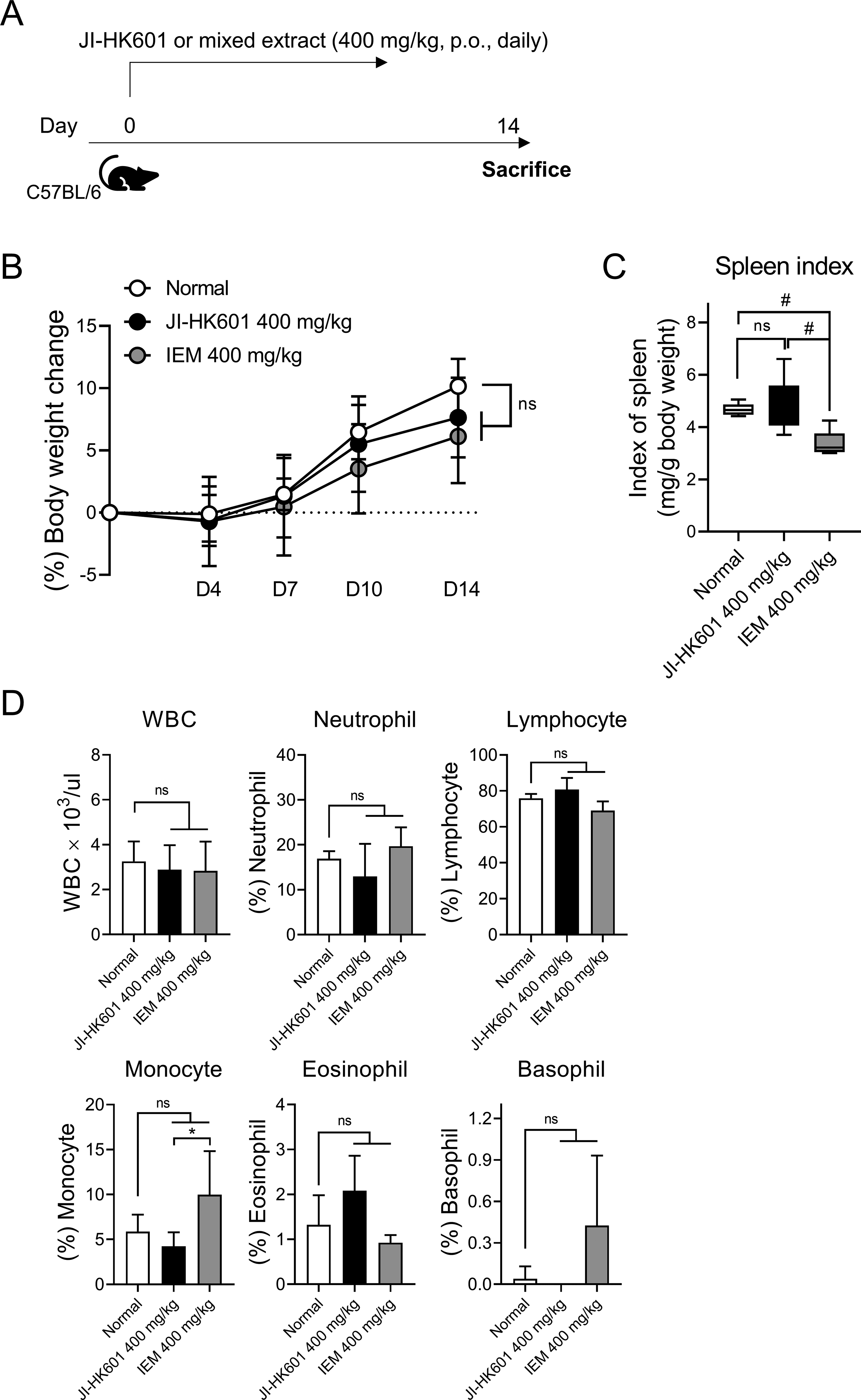
Safety evaluation of JI-HK601 and the individual extracts mixture (IEM) at a fixed dose.
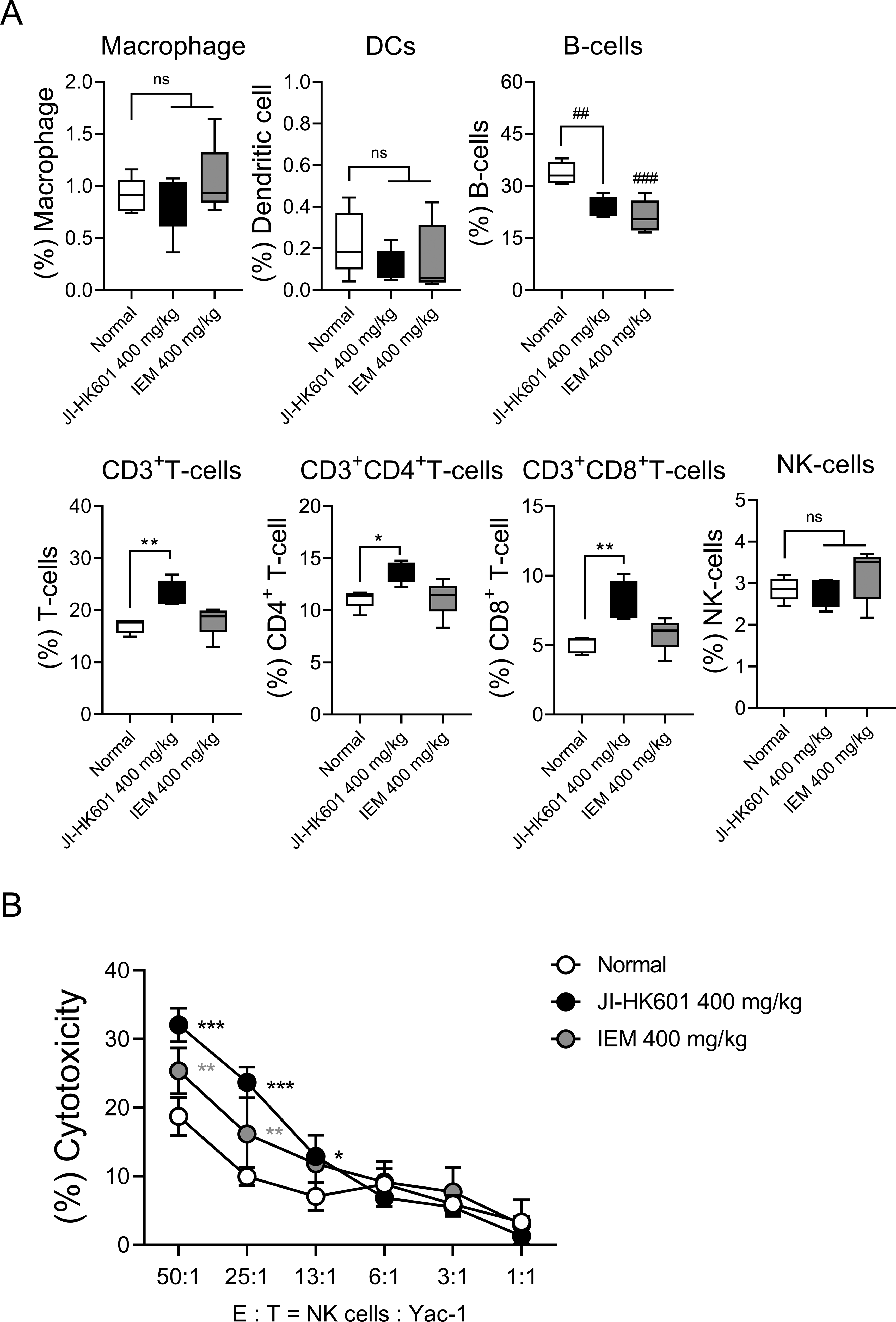
Effects of JI-HK601 and IEM on splenic immune cell composition and NK cell activity.
Based on these observations, subsequent experiments focused exclusively on JI-HK601 to determine its MED (Figs. 3 and 4). Mice were administered 100, 150, 200, or 400 mg/kg for 14 days. None of the doses affected body weight, spleen index, or hematological parameters, confirming a wide safety margin. Flow cytometric analysis showed that an increased proportion of T cells was observed only at 400 mg/kg. NK cell functional assays revealed dose-dependent effects. At a high effector-to-target ratio of 50:1, NK cytotoxicity increased at 150 mg/kg and above. However, at a lower ratio of 13:1, enhanced cytotoxicity was observed only at 400 mg/kg. To elucidate the immunological mechanism underlying these effects, transcriptional analysis of key T-cell regulators was performed. T-bet, the master transcription factor for Th1 differentiation and cell-mediated immunity, was significantly upregulated at 200 and 400 mg/kg. Consistent with the transcriptional upregulation of T-bet, administration of JI-HK601 was associated with increased production of Th1-related cytokines in splenocytes. Following ex vivo stimulation with Con A, splenocytes from the 400 mg/kg-treated group exhibited a marked increase in the secretion of IFN-γ and IL-2 compared with the normal control group. In contrast, GATA3 (Th2) and Foxp3 (Treg) remained unchanged, indicating that JI-HK601 selectively enhances Th1-type immune responses without promoting Th2-mediated humoral immunity or regulatory immune suppression. Th1 polarization is critical for antiviral, antibacterial, and antitumor immunity, and therefore this selectivity strongly supports the functional relevance of JI-HK601. Taken together, these data demonstrate that JI-HK601 enhances immune function primarily by boosting NK cell activity and promoting Th1-biased T-cell differentiation. Among the tested doses, 400 mg/kg produced the most consistent and biologically meaningful improvements across NK cytotoxicity, T-cell proportion, and T-bet expression. Previous studies have reported a maximum tolerated dose of 4800 mg/day in clinical settings and an animal-equivalent dose of approximately 558 mg/kg in tumor models.20,21 In this study, 400 mg/kg was identified as the MED, corresponding to a human-equivalent dose (HED) of approximately 3400–3500 mg/day for a 65 kg adult. These findings suggest that JI-HK601 can exert significant immunoenhancing effects within a clinically feasible intake range without detectable toxicity. This is particularly relevant for functional food or nutraceutical applications, in which long-term consumption requires both biological effectiveness and minimal adverse effects. Given its ability to enhance NK cell cytotoxic activity and promote Th1-biased immune responses, JI-HK601 may be considered a potential candidate for use as a functional food or dietary supplement for populations requiring immune support, such as elderly individuals or those recovering from chemotherapy-induced immunosuppression.
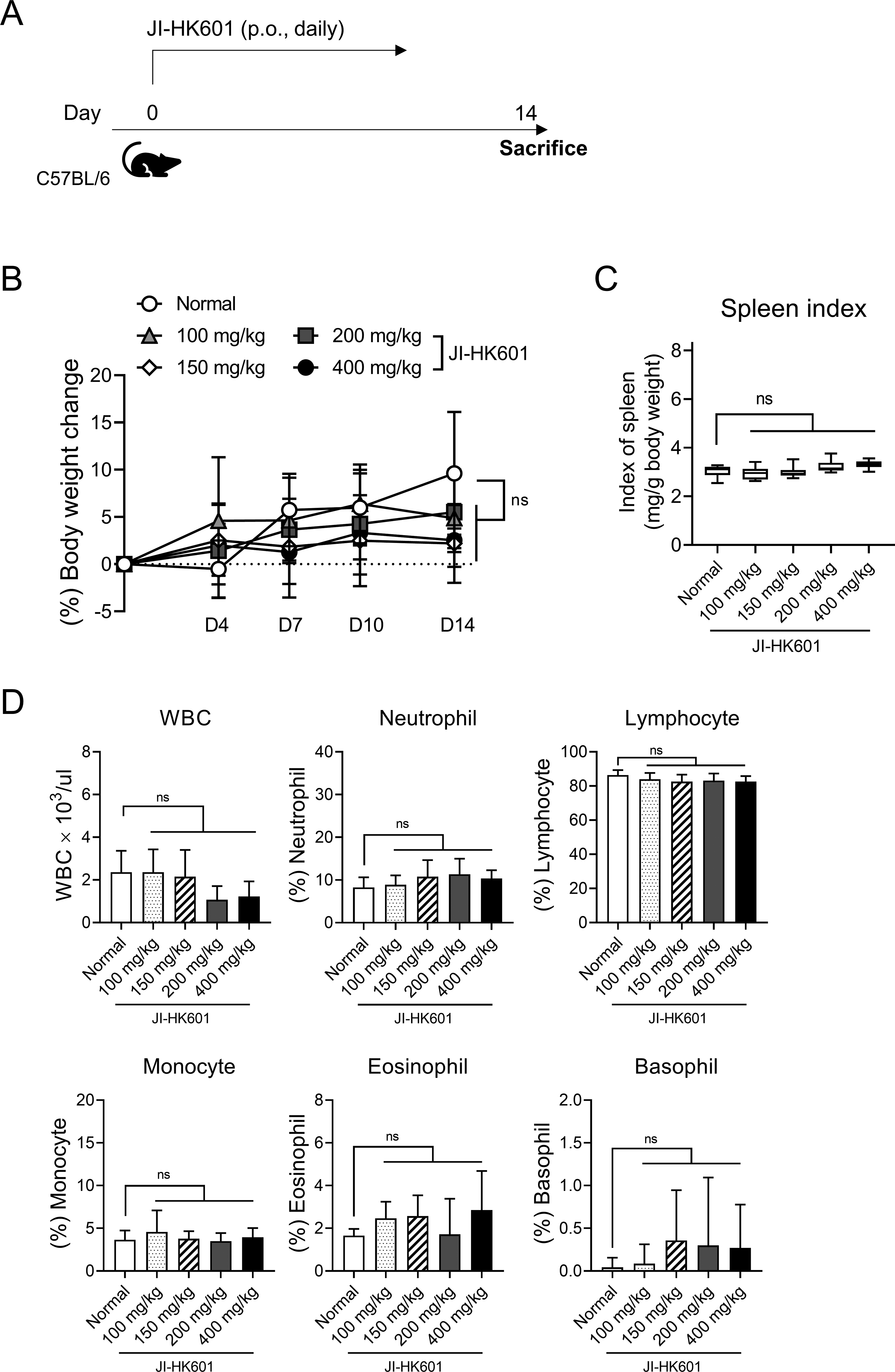
Dose-dependent safety evaluation of JI-HK601.
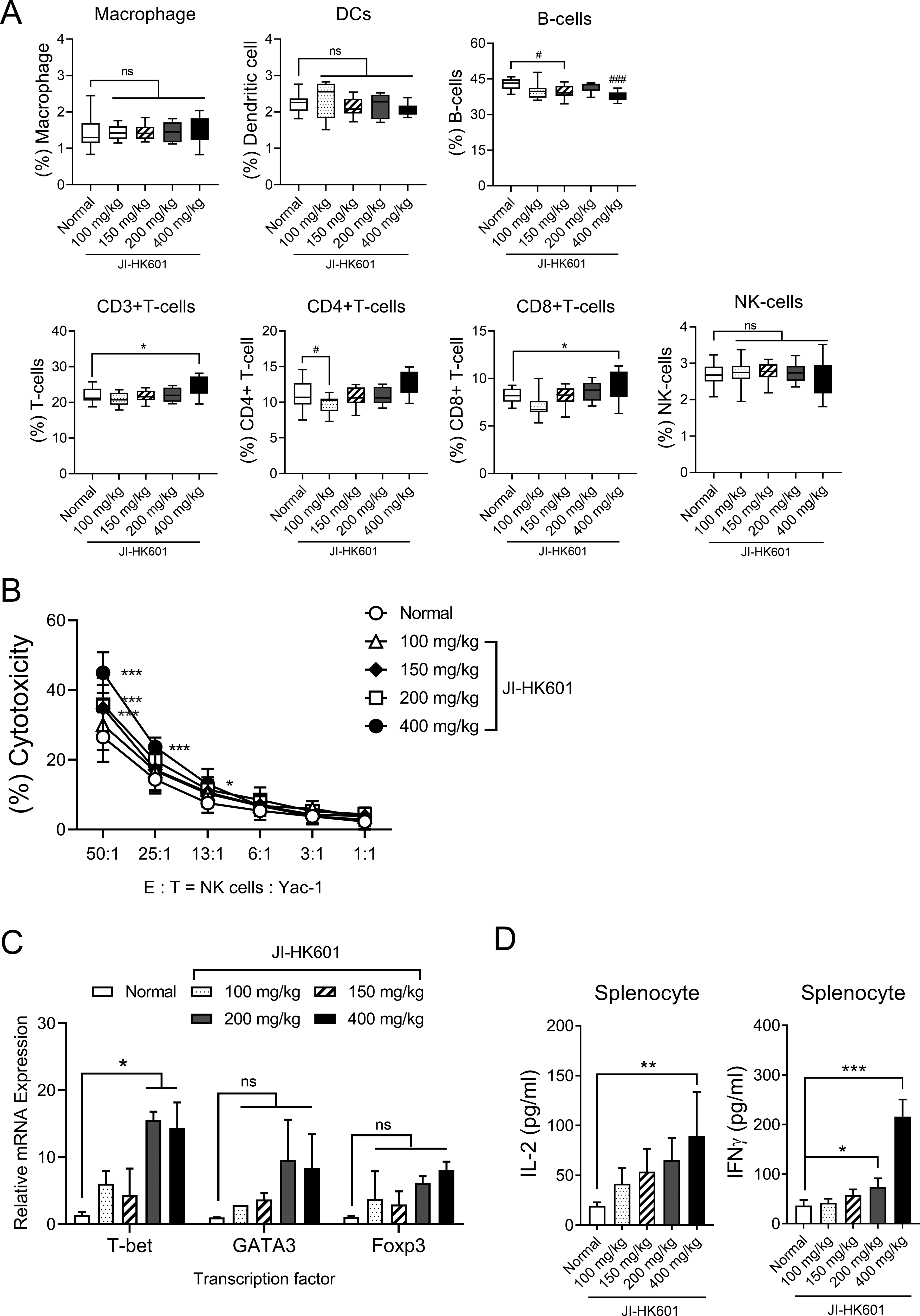
Dose-dependent immunomodulatory effects of JI-HK601.
Despite these encouraging observations, several limitations must be considered. First, the mechanisms underlying the observed modulation of B-cell populations were not fully elucidated. Future studies incorporating plasma cell markers, germinal center B-cell markers, and serum immunoglobulin measurements will be necessary to determine whether the observed reduction reflects functional differentiation, cellular redistribution, or immune suppression. Second, as this study was limited to short-term exposure in healthy mice, the long-term effects and applicability of JI-HK601 under pathological conditions remain to be determined. Given that NK cells and Th1-related pathways are pivotal in early host defense, evaluation in disease-relevant models, including viral infection models such as influenza and immunosuppression models induced by chemotherapeutic agents such as cyclophosphamide, will be important, with relevant endpoints such as survival, NK cell activity, restoration of immune cell populations, and cytokine responses. Determining whether JI-HK601 can serve as a restorative agent in such immune-dysfunctional states will help clarify its practical host-defense capabilities and translational relevance. Third, as this study was conducted in healthy mice, there are inherent limitations in directly extrapolating these findings to human applications. While the HED estimation provides a useful reference point, further clinical investigations are required to determine whether comparable immunomodulatory effects can be achieved in humans and to establish optimal dosing strategies.
Taken together, the present study provides foundational evidence that JI-HK601 enhances NK cell cytotoxic activity and promotes Th1-biased immune responses without detectable toxicity. These findings highlight its potential as a functional immunomodulatory material and provide a basis for further investigation into its role in improving host defense and disease resistance.
AUTHORS’ CONTRIBUTIONS
S.-E.L.: Conceptualization, methodology, data curation, validation, investigation, and writing—original draft. Y.H.K.: Conceptualization, data curation, validation, and investigation. Y.-B.S.: Methodology, resources, and writing—review and editing. S.-G.K.: Resources, funding acquisition, writing—review and editing, and supervision.
Footnotes
AUTHOR DISCLOSURE STATEMENT
The authors have declared no conflicts of interest.
FUNDING INFORMATION
This work was supported by the National Research Foundation of Korea grant funded by the Korea government (No. RS-2020-NR049559) and by the Technology Development Program (S3302137) funded by the Ministry of SMEs and Startups (MSS,Korea).
Supplemental Material
References
Supplementary Material
Please find the following supplemental material available below.
For Open Access articles published under a Creative Commons License, all supplemental material carries the same license as the article it is associated with.
For non-Open Access articles published, all supplemental material carries a non-exclusive license, and permission requests for re-use of supplemental material or any part of supplemental material shall be sent directly to the copyright owner as specified in the copyright notice associated with the article.