






Abstract
Case series summary
This report represents a retrospective case series of feline palpebral apocrine cystadenomatosis (PAC). Between 2019 and 2024, four client-owned cats (three Persian cats, one Siberian cat) were referred to the ophthalmology and dermatology departments of a private veterinary hospital for one or more greyish lesions of the eyelids that had been evolving for 1 month to 1 year. Three cats underwent histological examination, with a diagnosis of PAC. In the fourth case, this could not be performed as the single lesion ruptured during surgery. All cats were treated by diode laser vaporisation under general anaesthesia. No recurrence was reported at any previously treated sites, including the first case, which was followed up for 6 years.
Relevance and novel information
PAC is a rare benign cutaneous proliferation of modified apocrine glands of the eyelid (glands of Moll), mainly described in Persian cats. It presents as one or more greyish cysts of varying sizes. To the authors’ knowledge, this case series is the first to report multiple feline cases of PAC treated with diode laser vaporisation. Diode laser surgery appeared safe, well tolerated and effective, with minimal postoperative pain or inflammation and no recurrence observed at treated sites. This technique represents a practical alternative to carbon dioxide laser treatment. However, its clinical use may be limited by equipment cost, the need for appropriate operator training and the risk of collateral thermal injury if inappropriate settings are applied.
Plain language summary
Palpebral apocrine cystadenomatosis is a rare, non-cancerous condition affecting the eyelids of cats, especially Persian cats. It causes one or more fluid-filled cysts to develop along the eyelid margin, which may interfere with blinking or cause ocular irritation. Several treatment options have been described, but information on the use of diode laser surgery for this condition remains very limited. This article describes four cats that were treated with diode laser surgery for eyelid cysts between 2019 and 2024. Three cats had a confirmed diagnosis based on laboratory examination of the tissue. In the fourth cat, the diagnosis was strongly suspected based on the appearance and history of the lesion, although laboratory confirmation was not possible. All cats underwent diode laser treatment under general anaesthesia. The treatment was well tolerated in all cases, and healing was uneventful. No recurrence of the lesions was observed at the treated sites, including in one cat that was followed up for 6 years after treatment. No significant complications were reported. These results suggest that diode laser surgery is a safe and effective option for treating eyelid cysts in cats. As diode lasers are increasingly available in veterinary practice, particularly in dermatology, this technique may represent a practical alternative to other treatment methods for managing eyelid lesions in cats.
Introduction
Apocrine cystadenomatosis (AC) is a rare benign skin condition characterised by cystic proliferation of modified apocrine sweat glands. It has been reported in humans and domestic animals, particularly in cats, in which it most frequently affects the eyelids, where apocrine glands correspond to the glands of Moll, as well as peri-auricular skin, external ear canals, lips and perianal margins. 1 The ear pinnae and canals are the most affected sites. 1 Lesions usually present as one or more greyish, translucent or hyperpigmented cysts of varying sizes.
Feline palpebral apocrine hydrocystoma (PAC) occurs predominantly in Persian cats, suggesting a breed-related genetic predisposition.2 –4 The aetiopathogenesis remains uncertain: some authors consider it a reactive process secondary to chronic inflammation or duct obstruction, whereas others consider it a true benign neoplasm.3,2,5
Various therapeutic options have been reported in cats, including surgical excision, cryotherapy, intralesional or topical trichloroacetic acid, and carbon dioxide (CO2) laser ablatio.5 –11 Laser surgery offers the advantages of excellent haemostasis, limited postoperative pain and precise control in delicate anatomical areas such as the eyelids. However, diode laser use for PAC has been described only anecdotally. 12
The purpose of the present report was to describe the clinical features, pathological findings, treatment procedure and outcomes of four feline cases of PAC managed using diode laser vaporisation.
Case series description
This retrospective case series includes all cases of PAC received in a private veterinary hospital between 2019 and 2024: four client-owned cats were referred to the ophthalmology department for the presence of one or more greyish cutaneous cysts affecting the eyelids. The lesions had been evolving for periods ranging from 1 month to 1 year before presentation. Three of the cats were Persians and one was a Siberian, aged 6–12 years at disease onset. All cases were subsequently referred to the dermatology department for further investigation and management. The clinical data of the patients are summarised in Table 1. All diagnostic and therapeutic procedures described below were performed under general anaesthesia following the protocol detailed in the ‘Procedure and technical details’ section, with informed owner consent obtained in all cases, as part of routine clinical care.
Signalment, diagnosis and outcome of four feline cases of palpebral apocrine cystadenomatosis treated with diode laser vaporisation
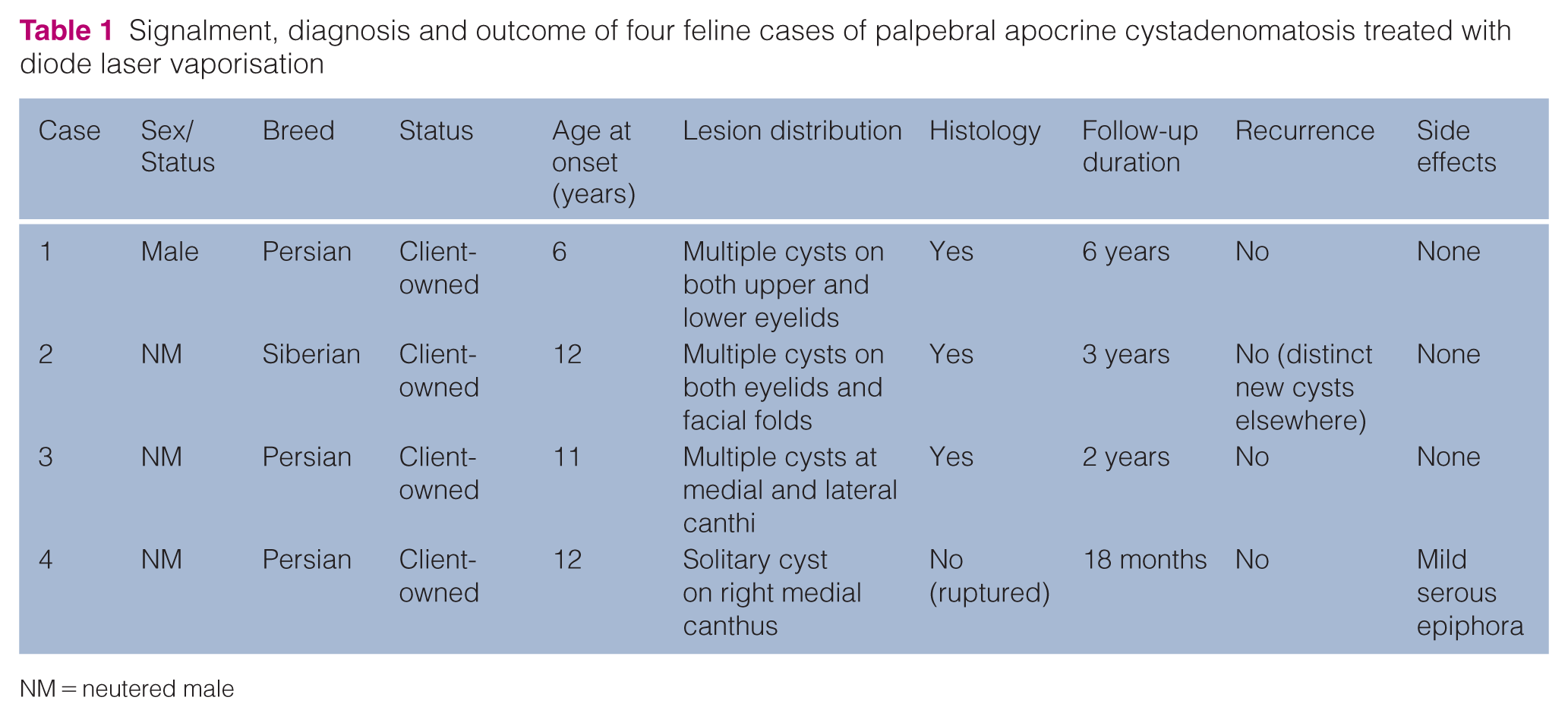
NM = neutered male
Case 1
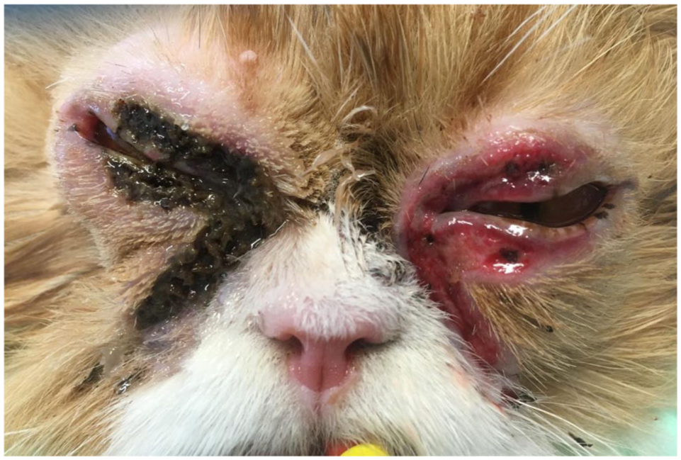
A 7-year-old, intact male Persian cat was referred in 2019 with multiple hyperpigmented cystic lesions of varying sizes affecting both upper and lower eyelids, extending to the facial folds (Figure 1). The lesions had been present for approximately 1 year and had recently increased in size. Prior topical ophthalmic treatment with chloramphenicol had been unsuccessful. The cat lived mainly indoors with occasional garden access and was otherwise healthy.

Case 1: multiple hyperpigmented cystic lesions affecting both eyelids and periocular folds
Clinical and histological findings
Dermatological examination revealed multiple pigmented, cystic nodules on the superior and inferior eyelids of both eyes and extending to the periocular region. No other abnormalities were detected. Pathological examination of three punch biopsy specimens (3–4 mm in diameter, depending on lesion size) confirmed the diagnosis of PAC.
Treatment and outcome
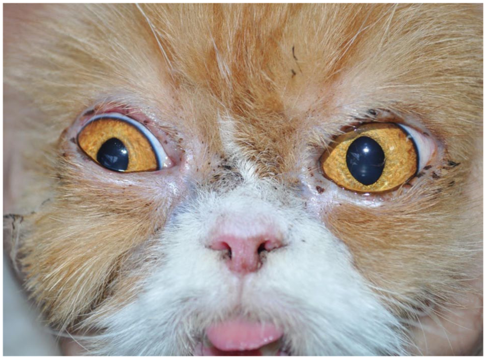
Given the bilateral extent, diode laser vaporisation was performed in two sessions 2 weeks apart, under general anaesthesia. An ocular protector and lubricant (Ocrygel; Dômes Pharma)-impregnated compress protected the cornea throughout the procedure. Postoperative medication consisted of dexamethasone–neomycin–polymyxin B ophthalmic ointment (Maxidrol; Novartis Pharma) q12h for 7 days, followed by fusidic acid ointment (Isathal; Dechra) for 15 days. The same protocol was applied to the contralateral eye: dexamethasone–neomycin–polymyxin B ointment q12h for 7 days, followed by fusidic acid ointment for a further 15 days. After each procedure, the cat was hospitalised for 24 h to monitor postoperative pain and discomfort. Analgesia consisted of morphine (Morphine; CDM Lavoisier) q4–6h until discharge. During hospitalisation, the cat showed no clinical signs of pain or discomfort. In view of this favourable postoperative course, no systemic anti-inflammatory or analgesic treatment was prescribed at discharge. However, the owners were informed of the possibility to contact the veterinary hospital and return for reassessment should any signs of pain or discomfort develop. Figure 2 shows the appearance of the periocular regions, 2 weeks after surgery for the left eye and immediately after surgery for the right eye. Healing was complete 1 month after the second procedure (Figure 3). No recurrence was observed during a 6-year follow-up.
Case 1: left eye 2 weeks postoperatively; right eye immediately postoperatively
Case 1: 1 month postoperatively, showing complete healing
Case 2
A 12-year-old, neutered male Siberian cat was presented in 2022 with multiple hyperpigmented cystic lesions on the upper and lower eyelids bilaterally extending to the facial folds, associated with moderate to severe bilateral epiphora. The lesions had developed 1 month before presentation.
Previous oral systemic antibiotics (amoxicillin–clavulanic acid) and chloramphenicol ophthalmic ointment had been ineffective.
Clinical and histological findings
Examination confirmed multiple pigmented cystic nodules and bilateral epiphora (Figure 4a). No other abnormalities were detected. Pathological examination of three punch biopsy specimens (3–4 mm in diameter, depending on lesion size) had been performed by the ophthalmology department before the dermatology consultation. It revealed one nodular hidradenoma and two lesions consistent with PAC.
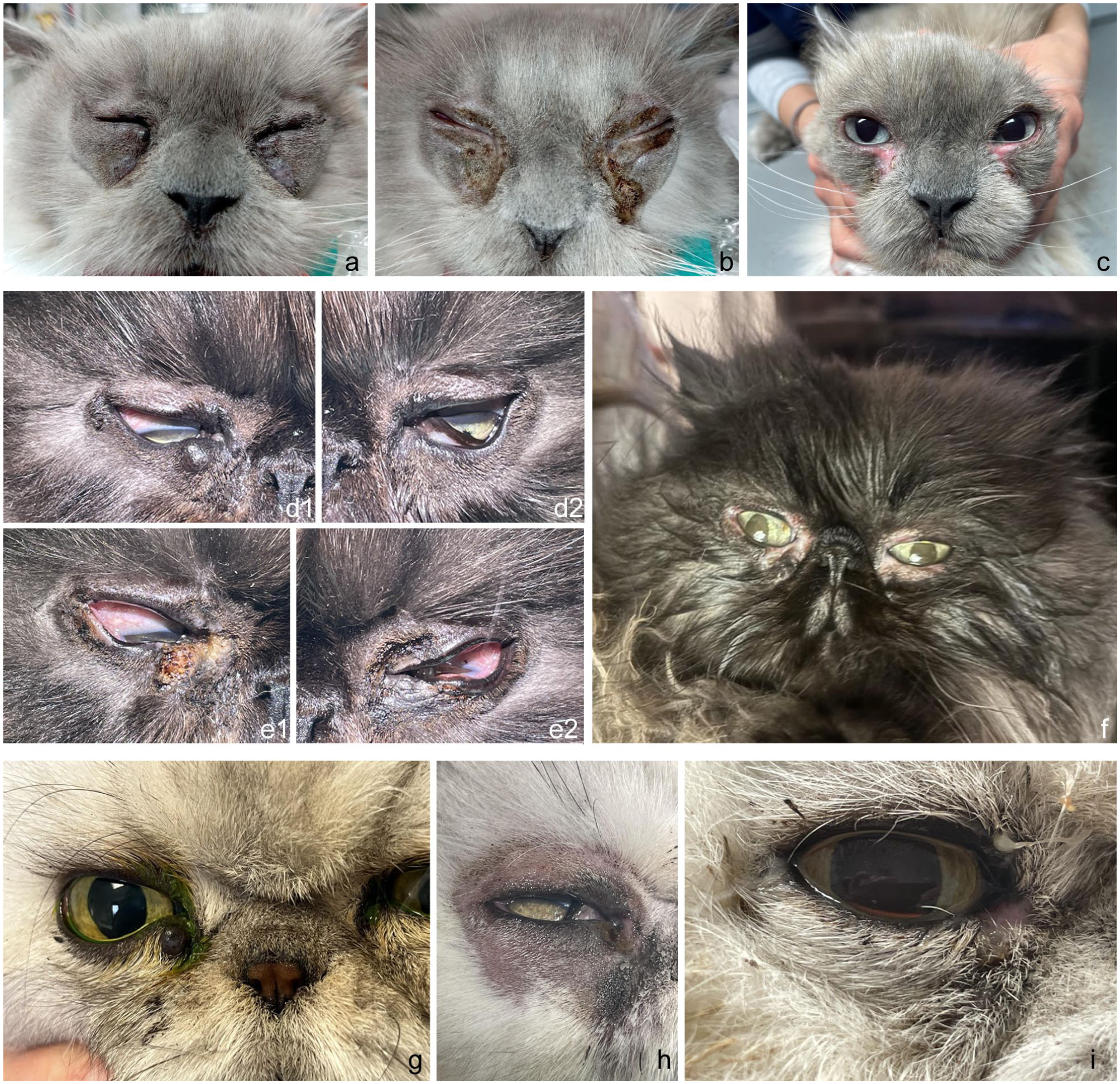
(a–c) Case 2, (d–f) case 3 and (g–i) case 4: before surgery (a, d, g), immediately after diode laser vaporisation (b, e, h) and 4 weeks after laser vaporisation (c, f, i)
Treatment and outcome
Diode laser vaporisation was performed under general anaesthesia, using corneal protection with an ocular protector and lubricant (Ocrygel; Dômes Pharma)-impregnated compress. Figure 4b shows the immediate postoperative appearance of the periocular regions. The cat was hospitalised for 24 h after the procedure for postoperative monitoring. Pain assessment during hospitalisation was based on clinical parameters including behaviour, appetite and periocular palpation. Mild signs of discomfort were observed, without marked inflammation. Consequently, a short course of systemic analgesia with tramadol (Tralieve; Dechra Veterinary Products) was prescribed for 3 days at discharge, together with a dexamethasone–neomycin–polymyxin B ophthalmic ointment (Maxidrol; Novartis Pharma) q12h for 8 days and an Elizabethan collar for 15 days. Complete healing was observed after 1 month (Figure 4c). Seven months later, mild blepharitis, which successfully resolved with a 15-day course of a chloramphenicol-containing ophthalmic ointment (Ophtalon; Dômes Pharma), and persistent bilateral serous epiphora were noted, together with two small cystic lesions at the left medial canthus, distinct from the previously treated sites. As these lesions remained small, stable and non-progressive, no further therapeutic intervention was deemed necessary.
Case 3
An 11-year-old, neutered male Persian cat was examined in 2023 for multiple hyperpigmented palpebral cystic nodules on the right lateral canthus and both medial canthi. The lesions had progressively enlarged over several months. The cat had a lifelong mild bilateral epiphora, but no other medical history.
Clinical and histopathological findings
Multiple, non-ulcerated, soft, pigmented cystic nodules were observed without associated pain or inflammation on the right lateral canthus and both medial canthi. Figure 4d1,d2 shows the appearance of the eyelids before intervention. No other abnormalities were observed. Pathological examination of one punch biopsy specimen (4 mm in diameter) confirmed the diagnosis of PAC.
Treatment and outcome
Diode laser vaporisation was performed under general anaesthesia, with an ocular protector and lubricant (Ocrygel; Dômes Pharma)-impregnated compress. Figure 4e1,e2 shows the appearance of the eyelids just after laser vaporisation. Postoperative surveillance was ensured through a 24-h hospital stay, during which pain and comfort were closely assessed. Analgesic management relied on repeated administrations of morphine (Morphine; CDM Lavoisier) q4–6h until discharge. Throughout hospitalisation, the cat remained clinically comfortable, with no observable signs of pain. Given this uneventful postoperative course, no systemic anti-inflammatory or analgesic medication was prescribed at discharge; nevertheless, the owners were advised to contact the referral centre and return for reassessment should any signs of discomfort arise. Postoperative treatment consisted of a dexamethasone–neomycin–polymyxin B ophthalmic ointment (Maxidrol; Novartis Pharma) q12h for 10 days, followed by gentle cleansing with sterile saline until full epithelialisation. Healing was complete within 1 month (Figure 4f) and no recurrence has been observed to date.
Case 4
A 12-year-old, neutered male Persian cat was referred in 2024 for a solitary 4 mm greyish cyst on the right medial canthus that had been enlarging for 6 months. The lesion was asymptomatic and non-ulcerated. Prior treatment with framycetin–dexamethasone ophthalmic ointment had no effect. The cat was otherwise healthy.
Clinical findings and presumptive diagnosis
A well-defined, greyish cystic lesion was observed on the right medial canthus (Figure 4g). The cyst was fluctuant and non-painful, and was not interfering with eyelid motion. During sampling with a 6 mm biopsy punch, the cyst ruptured spontaneously, precluding histological analysis; however, the clinical appearance, localisation and breed supported a presumptive diagnosis of PAC. No other abnormalities were detected.
Treatment and outcome
Residual tissue was vaporised with a diode laser under general anaesthesia using an ocular protector and lubricant (Ocrygel; Dômes Pharma)-impregnated compress. Figure 4h shows the right periocular region immediately after diode laser vaporisation. Because the lesion was solitary and the surgical site limited in size, postoperative monitoring was restricted to a short hospital stay, during which the cat remained comfortable and showed no clinically significant signs of pain. Postoperative therapy included fusidic acid ointment (Isathal; Dechra) q12h for 10 days. Healing was complete after 1 month (Figure 4i). A mild unilateral serous epiphora was observed postoperatively but remained stable thereafter. No recurrence was detected at 18-month follow-up.
Procedure and technical details
Anaesthetic management
All cats underwent diode laser treatment under general anaesthesia according to an identical protocol. Premedication consisted of morphine (0.1 mg/kg SC, Morphine; CDM Lavoisier) combined with diazepam (0.2 mg/kg IV, Valium; Atnahs Pharma Netherlands BV). Anaesthesia was induced with propofol (2–4 mg/kg IV, Proposure; Axience) to effect and maintained with isoflurane (ISO-VET; Piramal Critical Care) in oxygen.
Biopsy sampling strategy
Incisional punch biopsies were performed to obtain tissue for pathological examination. Except for case 2, biopsy sampling and diode laser vaporisation were carried out during the same anaesthetic episode. All biopsies were performed under general anaesthesia, with owner consent obtained as part of routine clinical care.
Laser system and fibre selection
A Lumix Surgery Dual 29 W diode laser (Fisioline) was used in all cases. The system was equipped with either a 400 µm or a 600 µm direct-contact optical fibre. Fibre diameter was selected according to lesion characteristics: the 400 µm fibre was preferentially used for smaller or more delicate periocular lesions requiring high precision, whereas the 600 µm fibre was chosen for larger or more extensive cystic areas.
The laser operated in continuous-wave mode at wavelengths in the range of 800–1064 nm, with mean power settings of 3–5 W.
Laser vaporisation technique
Laser vaporisation was initiated at higher power settings to allow rapid and effective tissue ablation. Power output was then progressively reduced as the procedure approached adjacent normal tissues or the tarsal plate to minimise collateral thermal injury. Each cystic lesion was vaporised for approximately 5–10 s, depending on lesion size and tissue thickness. As a result, total delivered fluence varied between lesions and was not standardised.
During vaporisation, visible tissue carbonisation was deliberately avoided to limit excessive heat accumulation and thermal diffusion.
Ocular protection and safety measures
Throughout the procedure, a sterile non-woven compress impregnated with an ocular protective and lubricating gel (Ocrygel; Dômes Pharma) was systematically placed over the cornea to protect adjacent ocular structures from direct laser exposure and secondary thermal effects. In addition, all personnel present in the operating theatre wore wavelength-specific protective eyewear.
Postoperative management and complications
Immediate postoperative management included topical corticosteroid–antibiotic ophthalmic combinations, with or without systemic analgesia depending on individual postoperative assessment. None of the cats required systemic anti-inflammatory medication. No intraoperative complications were recorded.
Operator consistency
All procedures were performed by the same surgeon.
Discussion
Diagnosis and histopathology
AC is a rare, benign proliferative skin condition affecting modified apocrine glands, often forming multiple cystic structures lined by secretory epithelium. 13 In cats, the condition predominantly affects Persians and shows a predilection for the eyelids, where the modified glands correspond to the glands of Moll.1 –4 Although the clinical appearance is often distinctive, differential diagnoses should include apocrine cysts as well as neoplastic conditions such as basal cell carcinoma, squamous cell carcinoma, melanoma, mast cell tumour and apocrine cystadenocarcinoma. 6 Histopathologically, PAC is characterised by cystic structures lined by a single layer of cuboidal-to-columnar epithelial cells, occasionally forming papillary projections extending into the lumen. 13 The cysts contain eosinophilic secretory material, and nuclear polarity is maintained, with minimal to absent mitotic activity (Figure 5). 13 These features distinguish PAC from apocrine cystadenocarcinoma, which exhibits nuclear pleomorphism, loss of polarity and increased mitotic rate. 13 In the present series, pathological confirmation was obtained in three cats. The fourth case lacked histological evaluation because of cyst rupture during sampling, but the diagnosis was strongly supported by the cat’s breed, age, lesion localisation and clinical presentation. Although cytology can assist in differentiating cystic adnexal proliferations, definitive diagnosis still requires pathological assessment.2,13
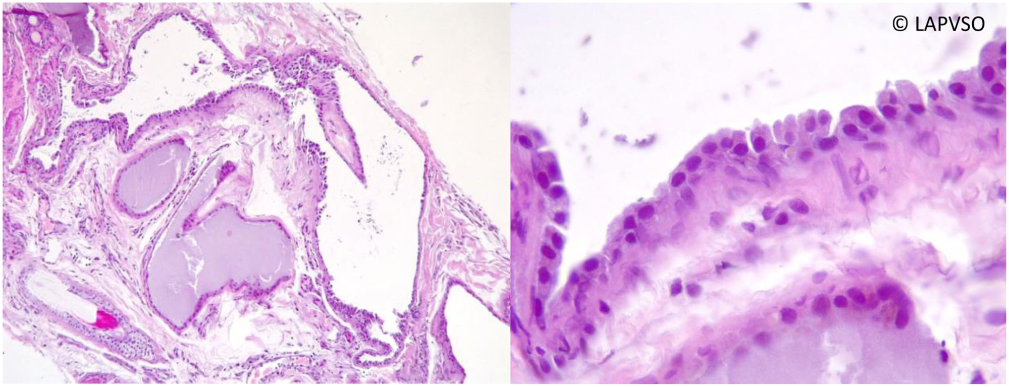
Histological section of feline palpebral apocrine cystadenomatosis showing multiple cystic structures lined by cuboidal epithelial cells containing eosinophilic secretory material (haematoxylin and eosin stain). Courtesy of LAPVSO, France
Therapeutic approaches
Various therapeutic strategies for feline PAC have been reported, ranging from conservative management to chemical, surgical or laser ablation. In mild or asymptomatic cases, observation may suffice, as the lesions are benign and slowly progressive. 1 However, when cysts enlarge, ulcerate or cause discomfort by interfering with eyelid motion, active treatment becomes necessary.
In human medicine, numerous therapeutic approaches have been described for the management of apocrine cystadenoma. These include destructive or surgical procedures such as simple excision, en bloc resection, CO2 laser, pulsed dye laser, incision and drainage, excision combined with adjuvant cryotherapy, cryotherapy alone, electrosurgery, diode laser
14
and argon laser.
15
Skin-directed therapies have also been reported, including botulinum toxin injections, intralesional trichloroacetic acid
16
or hypertonic glucose, photodynamic therapy with sodium chloride, topical retinoic acid and topical nystatin combined with triamcinolone.
15
Systemic medical treatments, such as oral anticholinergics, nifedipine and bromocriptine, have been used in selected cases.
15
Finally, combined protocols have been described, notably erbium:yttrium–aluminium–garnet (Er:YAG) laser therapy followed by oral isotretinoin, or oral isotretinoin administration followed by CO2 laser treatment.
15
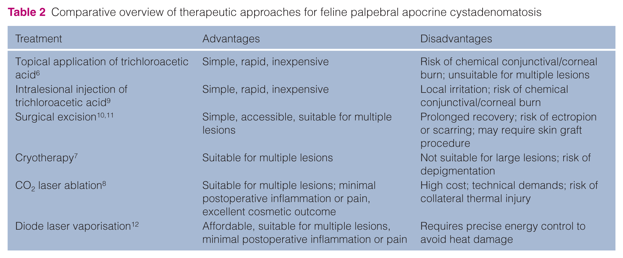
In veterinary medicine, reported treatment options for PAC include topical or intralesional trichloroacetic acid,7,10 surgical excision,5,11 cryotherapy 8 and CO2 laser ablation.9,6 –11 These modalities differ in terms of invasiveness, risk of haemorrhage, need for specialised equipment, postoperative inflammation and cosmetic outcome, as described in the literature. More recently, diode laser therapy has emerged as an effective alternative for the treatment of PAC, with excellent cosmetic outcomes and no adverse effects or recurrence reported 7 months after the procedure. 12 Based on previously published data, the advantages and disadvantages of the different therapeutic options are summarised in Table 2. The decision to intervene or to adopt a conservative approach should primarily depend on the animal’s degree of discomfort and the owner’s preference. As no universally applicable timing exists, management should be tailored to each individual case. When treatment is chosen, the treatment modality should be selected according to the size and number of lesions, and the availability of appropriate surgical equipment. In a retrospective clinical setting, topical treatment selection may reflect drug availability and clinician preference rather than a strictly standardised protocol. Despite differences in postoperative topical treatment, healing was uneventful and clinical outcomes were favourable in all cases.
Comparative overview of therapeutic approaches for feline palpebral apocrine cystadenomatosis
Diode and CO2 lasers are both widely used in veterinary surgery. CO2 lasers are generally more expensive, bulkier and less portable, and their operation requires greater technical expertise and training. In addition, the use of an oxygen-rich environment carries a small but recognised risk of combustion. In contrast, diode lasers are compact, user-friendly and relatively inexpensive, which makes them more accessible in general practice.
Despite these practical advantages, the interaction between laser type and tissue differs markedly. Both systems provide excellent haemostasis and minimise postoperative pain and inflammation, making them suitable for delicate areas such as the eyelids. However, CO2 lasers (10,600 nm) deliver shallower energy penetration and offer superior control of superficial tissues, whereas diode lasers (800–1064 nm) penetrate deeper, potentially increasing the risk of collateral thermal injury. CO2 lasers may shorten surgical time owing to their broader beam diameter compared with the optical fibre of diode units, although this may reduce precision when treating small, localised lesions.
Despite these differences, diode lasers have shown excellent efficacy and safety in dermatological and ophthalmological applications.12,17,18
Complications and prognosis
Potential complications of eyelid laser surgery include ectropion or entropion due to excessive damage to the tarsal plate, lacrimal duct injury causing chronic epiphora and, rarely, corneal burns if protection is inadequate. In this series, only one cat (case 4) developed mild unilateral serous epiphora after treatment of a cyst located at the medial canthus. This was likely related to excessive energy exposure near the nasolacrimal duct, highlighting the need for precise control of laser parameters (time exposure and power).
Only mild and transient postoperative local inflammation was observed clinically in some cases, without persistent scarring or eyelid deformities. No recurrence was observed at any previously treated site, including the earliest case, which was followed up for 6 years.
The technique’s safety and efficacy in this small cohort align with findings from a single previous report of feline PAC successfully managed by diode laser. 12 Moreover, postoperative cosmetic results were excellent, with minimal discomfort and rapid epithelialisation.
Although AC is a benign condition, untreated lesions may gradually enlarge and interfere with normal eyelid function. 9 To date, malignant transformation has not been documented; however, chronic irritation or recurrent cyst formation may lead to persistent ocular discomfort. 9 Early therapeutic intervention is therefore advisable in symptomatic or progressive cases.
Limitations
The main limitations of this case series are the small sample size and the absence of histological confirmation in one cat. Importantly, the retrospective nature of this case series constitutes an inherent limitation, as it reflects routine clinical management of client-owned cats rather than a prospectively designed protocol. Consequently, postoperative medical treatments were not standardised, and pain assessment relied on subjective clinical observation rather than on a validated pain-scoring system. In addition, follow-up duration varied among patients, with a range of 18 months to 6 years, which may have led to under-detection of late recurrences. No objective evaluation of postoperative pain, cosmetic outcome or owner satisfaction was performed. Despite these constraints, the consistency of clinical presentation, procedural standardisation and long-term success observed in the earliest case reinforce the clinical relevance of these findings.
Conclusions
PAC is a benign proliferative disorder of the modified apocrine glands of the eyelid in cats, most often seen in Persian cats. Although generally non-painful, lesions may enlarge and interfere with eyelid function, warranting intervention in symptomatic cases. The present series confirms that, when used carefully, diode laser vaporisation provides complete removal of cystic lesions with rapid healing and no recurrence over prolonged follow-up periods.
Diode laser vaporisation appears to be a safe, effective and minimally invasive technique for the removal of PAC lesions in cats when performed by an experienced operator. In this context, an ‘experienced operator’ refers to a clinician who has received structured and supervised training in laser surgery, has a sound understanding of laser–tissue interactions, and is able to adapt laser settings according to lesion characteristics and anatomical location.
The procedure offers an accessible alternative to CO2 laser ablation, combining affordability, ease of handling and excellent cosmetic results. Further studies involving larger cohorts and longer follow-up would help confirm its long-term efficacy and optimal energy parameters.
Footnotes
Conflict of interest
The authors declared no potential conflicts of interest with respect to the research, authorship, and/or publication of this article.
Funding
The authors received no financial support for the research, authorship, and/or publication of this article.
Ethical approval
The work described in this manuscript involved the use of non-experimental (owned or unowned) animals. Established internationally recognised high standards (‘best practice’) of veterinary clinical care for the individual patient were always followed and/or this work involved the use of cadavers. Ethical approval from a committee was therefore not specifically required for publication in JFMS. Although not required, where ethical approval was still obtained, it is stated in the manuscript.
Informed consent
Informed consent (verbal or written) was obtained from the owner or legal custodian of all animal(s) described in this work (experimental or non-experimental animals, including cadavers, tissues and samples) for all procedure(s) undertaken (prospective or retrospective studies). No animals or people are identifiable within this publication, and therefore additional informed consent for publication was not required.
