
Abstract
Preterm infants are at risk of brain injury and poor neurodevelopmental outcomes including impairments in cognition, behavioral functioning, sensory perception, and motor performance. Systemic inflammation has been identified as an important, potentially modifiable precursor of neurologic and neurodevelopmental impairments. Inflammation is typically measured by quantifying circulating cytokines and chemokines. However, it is unclear which specific cytokines/chemokines most consistently predict neurodevelopment in preterm infants. In this integrative review, we evaluated and analyzed the literature (N = 37 publications) to determine the cytokines/chemokines most predictive of neurodevelopment in preterm infants, the optimal timing for these measurements, and the ideal source for collecting cytokines/chemokines. Synthesis of the findings of these studies revealed that interleukin (IL)-6, IL-1β, IL-8, and tumor necrosis factor (TNF)-α collected during the first 3 weeks of life are most predictive of subsequent neurodevelopment. Methodological variation among studies hinders more specific analysis, including the evaluation of cytokine thresholds and meta-analyses, that would allow for the use of cytokines/chemokines to predict neurodevelopment. Future research should focus on identifying explicit cytokine values, specifically for IL-6, IL-1β, IL-8, and TNF-α, that are most predictive for identifying preterm infants most at risk of impairment, keeping in mind that longitudinal measures of cytokines/chemokines may be more predictive of future outcomes than single-time point measures.
Preterm birth is associated with neurodevelopmental impairments in cognition (Ionio et al., 2016; Kallankari, Kaukola, Olsen, Ojaniemi, & Hallman, 2015), behavioral and social function (Cassiano, Gaspardo, & Linhares, 2016), motor performance (Pitcher et al., 2012; Romeo et al., 2016), and sensory processing (Cabral, da Silva, Martinez, & Tudella, 2016; Wickremasinghe et al., 2013). One possible mediating mechanism of neurodevelopmental impairment is inflammation; critical illness during the neonatal period, as occurs with preterm birth, is associated with elevated systemic inflammation. For example, infants with necrotizing enterocolitis and bacteremia and those requiring prolonged mechanical ventilation demonstrate higher levels of pro-inflammatory cytokines and chemokines, including interleukin (IL)-8, IL-6, IL-1β, tumor necrosis factor (TNF)-α, and monocyte chemoattractant protein (MCP)-1, than infants with a less complex clinical course (Bhatia, Stoll, Cismowski, & Hamrick, 2014; Bose et al., 2013; Leviton et al., 2012).
Cytokines, small circulating proteins, act by binding cell surface receptors and contributing to the initiation, maintenance, and regulation of inflammatory immune responses (Kahn, 2016). They are an easily accessible measure of inflammation known to communicate with the central nervous system. Inflammatory cytokines/chemokines in peripheral circulation may affect the developing brain through a number of different mechanisms including (1) direct crossing of the blood–brain barrier at sites of increased permeability (Capuron & Miller, 2011; McAdams & Juul, 2012; Slavich & Irwin, 2014), (2) activation of vascular endothelial cells at the blood–brain barrier (Capuron & Miller, 2011; Slavich & Irwin, 2014), (3) cellular transport across the vascular endothelium (Capuron & Miller, 2011; McAdams & Juul, 2012; Slavich & Irwin, 2014), (4) recruitment of monocytes to the brain parenchyma via MCP-1 signaling (Capuron & Miller, 2011), and (5) stimulation of afferent nerve fibers in the periphery (Capuron & Miller, 2011; Slavich & Irwin, 2014). Once cytokines have gained access to the central nervous system, inflammatory signaling can be propagated by cells of the brain parenchyma (Banks & Erickson, 2010). The overall effect of systemic inflammation is a propagation of inflammatory signaling and damage to the immature brain, as evidenced by abnormalities visible on cranial ultrasound and magnetic resonance imaging (Basu, Agarwal, Anupurba, Shukla, & Kumar, 2015; Inomata et al., 2014).
Cytokines have been studied in diverse inflammatory disease states affecting preterm infants including necrotizing enterocolitis (Maheshwari et al., 2014; Ng et al., 2003), bronchopulmonary dysplasia (Leroy et al., 2018), and sepsis (Ng et al., 2003). Although researchers are beginning to study cytokines as predictors of neurodevelopmental outcomes in preterm infants, much about cytokine inclusion in such research remains unclear. The purpose of this integrative review was to identify important cytokine predictors of neurodevelopment in preterm infants. Its specific goals were to (1) determine the most significant cytokines/chemokines for predicting neurodevelopmental outcomes, (2) determine the optimal timing for cytokine/chemokine measures, and (3) determine the best source of cytokines/chemokines for predicting outcomes.
Method
We conducted this review using the guidelines described by Whittemore and Knafl (2005), which include the following steps: (1) problem identification, including identification of the population and sampling frame; (2) literature search using multiple approaches; (3) data evaluation for study quality; and (4) data analysis via data reduction, display, and comparison.
Problem Identification
Although systemic inflammation is recognized as a probable antecedent of neurodevelopmental impairment in preterm infants (Van Steenwinckel et al., 2014), there is little data to guide the selection of cytokines for studies of inflammation and neurodevelopment. We limited the sampling frame for this review to primary experimental and nonexperimental studies and meta-analyses. To facilitate assessment of data quality, we did not include literature reviews, editorials, or commentaries.
Literature Search
We searched PubMed, CINAHL, and PsycINFO using the key words “cytokine OR chemokine” AND “brain OR neuro* OR grey matter OR gray matter OR white matter” AND “preterm infant” and limited the search to articles published in English between January 2008 and February 2018. Additionally, we searched reference lists of all articles in the initial full-text review for additional articles. We included publications if they (1) measured cytokines and/or chemokines in neonatal fluid or tissue, (2) measured a neurologic or neurodevelopmental outcome, and (3) included infants born at less than 37 weeks postmenstrual age (PMA). We excluded publications if they (1) included full-term infants such that measures and outcomes for preterm infants could not be distinguished from those of full-term infants, (2) measured cytokines/chemokines only in postmortem samples, (3) measured only cytokines not typically associated with inflammatory responses (e.g., erythropoietin), or (4) included only nonhuman subjects. In our initial screening, we examined titles and abstract. We reviewed publications without abstracts during full-text screening. We subsequently reviewed the full text for articles that met inclusion criteria by abstract review.
Data Evaluation
We evaluated data quality using the National Heart, Lung, and Blood Institute’s (2014) Quality Assessment Tool for Observational Cohort and Cross-Sectional Studies. This tool is used to evaluate nonexperimental studies on 14 criteria, with scores ranging from 0 (poor) to 14 (excellent). We assigned scores to each article based on these criteria as they pertained to the predictor (cytokines/chemokines) and outcome of interest. In some cases, the predictors and outcomes of interest for this review were not measured as a primary aim of the included article, which may have affected quality scores.
Analysis
We extracted the following data points for all articles in the final sample: (1) sample characteristics, (2) cytokines/chemokines measured, (3) cytokine/chemokine measurement time points, (4) type of neonatal specimen analyzed, (5) neurologic and/or neurodevelopmental outcomes, (6) infant age at outcome assessment, (7) analytic method, and (8) main findings. Table 1 and Supplemental Table A provide a synopsis of all included studies. We categorized graphic displays of associations among individual cytokines/chemokines and outcomes by (1) specific outcome and (2) cytokine/chemokine time point to visualize trends in the data and compare results across studies (Supplemental Tables B and C). We considered cytokines to be significant predictors of outcomes if results were replicated in two or more studies.
Brief Overview of Studies Included in Integrative Review.
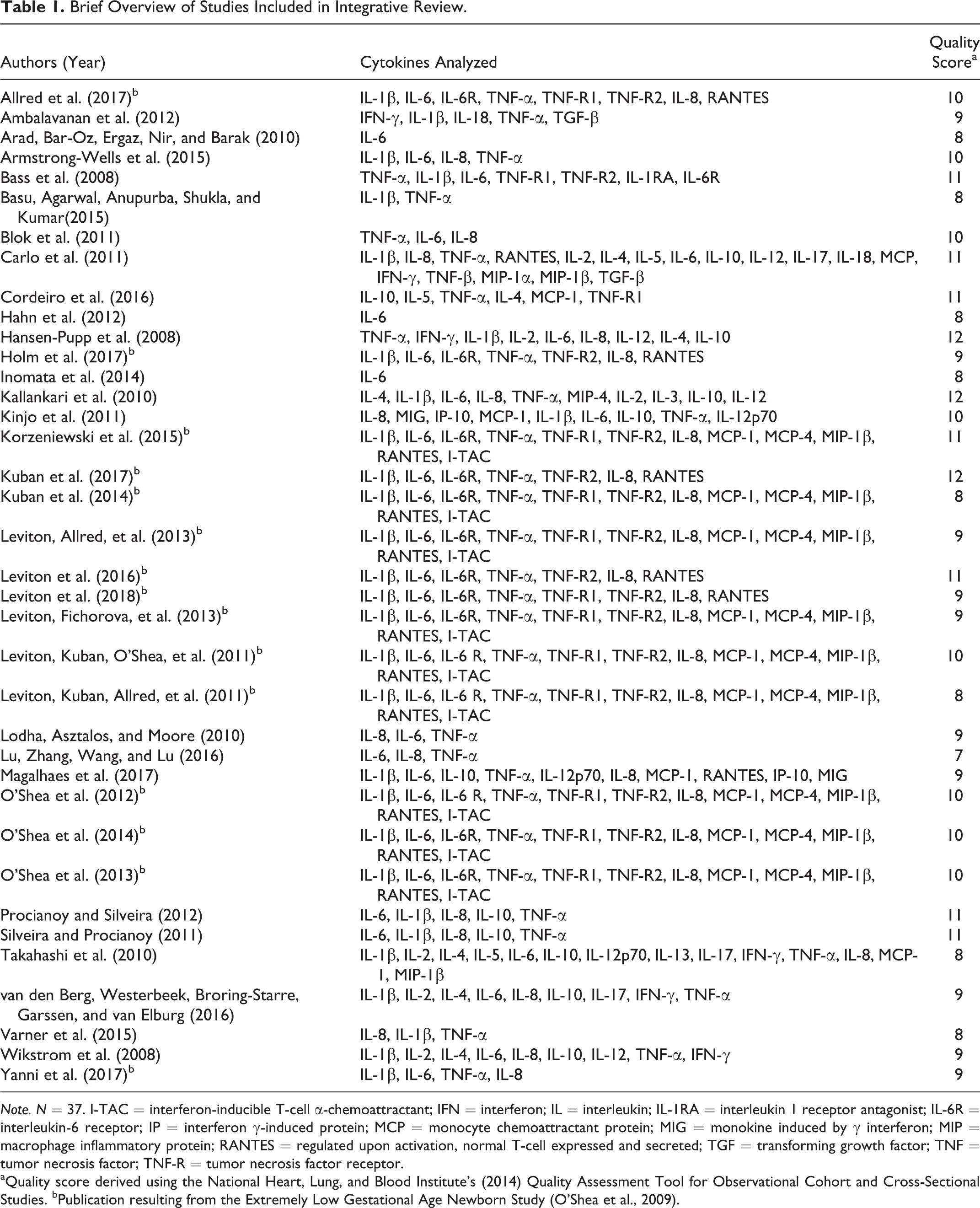
Note. N = 37. I-TAC = interferon-inducible T-cell α-chemoattractant; IFN = interferon; IL = interleukin; IL-1RA = interleukin 1 receptor antagonist; IL-6R = interleukin-6 receptor; IP = interferon γ-induced protein; MCP = monocyte chemoattractant protein; MIG = monokine induced by γ interferon; MIP = macrophage inflammatory protein; RANTES = regulated upon activation, normal T-cell expressed and secreted; TGF = transforming growth factor; TNF = tumor necrosis factor; TNF-R = tumor necrosis factor receptor.
aQuality score derived using the National Heart, Lung, and Blood Institute’s (2014) Quality Assessment Tool for Observational Cohort and Cross-Sectional Studies. bPublication resulting from the Extremely Low Gestational Age Newborn Study (O’Shea et al., 2009).
Results
Search Results
The database search yielded 201 publications. After removal of duplicates, we screened abstracts of 168 unique publications for inclusion. Of these, we excluded 125 records (see Figure 1). The majority were excluded because they did not contain primary data and were categorized as reviews, commentaries, or editorials (n = 42); consisted of only nonhuman subjects (n = 33); or did not include cytokine or chemokine measures (n = 24). We conducted full-text review on the remaining 43 publications. From review of reference lists of included publications, we identified an additional 15 publications. Of the resulting 58 publications, we excluded 21 because they did not include primary data (n = 10), did not measure a neurologic or neurodevelopmental outcome (n = 5), did not include a cytokine or chemokine measure (n = 2), did not provide data for individual cytokines/chemokines (n = 1), did not provide data for term and preterm infants separately (n = 2), or included only postmortem samples (n = 1), leaving 37 publications in the final data set.
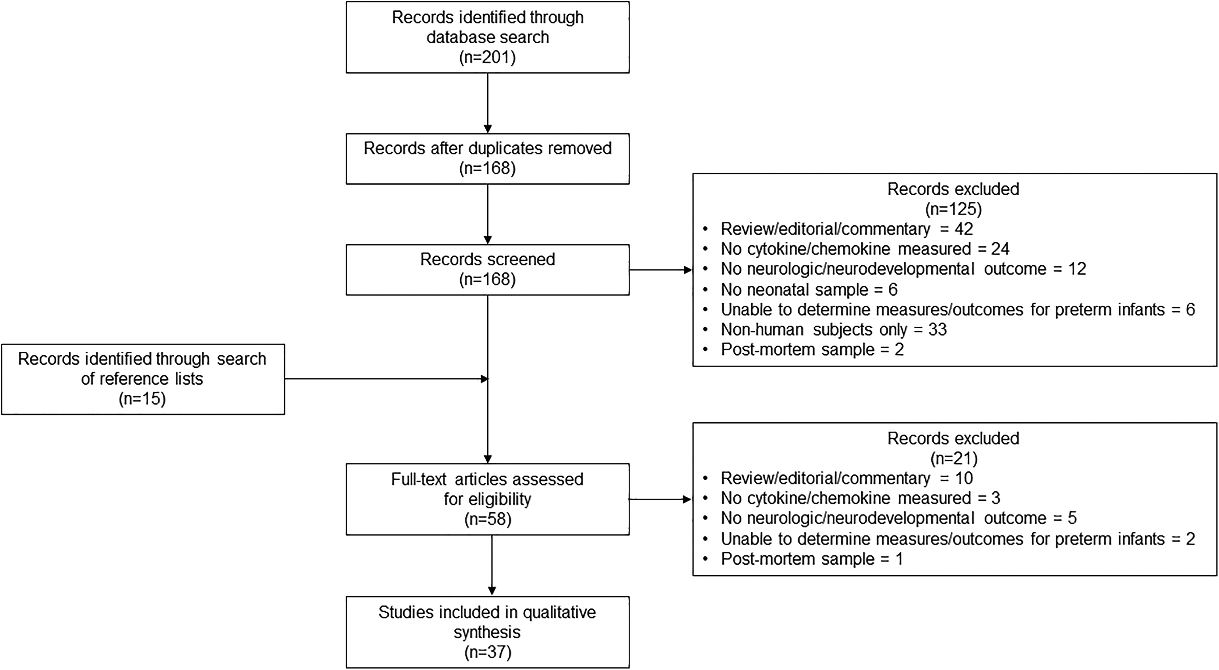
Preferred Reporting Items for Systematic Reviews and Meta-Analyses diagram for the selection of included studies.
Data Set
The 37 publications included in this review represent 20 unique studies, one study with two separate reports (Procianoy & Silveira, 2012; Silveira & Procianoy, 2011), and one study, the Extremely Low Gestational Age Newborn (ELGAN) Study, contributing 15 publications (O’Shea et al., 2009). Quality scores ranged from 7 (low) to 12 (high; median = 9; Table 1). Deductions were most commonly made for failure to provide a power analysis or sample size justification (n = 35), attrition greater than 20% (n = 27), failure to report the participation rate of eligible participants or participation <50% of those eligible (n = 23), and categorization of a continuous predictor (n = 19). Supplemental Tables B and C include all relevant results from the individual studies. In the following text, we summarize findings without attribution to any one study. For cases where there are conflicts or unique findings, we have included the citation.
Researchers of included studies measured 29 different cytokines/chemokines and their soluble receptors (Table 1, Supplemental Table A). The most commonly measured cytokines/chemokines include TNF-α (n = 35), IL-6 (n = 34), IL-8 (n = 31), and IL-1β (n = 31). These cytokines/chemokines remained the most commonly measured when we excluded multiple reports from the same study. Many researchers divided cytokine/chemokine data into empirical quartiles and examined the highest quartile compared to the lower three quartiles (n = 16). Others dichotomized measures into “high” and “low” based on a theoretical cut point (n = 2) or categorized levels based on distance from the mean (n = 1). Some used continuous measures of cytokines/chemokines (n = 18) in analysis of variance, linear regression, or other parametric or nonparametric statistical analyses. Although most researchers examined the effect of individual cytokines/chemokines on neurodevelopmental outcomes, a few researchers used methods to describe the effect of the inflammatory milieu. Bass et al. (2008) found the interactions of TNF-α, IL-6, and IL-1β with their receptors to be more predictive of outcomes than individual levels of cytokines/chemokines, while Cordeiro et al. (2016) used advanced statistical methods to determine the cytokine/chemokine profiles most related to outcomes. Many researchers, including those from the ELGAN Study, investigated the effect of repeated or sustained elevations of cytokines/chemokines on outcomes (Blok et al., 2011; Carlo et al., 2011; O’Shea et al., 2009).
Cytokine Predictors
To address the first aim, we organized findings by outcome, including brain injury, behavioral functioning, cognition, motor functioning, cerebral palsy (CP), composite neurodevelopment (i.e., motor, cognitive, language, social), ophthalmologic, and electroencephalography (Table 2, Supplemental Table B). Brain injury, including intraventricular hemorrhage, periventricular leukomalacia, ventriculomegaly, and microcephaly, was the most commonly measured outcome (n = 20). Elevations in IL-6, IL-1β, IL-8, TNF-α, MCP-1, MCP-4, TNF receptor 1 (R1), TNF-R2, macrophage inflammatory protein (MIP)-1β, and T-cell α chemoattractant (I-TAC) were associated with an increased risk of brain injury. Conversely, elevated levels of IL-6R and regulated on activation, normal T-cell expressed and secreted (RANTES) were inversely associated with brain injury in preterm infants.
Cytokine/Chemokine Predictors of Neurodevelopment by Outcome.
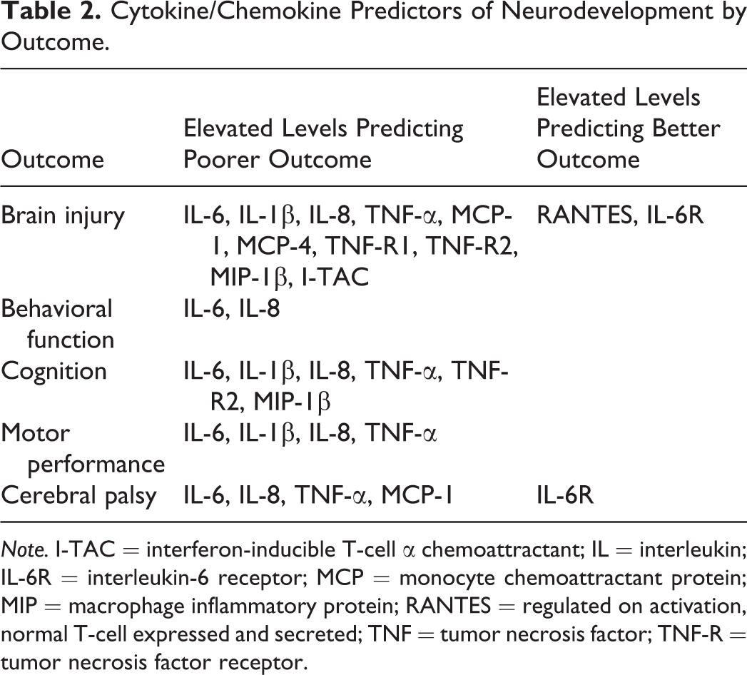
Note. I-TAC = interferon-inducible T-cell α chemoattractant; IL = interleukin; IL-6R = interleukin-6 receptor; MCP = monocyte chemoattractant protein; MIP = macrophage inflammatory protein; RANTES = regulated on activation, normal T-cell expressed and secreted; TNF = tumor necrosis factor; TNF-R = tumor necrosis factor receptor.
Behavioral function outcomes included symptoms of attention deficit hyperactivity disorder and scores on the Language Social subscale of the Kyoto Scale of Psychological Development (KSPD) for Japanese children. Impairments in behavioral functioning were reported following neonatal elevations in IL-6 and IL-8. There were no protective cytokines/chemokines reported.
Cognitive outcomes were assessed in three studies using the Griffith’s Mental Development Scale (Blok et al., 2011), the Cognitive-Adaptive subscale of the KSPD (Kinjo et al., 2011), and a composite cognitive test that measures overall intelligence quotient (Kuban et al., 2017). The remaining 11 publications that measured cognitive outcomes used the Bayley Scales of Infant Development (BSID). Elevations in IL-6, IL-1β, IL-8, TNF-α, TNF-R2, and MIP-1β were predictive of future cognitive impairment. There was one conflicting finding; five publications from the ELGAN Study (Korzeniewski et al., 2015; Kuban et al., 2017; Leviton et al., 2016; O’Shea et al., 2012, 2013) found elevations of TNF-α to be associated with poorer cognitive function, while Carlo et al. (2011) found that elevations in TNF-α predicted better performance on the BSID Mental Development Index (MDI) at 18–22 months’ corrected age. Leviton, Fichorova, et al. (2013) found that TNF-α predicted cognitive impairment but only when infants were also small for gestational age. No cytokines/chemokines were associated with improved cognitive functioning in more than one publication.
Motor performance was most commonly measured with the BSID Psychomotor Development Index, though studies also used the KSPD Postural Motor subscale (Kinjo et al., 2011) and the Test of Infant Motor Performance (TIMP; Magalhaes et al., 2017). Elevations in IL-6, IL-1β, and IL-8 were generally associated with poorer motor performance, except for in Magalhaes et al. (2017), where researchers found elevations in IL-1β and IL-8 during the first days of life to be predictive of normal performance on the TIMP at 34 weeks PMA. Differences in study design could account for this discrepancy: Magalhaes et al. administered the TIMP earlier than researchers in other studies administered the BSID and KSPD (34 weeks PMA for TIMP vs. at least 6 months corrected age for BSID), and Magalhaes et al. measured cytokines/chemokines in urine rather than plasma as in the other studies. There were no cytokines/chemokines that, when elevated, consistently predicted normal neurodevelopment.
A diagnosis of CP at 6–24 months corrected age was associated with elevations in IL-6, IL-8, TNF-α, and MCP-1. Elevations in IL-6R were inversely related to the odds of a CP diagnosis. While single reports showed protective effects against CP of neonatal elevations in RANTES, IL-12, and IL-17 (Carlo et al., 2011), no other studies replicated these results.
There were insufficient data to analyze results for composite neurodevelopmental outcomes, ophthalmologic outcomes, or electroencephalography. No significant cytokine/chemokine predictors of these outcomes were reported in more than one publication.
Timing of Measurements
To address the second aim, we organized findings from included studies into tables by cytokine/chemokine measurement time point (Table 3, Supplemental Table C). Among cytokines/chemokines measured at delivery (i.e., cord blood), IL-6, IL-8, and TNFα were predictive of impaired neurodevelopment. In samples collected within the first week of life, IL-6, IL-1β, IL-8, TNF-α, MCP-1, and MIP-1β predicted neurodevelopmental impairment. However, researchers reported conflicting results for TNF-α, IL-8, and IL-1β: In contrast to four other reports, Carlo et al. (2011) reported higher scores on the BSID MDI for those with higher TNF-α. Magalhaes et al. (2017) reported normal TIMP outcomes for infants with elevations in IL-8 and IL-1β. These conflicting findings could be the result of differences in outcome measure, infant age at the time of outcome assessment, or source of cytokines (i.e., urine). In two publications, authors reported that elevations in RANTES were protective in the first week of life, which conflicted with other findings. As with samples collected at delivery, the results for samples collected in the first week of life were also inconsistent, with multiple studies reporting no effect.
Cytokine/Chemokine Predictors of Neurodevelopment by Timing of Measurement.
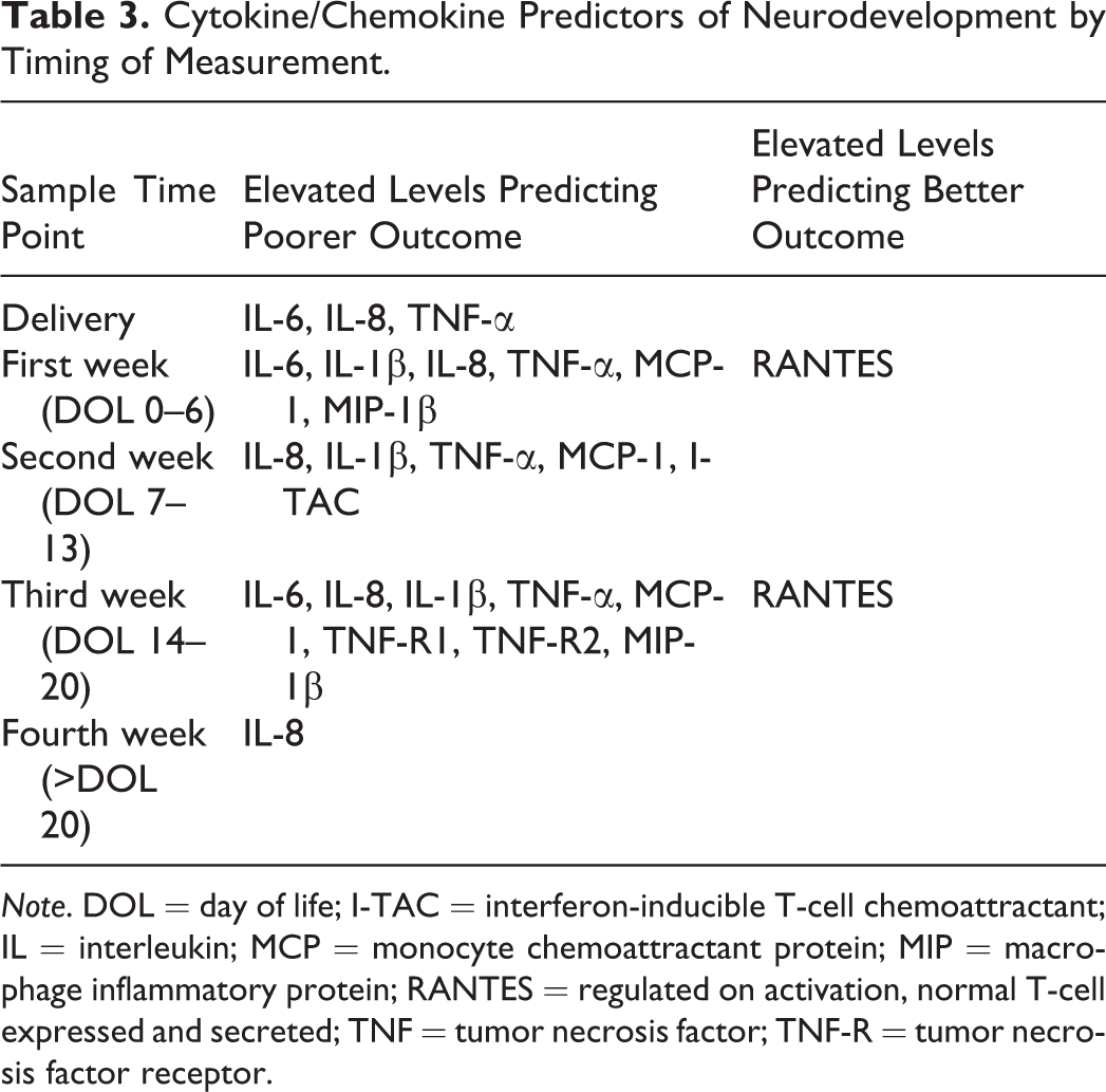
Note. DOL = day of life; I-TAC = interferon-inducible T-cell chemoattractant; IL = interleukin; MCP = monocyte chemoattractant protein; MIP = macrophage inflammatory protein; RANTES = regulated on activation, normal T-cell expressed and secreted; TNF = tumor necrosis factor; TNF-R = tumor necrosis factor receptor.
The majority of reports for samples collected during the second week of life and later came from the ELGAN Study, with IL-1β, IL-8, TNF-α, MCP-1, and I-TAC predicting impaired neurodevelopment. These findings were inconsistent with multiple reports of no effect. In samples collected in the third week of life, elevations in IL-6, IL-1β, IL-8, TNF-α, MCP-1, TNF-R1, TNF-R2, and MIP-1β predicted impaired neurodevelopment, while elevations in RANTES predicted more favorable outcomes. In samples collected during the fourth week of life, only elevations in IL-8 predicted poorer neurodevelopment in more than one study.
Several publications from the ELGAN Study (O’Shea et al., 2009) examined the effect on neurodevelopment of sustained levels of cytokines/chemokines defined as levels in the highest empirical quartile on two or more occasions. In these studies, sustained elevations in IL-6, IL-1β, IL-8, TNF-α, MCP-1, TNF-R1, TNF-R2, and MIP-1β during the first 3 weeks of life were associated with neurodevelopmental impairment. Sustained elevations in RANTES and IL-6R were associated with normal neurodevelopment.
Source of Cytokines
There were insufficient data to draw conclusions regarding the optimal source for cytokine samples. Most studies used cord or peripheral blood to measure plasma cytokine/chemokine levels. Basu, Agarwal, Anupurba, Shukla, and Kumar (2015) quantified IL-1β and TNF-α in the cerebrospinal fluid of infants with early-onset sepsis and compared mean levels between infants with and without brain injuries. Among infants with sepsis, cerebrospinal fluid levels of IL-1β and TNF-α were higher than plasma levels, although no infant was diagnosed with meningitis. Magalhaes et al. (2017) quantified multiple cytokines/chemokines within the first 3 weeks of life in plasma and urine of infants born at 28–32 weeks PMA. In contrast to multiple other studies reporting elevations in IL-1β and IL-8 to be predictive of poorer motor performance, Magalhaes et al. (2017) found that elevations in urinary levels of these cytokines/chemokines predicted normal performance on the TIMP. There were no other reports of alternative sources of cytokines/chemokines.
Discussion
The findings from the present review suggest that elevated levels of IL-6 and IL-8 and, to a lesser extent, TNF-α and IL-1β measured during the first 3 weeks of life most consistently predict poorer performance across neurodevelopmental outcomes. These cytokines/chemokines were also the four most commonly measured inflammatory biomarkers, thus providing the most opportunity for replication across studies. The identification of IL-6, IL-8, TNF-α, and IL-1β as important predictors of neurodevelopment is based on their consistent performance across the domains of neurodevelopment (e.g., motor, cognitive) and time points (e.g., birth, first week of life). While elevations in RANTES and IL-6R appeared to be protective against early brain injury, there are no cytokines/chemokines that consistently predict better outcomes across the various neurodevelopmental domains.
The selection of cytokines/chemokines for the operationalization of inflammation in future studies should be guided by theoretical support and empirical evidence. Based on the results of the present review, we suggest that future studies of inflammation and neurodevelopment include IL-6, IL-8, TNF-α, and IL-1β as measures of inflammation. Although we are unable to suggest the inclusion of other cytokines/chemokines due to inconsistencies in published studies, there may be theoretical reasons for including other cytokines/chemokines. For example, inflammation is reciprocally regulated by pro- and anti-inflammatory factors that establish a relative level of systemic inflammation. Research included in this review reported primarily on pro-inflammatory factors, specifically IL-6, IL-8, IL-1β, and TNF-α. Anti-inflammatory factors, including IL-10, IL-4, and transforming growth factor-β, were less commonly measured and, on average, were not significantly different between infants with impairment and infants without, but there were insufficient data to draw accurate conclusions.
Combinations of cytokines/chemokines are more likely to describe the inflammatory milieu and predict neurodevelopment than individual measures. Researchers should use analytic methods to describe the full inflammatory milieu while accounting for the high level of multicollinearity among immune biomarkers. A few investigators have used multiple cytokines/chemokines in the same statistical model. For example, Hansen-Pupp et al. (2008) found associations between high levels of TNF-α, IL-6, or IL-8 and neurodevelopmental impairment in their univariate analyses; however, with multivariate analysis, many of these associations were no longer statistically significant. Cytokines and chemokines are highly correlated, with empirically determined correlations higher than 0.9 for multiple cytokine pairs (i.e., TNF-α and IL-1β, interferon-γ and IL-1β, IL-8 and IL-6, and IL-4 and interferon-γ; Takahashi et al., 2010). Therefore, a loss of statistical significance when multiple cytokines are modeled together is not surprising. Newer methods of operationalizing inflammation, including descriptions of cytokine “networks” associated with disease states (Bhavnani et al., 2011) and composite scores of inflammation (Beenakker et al., 2013; Heringa et al., 2014; von Scholten et al., 2016), may be useful methods for the study of systemic inflammation in preterm infants.
The results of this review also suggest that between the first and third weeks of life, a number of cytokines/chemokines may be predictive of neurodevelopment; however, only IL-8 remained an important predictor after the 20th day of life. In contrast, RANTES may be an important predictor of better neurodevelopment over time, although this cytokine failed to perform consistently across various neurodevelopmental domains.
An important moderator of the relationship between inflammation and neurodevelopment is likely the duration of exposure of the preterm infant brain to systemic inflammation and not simply the presence of inflammation in the days after birth. The duration of exposure to inflammation may be increased through one of the two mechanisms: (1) infants exposed to significant, prolonged inflammation in utero as a result of maternal conditions such as chorioamnionitis or maternal stress or (2) sustained inflammation due to chronic stress or critical illness during the neonatal period. Association between inflammatory markers at birth and impaired neurodevelopment may reflect chronic inflammation due to maternal infection; many studies did not control for maternal chorioamnionitis. Chorioamnionitis, a common cause of prematurity (Galinsky, Polglase, Hooper, Black, & Moss, 2013), affects as many as 28% of pregnancies that result in preterm birth before 32 weeks’ PMA (Manuck et al., 2016) and is associated with a fetal inflammatory response that includes elevated levels of TNF-α, IL-6, IL-8, and IL-1β and subsequent brain injury in very preterm infants (Galinsky et al., 2013; Lu, Zhang, Wang, & Lu, 2016; Takahashi et al., 2010). The most compelling evidence for an association between neonatal inflammation and impaired neurodevelopment comes from studies of very preterm infants with sustained inflammation through at least 2 weeks of life (O’Shea et al., 2009). Single inflammatory episodes, such as those accompanying neonatal sepsis, rarely represent sustained inflammation in preterm infants, as cytokine levels tend to decrease within 1 week (Segura-Cervantes et al., 2016).
There were insufficient data to draw accurate conclusions regarding the best source of neonatal cytokines for the prediction of outcomes. Only two studies reported measuring cytokines/chemokines in sources other than blood. Future research should examine the correlations between blood levels of cytokines/chemokines and levels in other sources, such as urine, which is noninvasive.
Limitations
There are several limitations to this review. The sampling frame was limited to studies published in English; there may be important findings from studies published in other languages that were not included. Second, although we used multiple methods to find relevant publications, we may have missed publications. Third, of the 37 publications included in our review, 15 grew out of the ELGAN Study (O’Shea et al., 2009), introducing potential bias into our results. During the data analysis, we attempted to control for this possibility by clustering the ELGAN Study publications together. This allowed us to visualize the entire data set and the subset contributed by ELGAN researchers (Supplemental Tables B and C). Finally, there were insufficient data to conduct subgroup analyses based on infant PMA at birth, time between cytokine measurement and neurodevelopmental assessment, and infant sex. In most cases, researchers did not provide these details in their study reports.
Future Research Implications
Despite inconsistencies in findings from different studies on the important cytokine/chemokine predictors of neurodevelopment in preterm infants and the optimal timing of such measures, the ability to operationalize systemic inflammation for its potential effect on the developing brain remains an important research goal. Future research should focus on determining cytokine/chemokine levels that predict outcomes with a high degree of sensitivity and specificity. Some researchers have reported preliminary levels that could be tested in different samples to determine their reproducibility and utility in clinical practice (Procianoy & Silveira, 2012). Investigators using empirical quartiles who have also reported the protein levels used in creating these quartiles have provided useful information for determining thresholds associated with impairment (Leviton et al., 2011).
The results of studies demonstrating a relationship between inflammation and neurodevelopment need to be replicated in high-quality systematic studies that will allow for meta-analysis of the data. Based on the NHLBI quality tool we used, studies included in this review were of moderate to high quality; however, the overall quality could be improved by including more detailed information in the reports that would allow for future subgroup analysis and pooling of the data. Additionally, findings from the ELGAN researchers reveal that sustained inflammation more reliably predicts long-term neurodevelopmental impairment than transient inflammation. Thus, studies of inflammation and neurodevelopment should assess systemic inflammation over time rather than relying solely on single, early time point measures.
Finally, there may be important demographic or clinical variables that interact with cytokine/chemokine levels to predict neurodevelopment. For example, the preterm infant’s developing brain may be more or less vulnerable to inflammatory insults depending on developmental stage (Dammann & O’Shea, 2008). Therefore, future studies should analyze the moderation of cytokine/chemokine effects on neurodevelopment by gestational age. In the majority of studies included in this review, researchers controlled for gestational age in the analysis. However, we were unable to determine the moderation effect of gestational age on the relationship between cytokine/chemokine levels and neurodevelopment based on the available data. Interactions between gestational age and cytokine/chemokine levels should be included in future studies to more fully explain the effect of inflammation on the developing brain.
Conclusion
The ability to operationalize inflammation and determine thresholds of inflammatory biomarkers that predict subsequent neurodevelopmental impairment in preterm infants is an important research goal that has the potential to guide clinical practice. Elevations of IL-6, IL-1β, IL-8, and TNF-α during the first 3 weeks of life may be important, independent predictors of brain injury and neurodevelopmental impairment. Future research on inflammatory processes and long-term outcomes in preterm infants should continue to include these cytokines/chemokines as biomarkers of inflammation.
Supplemental Material
Supplemental Material, Nist_18100101_toSage_SuppTbls - An Integrative Review of Cytokine/Chemokine Predictors of Neurodevelopment in Preterm Infants
Supplemental Material, Nist_18100101_toSage_SuppTbls for An Integrative Review of Cytokine/Chemokine Predictors of Neurodevelopment in Preterm Infants by Marliese Dion Nist and Rita H. Pickler in Biological Research For Nursing
Footnotes
Authors’ Note
The content is solely the responsibility of the authors and does not necessarily represent the official views of the National Institutes of Health.
Author Contributions
M.D. Nist contributed to conception design, acquisition, analysis, and interpretation; drafted the manuscript; gave final approval; and agreed to be accountable for all aspects of work ensuring integrity and accuracy. R. H. Pickler contributed to conception, design, analysis, and interpretation; critically revised the manuscript; gave final approval; and agreed to be accountable for all aspects of work ensuring integrity and accuracy.
Declaration of Conflicting Interests
The author(s) declared no potential conflicts of interest with respect to the research, authorship, and/or publication of this article.
Funding
The author(s) disclosed receipt of the following financial support for the research, authorship, and/or publication of this article: This work was supported by the National Institute of Nursing Research of the National Institutes of Health under award numbers F31NR017321 (Nist, principal investigator), T32NR014225 (Melnyk & Pickler, MPI; Nist, Fellow), and R01NR012307 (Pickler, PI).
Supplemental Material
Supplemental material for this article is available online.
References
Supplementary Material
Please find the following supplemental material available below.
For Open Access articles published under a Creative Commons License, all supplemental material carries the same license as the article it is associated with.
For non-Open Access articles published, all supplemental material carries a non-exclusive license, and permission requests for re-use of supplemental material or any part of supplemental material shall be sent directly to the copyright owner as specified in the copyright notice associated with the article.