
Abstract
Chronic inflammation coupled with cardiovascular disease (CVD) risk factors influences the progression of atherosclerosis in systemic lupus erythematosus (SLE). High-sensitivity C-reactive protein (hs-CRP) and homocysteine (Hcy) are associated with the risk of CVD in the general population, but their associations with CV risk and disease activity in SLE are unclear. In this cross-sectional study (N = 139 SLE patients, mean age = 45.27 ± 13.18 years), we investigated associations between hs-CRP and Hcy levels and disease activity, damage accrual, and CVD risk in SLE. Disease activity and damage accrual were measured with the SLE Activity Index 2000 (SLEDAI-2K), the Systemic Lupus Erythematosus International Collaborating Clinics Group/American College of Rheumatology damage index (SDI), and anti-double-stranded DNA antibodies (anti-dsDNA). CVD risk factors of obesity, diabetes mellitus, hypertension, blood lipids, and ankle–brachial index were collected. Linear regression analysis and one-way analysis of variance were used to analyze relationships of hs-CRP and Hcy with SLE activity, damage accrual, and CVD risk factors.
Results:
hs-CRP correlated significantly with SLEDAI-2K (p = .036), SDI (p = .00), anti-dsDNA titers (p = .034), diabetes (p = .005), and obesity (p = .027). hs-CRP and Hcy correlated with triglyceride (TG) levels (p = .032 and p < .001, respectively), TG/high-density lipoprotein cholesterol index (p = .020 and p = .001, respectively), and atherogenic index of plasma (p = .006 and p = .016, respectively). hs-CRP levels >3 mg/L correlated with SDI score (p = .012) and several CVD risk factors.
Discussion:
Findings suggest SLE patients with elevated hs-CRP and/or Hcy have a higher prevalence of CVD risk factors.
Systemic lupus erythematosus (SLE) is one of the most common autoimmune diseases, yet it remains poorly understood (Selmi et al., 2015). Although several complications arise from the inflammatory process and the impairment of the immune response in SLE, cardiovascular disease (CVD) due to an accelerated atherosclerosis process is the main cause of morbidity and mortality in SLE patients (Abu-Shakra & Novack, 2012; McMahon, Hahn, & Skaggs, 2011). Although the etiology and pathogenesis of accelerated atherosclerosis in SLE have not been fully elucidated, it seems to be the result of traditional and/or classic CVD risk factors (smoking, diabetes mellitus, hypertension, obesity, and dyslipidemia) in combination with specific events in SLE patients such as the chronic inflammatory process, the presence of autoantibodies, and the negative impact of multidrug therapy (Esdaile et al., 2001; Magder & Petri, 2012). The chronic inflammatory process is characterized by the release of multiple inflammatory mediators that create a hostile environment, which promotes atherosclerosis development, endothelium damage, and SLE progression (Bugała et al., 2018).
Levels of C-reactive protein (CRP), one of the primary acute-phase reactants that participate in the systemic response to inflammation, are often elevated in chronic inflammatory conditions including both cardiovascular and autoimmune rheumatic diseases (Thiele et al., 2015). CRP levels appear to correlate with flare-ups and activity (Rhodes, Furnrohr, Vyse, Fürnrohr, & Vyse, 2011). In lupus patients, the role of CRP is yet to be fully clarified. Levels of CRP are significantly higher in lupus patients compared to the general population, but they are often only slightly elevated (Barnes et al., 2005; Rezaieyazdi et al., 2011). Authors have suggested that high levels of CRP in SLE may be more strongly associated with infections or traumas that often occur during SLE, rather than with SLE, itself (Rezaieyazdi et al., 2011). Regarding the role of CRP as a marker of SLE activity and progression, results have been mixed (Barnes et al., 2005; Bertoli, Vilá, Reveille, Alarcón, & Systemic lupus erythaematosus in a multiethnic US cohort (LUMINA) Study Group, 2008; Lee, Singh, Link, & Petri, 2008; Mok, Birmingham, Ho, Hebert, & Rovin, 2013). Although elevated high-sensitivity (hs)-CRP levels are considered an independent risk factor for CVD in the general population across different countries and ethnicities (Fonseca & Izar, 2016; Parrinello et al., 2015), their role as a CVD risk factor in SLE is unclear (Barnes et al., 2005; Lee et al., 2008; Nikpour, Gladman, Ibanez, & Urowitz, 2009).
Elevated levels of homocysteine (Hcy), an amino acid metabolite, are associated with endothelial dysfunction and increased risk of CVD (Schaffer et al., 2014; Wierzbicki, 2007). Hcy levels are often elevated in lupus patients (Bonciani et al., 2016; Lertratanakul et al., 2014; Martínez-Berriotxoa, Ruiz-Irastorza, Egurbide Arberas, Rueda Gutiérrez, & Aguirre Errasti, 2003; McMahon et al., 2014; Sabio et al., 2014). Although Hcy has been associated with the presence and progression of atherosclerosis in SLE (Lertratanakul et al., 2014; McMahon et al., 2014), there is little evidence concerning its correlation with disease progression or CVD risk factors in adults with SLE (Bonciani et al., 2016; Martínez-Berriotxoa et al., 2003; Sabio et al., 2014).
Therefore, considering that CRP and Hcy are known biomarkers associated with the risk of CVD in the general population but their associations with cardiovascular risk and disease activity in SLE patients are unclear, in the present study, we aimed to investigate the possible correlations between hs-CRP and Hcy levels and disease activity, damage accrual, and CVD risk factors in adult patients with SLE.
Method
Study Population
We conducted the present cross-sectional study among 139 patients who had been diagnosed with SLE and were attending the Outpatient Clinic of the Systemic Autoimmune Diseases Unit across three public hospitals in the Andalusian region of Spain from January 2016 through October 2017. SLE patients between the ages of 18 and 80 years who were routinely seen in the Systematic Autoimmune Disease Units were approached regarding study participation and screened for eligibility. After medical consultation and according to the inclusion and exclusion criteria, a total of 139 patients were included in the study. All patients met the revised SLE criteria of Systemic Lupus Erythematosus International Collaborating Clinics Group (SLICC)/American College of Rheumatology (ACR) criteria (Hochberg, 1997; Petri et al., 2012). According to the SLICC criteria, a person is classified as having lupus if he or she has “lupus nephritis” in the presence of anti-double-stranded DNA antibodies (anti-dsDNA, an antinuclear antibody) or meets four criteria (with at least one criterion being clinical and one immunological) from a series of clinical and analytical manifestations characteristic of the disease (Petri et al., 2012). Participants had received a diagnosis of SLE at least 1 year prior to the study and were clinically stable, with no changes in their score on the Systemic Lupus Erythematosus Disease Activity Index 2000 (SLEDAI-2K; Gladman, Ibañez, & Urowitz, 2002) or medical treatment over the 6 months just prior to the study. Patients with serum creatinine ≥1.5 mg/dl, chronic renal failure, cerebrovascular disease, recent infections, major trauma or surgery in the 6 previous months, pregnancy, and/or other chronic and/or autoimmune systemic conditions not related with the main disease were excluded. We obtained written informed consent from each participant, and the local ethics committees approved the study. We conducted the study in accordance with the Declaration of Helsinki.
Each participant’s medical history, including medication use, was obtained during an in-person medical consultation. Following the medical consultation, information regarding smoking status was obtained through an in-person interview. We collected biological measures from the medical record.
Clinical Disease Activity
SLEDAI and SLICC/ACR damage index assessment
We assessed disease activity with the SLEDAI-2K (Gladman et al., 2002). The instrument comprises 24 items, 16 of which are clinical items and 8 of which are laboratory results. Items are scored based on whether these manifestations were present or absent in the previous 10 days. The total score of the SLEDAI-2K is the sum of the 24 descriptor scores and falls between 0 and 105; scores of 6 or more are considered clinically important. The SLEDAI-2K is a revision of the SLEDAI that allows for the documentation of ongoing disease activity for some clinical items such as skin rash, alopecia, mucosal ulcers, and proteinuria as opposed to only new manifestations, as defined in the original SLEDAI (Gladman et al., 2002). Meaningful improvement is best defined as a reduction in the SLEDAI-2K score of 4 or more (Mikdashi & Nived, 2015).
We assessed disease-related organ damage using the SLICC/ACR damage index (SDI; Gladman et al., 1996). This instrument assesses irreversible damage in SLE patients, independent of the cause of the damage (Griffiths, Mosca, & Gordon, 2005). Higher scores indicate more damage, and the maximum possible score is 47. The SDI damage score gradually increases over time in patients with SLE, and those with higher damage scores early in the course of disease have a poorer prognosis and increased mortality (Isenberg & Ramsey-Goldman, 1999).
Assay of anti-dsDNA and complement levels
Anti-dsDNA titers were measured using the commercially available BioPlex 2200 System (Bio-Rad, Hercules, CA), an automated analyzer that detects antibodies to several antigens in one tube. Results are expressed in IU/ml, and the cutoff values established by the manufacturer are 5–9 IU/ml (indeterminate) and ≥10 IU/ml (positive). Human complement components C3 and C4 were determined quantitatively in serum samples by immunoturbidimetric assay (Beckman Coulter AU System CRP Latex reagent) in a Beckman Coulter analyzer (AU5800 Analyzer, Beckman Coulter, CA). Normal ranges are as follows: 90–180 mg/dl for C3 and 10–40 mg/dl for C4.
Cardiovascular Risk Factors
Comorbidities
We collected data on the cardiovascular risk factors of presence of Type 2 diabetes, hypertension, and dyslipidemias. We defined the presence of Type 2 diabetes mellitus by clinician diagnosis based on a fasting blood glucose level of ≥126 mg/dl or use of drug therapy. We regarded patients as having hypertension by previous diagnosis by a clinician based on blood pressure ≥140/90 mmHg or when antihypertensive therapy had been initiated. We defined a patient as having dyslipidemia when blood lipid parameters were out of the suggested range (low-density lipoprotein cholesterol [LDL-C] ≥ 140 mg/dl, high-density lipoprotein cholesterol [HDL-C] < 40 mg/dL, triglycerides [TGs] ≥ 150 mg/dl) for at least two separate measures or if they had been previously diagnosed with dyslipidemia and were being treated with statins (Teramoto et al., 2013).
Blood lipid profile and hs-CRP/Hcy determination
Venous blood samples were collected between 07:30 and 10:00 following an overnight fast and then centrifuged for 15 min to obtain serum. The serum was analyzed immediately by standard laboratory methods to obtain levels of the biochemical markers used as cardiovascular risk factors in the present study: total cholesterol (TC), LDL-C, HDL-C, TGs, hs-CRP, and Hcy.
We calculated the atherogenic index of plasma (AIP), which is a strong marker for predicting the risk of atherosclerosis and coronary heart disease, according to the formula log(TG)/HDL-C (Dobiásová & Frohlich, 2001; Khazaál, 2013). An AIP value <0.11 is associated with a low risk of CVD, a value between 0.11 and 0.21 is associated with a medium risk, and an AIP above 0.21 indicates a high risk of CVD (Dobiásová, 2006; Niroumand et al., 2015).
Immunoturbidimetric assays (Beckman Coulter AU System CRP Latex reagent) were used to determine hs-CRP levels in a Beckman Coulter analyzer (AU5800 Analyzer, Beckman Coulter). The highly sensitive technique was used to measure hs-CRP levels in the range of 0.2–80 mg/L. According to the American Heart Association (AHA) guidelines for assessment of cardiovascular risk, we established two categories for hs-CRP levels: ≤3.0 mg/L for low-to-normal/average risk and >3.0 for a high risk of CVD (Pearson et al., 2003).
Hcy serum levels were measured with an enzymatic colorimetric assay using the Axis-Shield Liquid Stable (LS) 2-Part Homocysteine Reagent (Axis-Shield Diagnostics Ltd., Dundee, UK) in a Beckman Coulter analyzer (AU680, Beckman Coulter). This method measures Hcy levels in the range of 2–44 µmol/L. Normal laboratory reference levels range between 5 and 15 µmol/L; however, recommendations for ideal levels are values of <10 µmol/L in patients with an increased risk of CVD such as those with SLE (Malinow, Bostom, & Krauss, 1999).
Ankle–brachial index (ABI)
We determined the ABI, a validated and effective tool recognized as a prognostic factor for cardiovascular events and all-cause mortality, to assess subclinical atherosclerosis (Aboyans et al., 2012). We performed ABI measurement according to AHA recommendations (Aboyans et al., 2012). One researcher measured systolic blood pressures at the ankle level and in the brachial arteries on both arms and legs using a portable vascular Doppler (Hadeco Minidop ES-100VX. Kawasaki, Japan) and a manual sphygmomanometer (Riester 1312 minimus® II. Jungingen, Germany). We obtained three consecutive measures, waiting for at least 30 s between readings. In accord with AHA criteria, we used the lowest ABI value to classify patients as normal (ABI 1.00–1.40), positive for peripheral artery disease (PAD; ABI ≤ 0.9), borderline for PAD (ABI 0.91–0.99), or positive for calcinosis (noncompressible arteries; ABI > 1.40; Rooke et al., 2011).
Statistical Analysis
We used SPSS® Statistics Version 21.0 (SPSS, Chicago, IL) for all analyses. Continuous variables are presented as mean (M) ± standard deviation (SD) and categorical variables as frequencies and percentages (n [%]). We used the Kolmogorov–Smirnov test to verify data distribution normality before performing association analysis. Due to their skewed distributions, we log transformed the following variables before regression and analysis of variance (ANOVA): SLE duration, SLEDAI-2K and SDI scores, anti-dsDNA titers, complement C3 and C4, corticosteroid dose, hs-CRP, Hcy, and AIP. To aid interpretation, we back-transformed data from the log scale for presentation in the results. We used linear regression analyses to examine the relationships between the different variables (clinical disease activity and cardiovascular risk factors) and hs-CRP and Hcy serum levels. The results are reported as percentage change (β) with 95% confidence intervals (95% CI). To analyze the correlations of cardiovascular risk factors and clinical disease activity with hs-CRP classification ranges (≤3.0 mg/L or >3.0), we used one-way ANOVA for both continuous and categorical variables. We considered age, sex, and medical treatment (prescription for immunosuppressants, antimalarials, corticosteroids, antihypertensives and/or statins) to be confounding factors and adjusted for them in the prior analysis. p Values <.05 were considered statistically significant.
Results
Table 1 lists participant characteristics. Most participants were female (90.6%), and the mean age of the study population was 45.27 (SD = 13.18) years. Note that the mean time since diagnosis with SLE was 9.52 years (range = 1–37) and most of the patients presented low-to-moderate disease activity (SLEDAI-2K, M = 3.37) and a low damage index (SDI, M = 1.19).
Demographic Characteristics, Medication Use, Cardiovascular Risk Factors, and Disease Activity Variables of Participants.
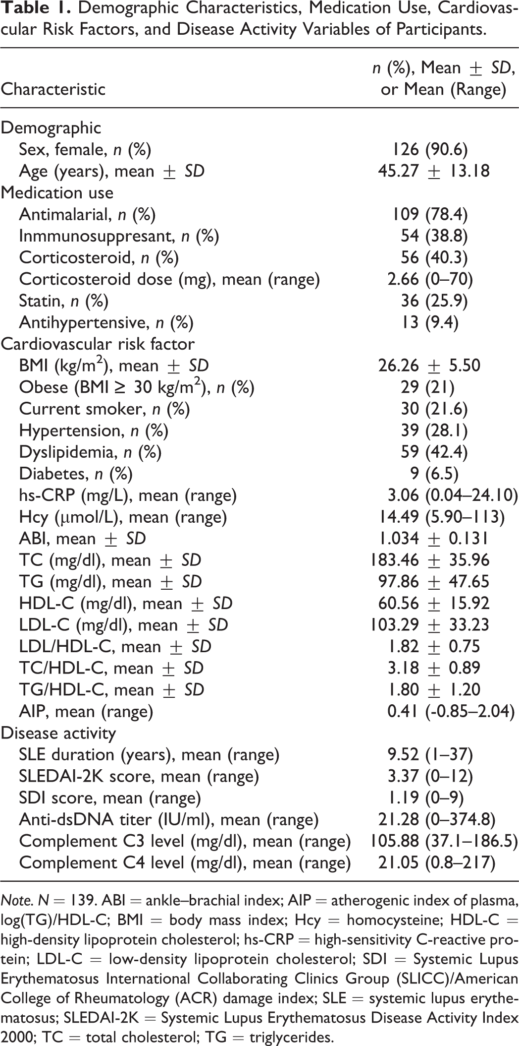
Note. N = 139. ABI = ankle–brachial index; AIP = atherogenic index of plasma, log(TG)/HDL-C; BMI = body mass index; Hcy = homocysteine; HDL-C = high-density lipoprotein cholesterol; hs-CRP = high-sensitivity C-reactive protein; LDL-C = low-density lipoprotein cholesterol; SDI = Systemic Lupus Erythematosus International Collaborating Clinics Group (SLICC)/American College of Rheumatology (ACR) damage index; SLE = systemic lupus erythematosus; SLEDAI-2K = Systemic Lupus Erythematosus Disease Activity Index 2000; TC = total cholesterol; TG = triglycerides.
The mean hs-CRP serum levels (3.06 mg/L) of the study population corresponded to a high risk of CVD according to AHA criteria (Pearson et al., 2003). The mean Hcy serum level was 14.49 µmol/L. Values ≥12 µmol/L are considered predictive of atherosclerosis in SLE (McMahon et al., 2014).
Based on classification by body mass index (BMI), 45.7% of patients were of normal weight, but a slight majority were either overweight (31.9%) or obese (21.0%). The mean BMI (M = 26.26, SD = 5.50 kg/m2) of the study population also exceeded indications for healthy weights.
The relationships of hs-CRP and Hcy with SLE clinical disease activity markers appear in Table 2. The linear regression analysis revealed significant correlations between SLEDAI-2K, SDI, and anti-dsDNA titers with hs-CRP serum levels after adjustment for age, sex, and medical treatment (p = .036, p = .001, and p = .034, respectively). We observed no significant correlations between serum levels of Hcy and any of the clinical disease activity markers analyzed.
Relationships Between Disease Activity and C-Reactive Protein and Homocysteine Levels.
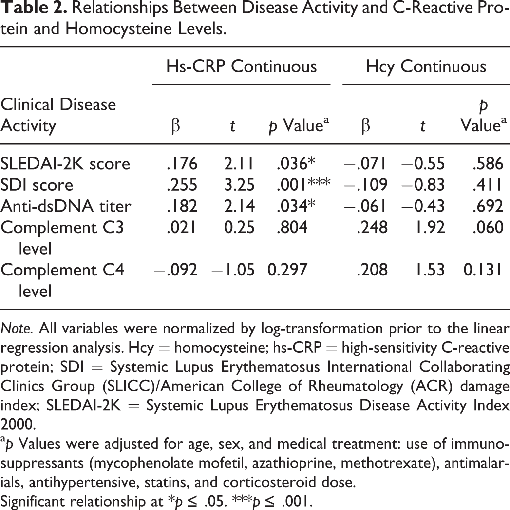
Note. All variables were normalized by log-transformation prior to the linear regression analysis. Hcy = homocysteine; hs-CRP = high-sensitivity C-reactive protein; SDI = Systemic Lupus Erythematosus International Collaborating Clinics Group (SLICC)/American College of Rheumatology (ACR) damage index; SLEDAI-2K = Systemic Lupus Erythematosus Disease Activity Index 2000.
a p Values were adjusted for age, sex, and medical treatment: use of immunosuppressants (mycophenolate mofetil, azathioprine, methotrexate), antimalarials, antihypertensive, statins, and corticosteroid dose.
Significant relationship at *p ≤ .05. ***p ≤ .001.
Table 3 shows the cardiovascular risk markers for the study participants according to hs-CRP and Hcy serum levels. After adjustment for confounding factors, the linear regression analysis revealed significant correlations between both hs-CRP and Hcy levels and serum TG levels (p = .032 and p = .000, respectively), the TG/HDL-C index (p = .020 and p = .001, respectively), and the AIP (p = .006 and p = .016, respectively). In addition, Hcy level was significantly correlated with TC/HDL-C (p = .034), and hs-CRP level was significantly correlated with diabetes, obesity, and higher BMI values (p = .005, p = .027, and p = .009, respectively). We found no significant correlations between either hs-CRP or Hcy level with ABI in any of the analyses.
Relationships Between Cardiovascular Risk Factors and C-Reactive Protein and Homocysteine Levels.
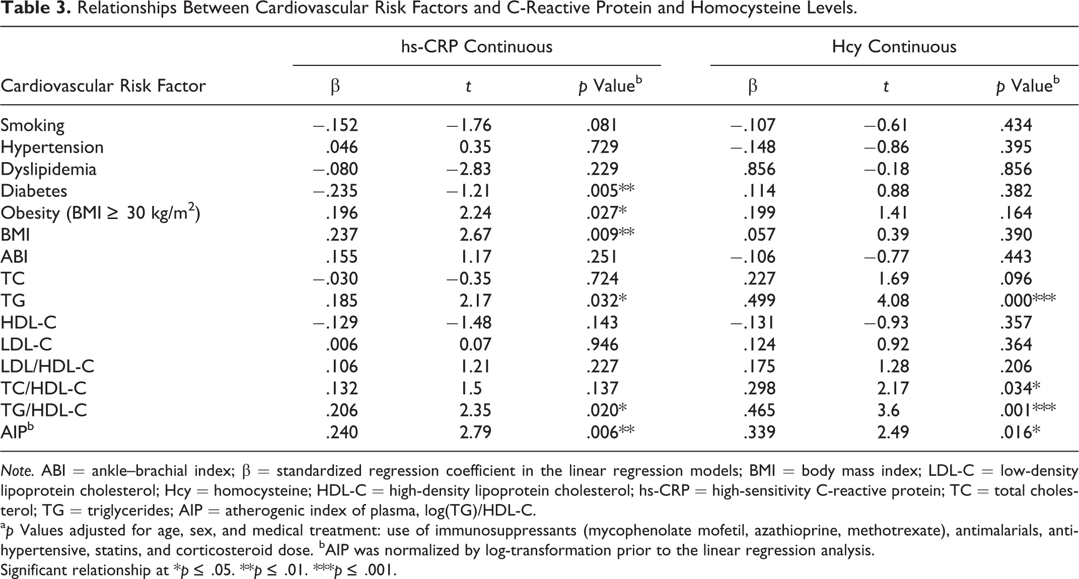
Note. ABI = ankle–brachial index; β = standardized regression coefficient in the linear regression models; BMI = body mass index; LDL-C = low-density lipoprotein cholesterol; Hcy = homocysteine; HDL-C = high-density lipoprotein cholesterol; hs-CRP = high-sensitivity C-reactive protein; TC = total cholesterol; TG = triglycerides; AIP = atherogenic index of plasma, log(TG)/HDL-C.
a p Values adjusted for age, sex, and medical treatment: use of immunosuppressants (mycophenolate mofetil, azathioprine, methotrexate), antimalarials, antihypertensive, statins, and corticosteroid dose. bAIP was normalized by log-transformation prior to the linear regression analysis.
Significant relationship at *p ≤ .05. **p ≤ .01. ***p ≤ .001.
We also performed an analysis of the SLE disease activity and cardiovascular risk factors by hs-CRP serum-level classification (Table 4). We identified a significant difference between hs-CRP categories for SDI score (p = .012), smoking (p = .024), BMI (p = .005), dyslipidemia (p = .027), obesity (p = .029), and several blood lipid parameters including TG (p = .02) and HDL-C (p = .049). TC/HDL-C, TG/HDL-C, and the AIP were significantly higher in patients with hs-CRP > 3.0 mg/L than in patients with hs-CRP ≤ 3.0 mg/L (p = .039, p = .003, and p = .001, respectively). We observed no significant differences for SLE disease activity/damage accrual or cardiovascular risk factors when we analyzed Hcy as a dichotomous variable (<12 or ≥12 μmol/L; data not shown).
Clinical Systemic Lupus Erythematosus (SLE) Disease Activity and Cardiovascular Risk Factors by High-Sensitivity C-Reactive Protein (hs-CRP) Classification Range.
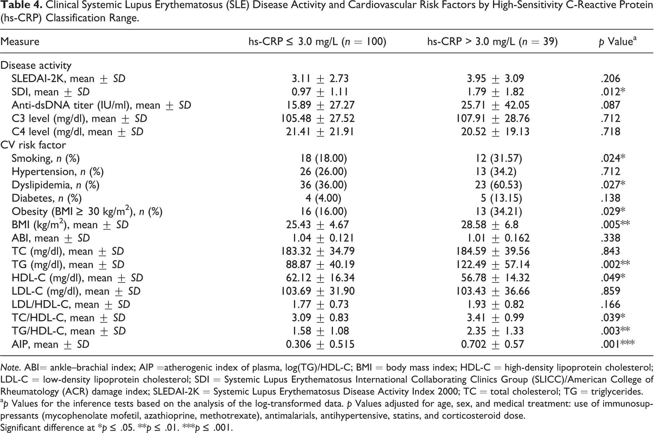
Note. ABI= ankle–brachial index; AIP =atherogenic index of plasma, log(TG)/HDL-C; BMI = body mass index; HDL-C = high-density lipoprotein cholesterol; LDL-C = low-density lipoprotein cholesterol; SDI = Systemic Lupus Erythematosus International Collaborating Clinics Group (SLICC)/American College of Rheumatology (ACR) damage index; SLEDAI-2K = Systemic Lupus Erythematosus Disease Activity Index 2000; TC = total cholesterol; TG = triglycerides.
a p Values for the inference tests based on the analysis of the log-transformed data. p Values adjusted for age, sex, and medical treatment: use of immunosuppressants (mycophenolate mofetil, azathioprine, methotrexate), antimalarials, antihypertensive, statins, and corticosteroid dose.
Significant difference at *p ≤ .05. **p ≤ .01. ***p ≤ .001.
Discussion
In the present study, we assessed the relationships between serum levels of hs-CRP and Hcy, two biomarkers of inflammation, and clinical disease activity, damage accrual, and cardiovascular risk factors in a European population of patients with SLE. Our findings show that hs-CRP levels, but not those of Hcy, correlated significantly with SLE activity, measured with the SLEDAI-2K and anti-dsDNA titers and with damage score, which was estimated using SDI. Furthermore, both hs-CRP and Hcy levels correlated significantly with several cardiovascular risk factors in these patients.
Our results agree with previous studies that revealed a correlation between levels of hs-CRP and SLE activity. A recent study conducted in an Asian population showed that hs-CRP levels correlated with SLEDAI-2K score (Mok et al., 2013). Additionally, Lee, Singh, Link, and Petri (2008) showed that hs-CRP levels correlated significantly with anti-dsDNA and SDI score and that these associations held true regardless of sociodemographic factors, disease activity, and medical treatment. Similarly, a prospective study investigating SLE in a multiethnic U.S. cohort reported that hs-CRP levels correlated with lupus activity and several domains of the damage index (Bertoli et al., 2008). A longitudinal study revealed the significant impact of total disease activity on the onset of damage in SLE patients measured over a 5-year period (Stoll, Sutcliffe, Mach, Klaghofer, & Isenberg, 2004). As authors have suggested that damage accrual in SLE is influenced and predicted by disease activity over time (Lee et al., 2008; Stoll et al., 2004), we might hypothesize that the association we found between hs-CRP levels and damage in patients in the present study was primarily due to the fact that hs-CRP levels reflect disease activity.
Regarding the relationship between hs-CRP and cardiovascular risk factors, our data revealed a correlation between this biomarker and lipid profile alterations, diabetes, obesity, and BMI. These results concur with previous studies (Barnes et al., 2005; Mok et al., 2013; Stoll et al., 2004). Another interesting finding is that serum levels of TG, the TG/HDL-C ratio, and AIP were significantly correlated with hs-CRP levels. Likewise, hs-CRP levels >3.0 mg/L correlated with higher levels of TG, lower levels of HDL-C, and worse TC/HDL-C, TG/HDL-C, and AIP ratios. Similarly, Mok et al. (2013) found that AIP and both TC/HDL-C and TG/HDL-C ratios were significantly higher in patients with hs-CRP levels >3 mg/L than in those with levels ≤3 mg/L and that hs-CRP levels >3.0 mg/L correlated with chronic smoking and diabetes mellitus (Mok et al., 2013). Additionally, prior research has found an independent correlation between obesity and markers of inflammation, including hs-CRP, in patients with SLE (Oeser, Chung, Asanuma, Avalos, & Stein, 2005). Thus, maintaining a healthy weight may be a strategy to reduce inflammation and CVD risk among SLE patients.
On the other hand, the mean Hcy level in our study (14.1 μmol/L) was at the upper limit of the normal range according to the AHA advisory statement (Malinow et al., 1999). This finding is in line with previous results in which SLE patients had high Hcy levels (Bonciani et al., 2016; Lertratanakul et al., 2014; Martínez-Berriotxoa et al., 2003; McMahon et al., 2014; Sabio et al., 2014). Interestingly, we also found that Hcy levels correlated significantly with total serum TG levels, both TC/HDL-C and TG/HDL-C ratios, and the AIP. Previous studies have reported similar findings in adult populations where there was a positive relationship between serum Hcy levels and blood lipid alterations in patients with SLE (Martínez-Berriotxoa et al., 2003; McMahon et al., 2014; Sabio et al., 2014). Similarly, higher Hcy levels have been independently linked to higher TG levels (Ardoin et al., 2010) and dyslipidemia in pediatric and juvenile patients with SLE (Ortiz et al., 2013). Investigators have reported that Hcy plays an important role in hepatic oxidative stress and the production of thiolactone, which may influence lipoprotein levels (Jakubowski & Głowacki, 2011; Lentz, 2005) by stimulating cholesterol production (Lynn et al., 1998). This mechanism could explain the relationship we observed between Hcy levels and a worse lipid profile.
Our findings, along with those of the abovementioned studies, suggest that patients with SLE who have elevated hs-CRP and/or Hcy levels also have a higher prevalence/presence of CVD risk factors. Therefore, promotion of strategies for controlling modifiable CVD risk factors, including weight control and the adoption of healthy lifestyle habits to improve blood lipid profile and reduce the inflammatory environment in patients with SLE, would likely be beneficial.
We did not find any correlation between Hcy levels and scores on either the SLEDAI-2K or the SDI, which suggests that Hcy level might not be a predictor of disease activity or damage accrual in patients with SLE. To date, only one similar study has found a significant correlation between Hcy levels and disease activity in a population of just 16 patients with SLE (Bonciani et al., 2016). Besides the study’s small sample size, the difference in SLEDAI-2K scores between the patients with SLE in the present study (SLEDAI-2K, M = 3.37) and Bonciani et al.’s study (SLEDAI-2K, M = 10.5) may explain the contrasting results. Another study performed in pediatric patients with SLE (mean age = 15.7 years) found that Hcy levels correlated with SLEDAI-2K score but also with age (Oeser et al., 2005), which could explain the difference with our results, since our population featured adult patients. Considering the contrasting findings and limited evidence regarding potential associations between Hcy levels and scores on the SLEDAI-2K and SDI, further studies are required to investigate the role of Hcy in SLE disease activity and damage accrual.
The present study has some limitations. First, due to its cross-sectional design, we can draw no conclusions about causality. Longitudinal studies that gather data across a prolonged period of time are therefore needed to confirm the relationships between serum hs-CRP and Hcy levels and SLE activity, cardiovascular risk factors, and especially damage accrual, which is better interpreted across time. In addition, authors have reported that anti-dsDNA is not the most important autoantibody in SLE because it may have limited value in clinical correlation with the disease and, therefore, should be considered just one among many antibodies in SLE (Fu, Dai, Zhao, & Gaskin, 2015). Nevertheless, we found a correlation between levels of anti-dsDNA and hs-CRP but not with Hcy. Further studies may be necessary to confirm our results.
The main strength of the present study is that it is the first to assess the relationships between serum levels of both hs-CRP and Hcy and disease activity, damage, and cardiovascular risk factors in European patients with SLE. In addition, our study sample comprised a well-characterized cohort of patients with lupus, as we chose patients with early-stage SLE and excluded those with lupus flare-ups or other associated autoimmune conditions. We also used a high-sensitivity assay to assess CRP values. Finally, we considered several medical treatments (statins, immunosuppressants, corticosteroids, and antihypertensives) as confounding factors in the statistical models. The present study thus provides preliminary data which could be strengthened by future research to develop new health care strategies for reducing the risk of CVD and other complications in patients with SLE. Nurses are involved in the implementation of preventive programs and could help improve patients’ prognosis and reduce their cardiovascular risk.
In summary, we found in the present study that serum levels of hs-CRP, but not Hcy, correlated significantly with scores on the SDI and SLEDAI-2K and levels of anti-dsDNA antibodies. Furthermore, serum levels of both hs-CRP and Hcy correlated with a worse lipid profile, but only those of hs-CRP correlated with smoking, diabetes mellitus, dyslipidemia, and obesity. Further studies are necessary to investigate the potentials of hs-CRP and Hcy as predictors of SLE prognosis and cardiovascular risk and develop early preventive strategies that reduce complications in SLE patients.
Footnotes
Author Contribution
G. Pocovi-Gerardino contributed to conception and design, acquisition, analysis, and interpretation; drafted the manuscript; gave final approval; and agrees to be accountable for all aspects of work ensuring integrity and accuracy. M. Correa-Rodríguez contributed to analysis and interpretation, drafted the manuscript, critically revised the manuscript, gave final approval, and agrees to be accountable for all aspects of work ensuring integrity and accuracy. J.-L. Callejas Rubio contributed to acquisition and interpretation, critically revised the manuscript, gave final approval, and agrees to be accountable for all aspects of work ensuring integrity and accuracy. R. Ríos Fernández contributed to acquisition and interpretation, critically revised the manuscript, gave final approval, and agrees to be accountable for all aspects of work ensuring integrity and accuracy. M. Martín Amada contributed to acquisition and interpretation, critically revised the manuscript, gave final approval, and agrees to be accountable for all aspects of work ensuring integrity and accuracy. M.-G. Cruz Caparros contributed to acquisition and interpretation, critically revised the manuscript, gave final approval, and agrees to be accountable for all aspects of work ensuring integrity and accuracy. B. Rueda-Medina contributed to conception and design and interpretation, critically revised the manuscript, gave final approval, and agrees to be accountable for all aspects of work ensuring integrity and accuracy. N. Ortego-Centeno contributed to conception and design and interpretation, critically revised the manuscript, gave final approval, and agrees to be accountable for all aspects of work ensuring integrity and accuracy. B. Rueda-Medina and N. Ortego-Centeno have contributed equally to this work.
Transparency Declaration
The corresponding author, on behalf of the other authors, guarantees the accuracy, transparency, and honesty of the data and information contained in the study, that no relevant information has been omitted and that all discrepancies between authors have been adequately resolved and described.
Declaration of Conflicting Interests
The author(s) declared no potential conflicts of interest with respect to the research, authorship, and/or publication of this article.
Funding
The author(s) disclosed receipt of the following financial support for the research, authorship, and/or publication of this article: This study was supported by grant PI0523-2016 from Consejería de igualdad, salud y políticas sociales (Junta de Andalucía).