






Abstract
Introduction
The population of the world is getting more aged, and it is expected that people over 60 years will more than double from 841 million in 2012 to more than 2 billion in 2050 (Nations, 2015). Loss of muscle mass and strength are signs of aging (Koopman & van Loon, 2009). Various strategies can be used to reduce disabilities in older age. Resistance training (RT) is one of the most recommended potent strategies to maintain lean tissue mass and physical function in the elderly (Rennie et al., 2004). Pieces of evidence indicates that regular RT in older adults induced improvements in muscle mass and strength, as well as the quality of life (Clark et al., 2016). The role of the transient increase in hormones and other signaling molecules in the adaptive response to exercise training is not well understood (Bagheri et al., 2020). It has been suggested that elevations in circulating concentrations of cell-signaling molecules may increase the likelihood of receptor interaction and thus enhance the probability of a physiological effect within skeletal muscle (Patel & Demontis, 2014). Among these cell-signaling molecules, there are several myokines involved in the inhibition of the muscular hypertrophic response (Pedersen et al., 2007). Muscle protein balance can be influenced by various hormones and myokines (Koopman & van Loon, 2009). Indeed, the assessment of different anabolic and catabolic muscular regulatory markers might help in the identification of the mechanisms for potential favorable changes in body composition and muscular performance (Bagheri et al., 2020).
Myostatin (GDF8) is an antianabolic and pro-catabolic peptide hormone that has a negative effect on skeletal muscle size and development secreted from skeletal muscle (Elliott et al., 2012; Hittel et al., 2010; McPherron et al., 1997). Myostatin is a member of the transforming growth factor-beta (TGF-β) family that acts by the activin type II A and B receptors; its inhibitory effect on the mechanistic target of rapamycin (mTOR) pathway and myogenesis suppresses protein synthesis in skeletal muscles (McCroskery et al., 2003; Winbanks et al., 2012). Follistatin, another member of the TGF-β family, is a glycosylated plasma protein that is abundant in skeletal muscles (Hansen et al., 2011, 2013) and prevents the binding of myostatin to the activin type II B (Hansen et al., 2011). The elimination of the follistatin and myostatin gene leads to the loss of skeletal muscle mass and excessive muscle growth, respectively (Lee, 2007). Another member of the TGF-β family is growth and differentiation factor 11 (GDF11), which is the nearest neighbor to myostatin, and has an important role in the aging process (Egerman et al., 2015). Higher circulation of GDF11 is a negative regulator of skeletal muscle (Egerman & Glass, 2019).
The mechanical tension and metabolic stress imposed on skeletal muscle via RT modalities, when combined with proper nutritional factors, can result in a net anabolic response in myofibrillar protein metabolism leading to myofiber hypertrophy and muscular growth over time (Bagheri et al., 2020; Damas et al., 2018). It is suggested that post-exercise protein ingestion leads to increased muscle protein synthesis (MPS) through the provision of sufficient amino acids, particularly leucine, thus activating the key anabolic signaling mechanism of the mechanistic target of rapamycin (mTOR) (Macnaughton et al., 2016; Moro et al., 2019). Consequently, dietary protein intake is considered an essential component in the optimization of skeletal muscle adaptations to RT (Bagheri et al., 2021). With repeated training sessions, this could result in muscle hypertrophy and improvements in physical performance (i.e., strength and power; Pourabbas et al., 2021). Recent evidence suggests that soy milk (a plant-based non-dairy beverage) can be considered to be a nutritionally adequate and complete protein because it is rich in isoflavones, particularly genistein (Eslami et al., 2019; Gardner et al., 2007; Phillips et al., 2009). Soy milk comes from plants and also is naturally free of cholesterol, low in saturated fat, and contains no lactose (Mohammad-Shahi, Mowla, Haidari, Zarei, & Choghakhori, 2016). Soy milk is a good source of protein, calcium, and potassium. Also, soy milk contains essential amino acids such as leucine, lysine, phenylalanine, and isoleucine (Keshavarz et al., 2012; Liao et al., 2019; Phillips et al., 2009).
To the best of the author’s knowledge, the effect of soy milk supplementation and RT on the biomarkers of follistatin, myostatin, and GDF11 has not yet been examined; however, limited and contradictory studies have been done on body composition (Liao et al., 2019). While the definitive occurrence of molecular interference based on soy milk combined with RT is not established yet, and the translation between acute findings and long-term adaptations is uncertain, the paucity of data indicates that additional investigation is needed. Therefore, we conducted the present study to evaluate the effects of 12 weeks of soy milk consumption combined with RT on body composition, physical performance, and skeletal muscle regulatory markers (Myostatin, follistatin, and GDF11) in older men. It was hypothesized that a 12-week regimen of soy milk consumption combined with RT will improve body composition and physical performance by increasing muscle-related anabolic markers and reducing muscle-related catabolic markers in aging males.
Method
Participants
Sixty healthy older men (age = 65.63 ± 3.16 years; body mass = 62.63 ± 3.86 kg) volunteered to take part in this study (Figure 1). The inclusion criteria were age 60–80 years and physically independent. Exclusion criteria were as follows: had current or previous history of significant cardiovascular, neurological, respiratory, muscular, metabolic, inflammatory, bone problems, joints, and movement disorders; were consuming nutritional supplements; were consuming drugs affecting muscle metabolism; were consuming alcohol or smoking for at least 1 year before enrolling in the study; having soy milk allergy/sensitivity, and history of regular physical activity at least in the past year. All these criteria were evaluated by a physician using the Physical Activity Readiness-Questionnaire (PAR-Q) and medical health/history questionnaire. Written informed consent was obtained from all participants. All experimentation was carried out following the Declaration of Helsinki. The present study was approved by the Sport Sciences Research Institute and registered at the Iranian Registry of Clinical Trials (IRCT20190731044398N3). Participant assignment and allocation. RT; Resistance Training, SMC; Soy Milk Consumption, RSM; Resistance Training + Soy Milk Consumption, CON; Control.
Study design
This study was a randomized, double-blind (for soy milk), and parallel prospective clinical trial. Before baseline measurements, all participants were familiarized with all testing and procedures. Subsequently, participants were randomly divided into 4 groups: RT + placebo (RT; n = 15), soy milk consumption (SMC; n = 15), RT + soy milk consumption (RSM; n = 15), or control (CON; n = 15). The allocation was performed by using a digital tool available at www.randomizer.org. Participants in the RT group performed an exercise program 3 times per week for 12 weeks and received a placebo; participants in the SMC group received 240 mL of soy milk daily, and the participants in the RSM group performed RT and consumed soy milk. Participants in the CON group were asked to maintain a normal daily life pattern for the duration of the study. Measurements were performed at baseline and the end of 12 weeks of interventions (approximately 48 h after the last training session). All measurements were recorded at the same time of day (within ∼1 hour) and under the same environmental conditions (∼20°C and ∼55% humidity). The participants were asked not to change their regular lifestyle and habitual dietary intake during the study period.
Measurements
Body Composition Assessments
Upon arriving at the laboratory, participants were asked to void completely within 30 min of the test. Body mass (BM) was measured with a digital scale (SECA, Germany) to the nearest 0.1 kg. The participant’s height was measured with a stadiometer (SECA, Germany) to the nearest 0.1 cm. Waist-hip ratio (WHR: to the nearest 0.01 m) was measured using tape. Body fat percent (BFP), body mass index (BMI), fat mass (FM), and muscle mass were evaluated by a multi-frequency bioelectrical impedance device (BIA; Jawon Medical X Contact-356, South Korea) as previously described (Moghadam et al., 2020). The test–retest reliability of the bioelectrical impedance method is high (R = 0.95 to 0.99; Ling et al., 2011).
Physical Performance Tests
By using a hydraulic hand dynamometer (YAGAMI, Japan), handgrip strength of the dominant hand was assessed. Upper and lower limbs anaerobic power was evaluated by the Wingate anaerobic cycle (Monarch 831E and 894 Ea, Varberg, Sweden) test as previously described (Beam & Adams, 2013). A doctor supervised the Wingate testing to monitor signs of cardiovascular discomfort. Maximal oxygen consumption (VO2max) of each participant was measured by modified Bruce protocol as previously described to determine fitness level by following equation (Bullock et al., 2018). Upper body strength (UBS) and lower body strength (LBS) were evaluated by one repetition maximum (1RM) on the chest press and leg press machines, respectively, as previously described (Timmons et al., 2018).
Blood Biochemistry
Fasting blood samples (∼8 mL) were collected from the antecubital vein using standard procedures ∼48 h before and after the last training session. Following the completion of blood sampling, the samples were centrifuged at 3000 rpm for 10 min, and serum was stored at −80°C until further analysis. Serum myostatin (kit: R & D, USA, sensitivity: 2.25 pg/ml), follistatin (kit: R & D, USA, sensitivity: 29 pg/ml), GDF11 (kit: Cusabio Co, sensitivity: less than 7 pg/ml) concentrations were measured using commercial human ELISA kits. The intra- and inter-assay coefficients for all factors were <8% and <10%, respectively.
Interventions
Exercise Training Program
Overview of the Training Intervention for the Different Groups.
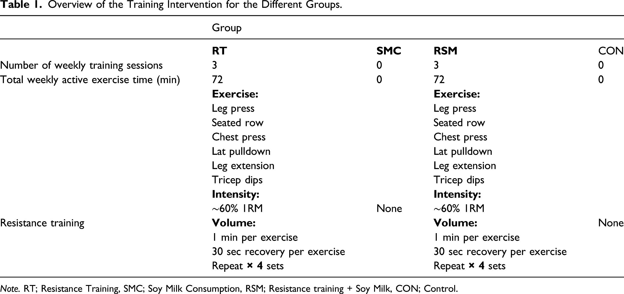
Note. RT; Resistance Training, SMC; Soy Milk Consumption, RSM; Resistance training + Soy Milk, CON; Control.
Supplementation Protocol
Participants in the RSM group received 240 ml of vanilla flavored non-dairy soy milk immediately after every training session and at the same time on non-training days for 12 weeks. Participants in the SMC group received 240 ml of vanilla flavored non-dairy soy milk daily at the same time as the RSM group. Participants in the RT group received daily the same amount of placebo (an artificially sweetened-water placebo); participants in the CON group did not either receive the soy milk or placebo (Liao et al., 2019). Soy milk and placebo were in opaque (masked) beverage containers and were similar in taste, smell, texture, and appearance (Pourabbas et al., 2021). The timing and dose of soy milk were chosen according to previous investigations (Eslami et al., 2019; Liao et al., 2019). Soy milk (dairy-, lactose-, and casein-free; absolutely no carrageenan, gluten, egg, or peanut) was supplied from Saina Ghaza Part Company, Iran and registered with the health ministry (No:56/16,554). The energy and macronutrient composition of this milk per 240 mL are as follows: energy, 99.6 kcal; protein, 6.75 g; carbohydrate, 9.15 g; fat, 4 g; calcium, 100 mg; sodium, 98 mg; cholesterol, 0 mg; iron, 1.1 mg; vitamin D 2.5 mcg; vitamin A, 150 mcg; vitamin B12, 1.5 mcg; potassium, 300 mg; and riboflavin, 0.4 mg. Researchers monitored the soy milk supplementation. Following the training sessions, the soy milk was given to the participants by a researcher who witnessed its consumption. On non-training days, the supplementation was verified by a phone call or text message. To evaluate compliance with supplementation on non-training days, participants delivered the empty boxes of the supplement to the research staff.
Nutrient Intake and Dietary Analysis
Participants were asked to maintain their normal dietary habits during the study. To minimize dietary variability, participants submitted 3-day (2 weekdays and 1 weekend day) food records at three times (before and at the end of week 6 and week 12). Each item of food was individually entered into Diet Analysis Plus version 10 (Cengage, Boston, MA, USA), and total energy consumption and the amount of energy derived from proteins, fats, and carbohydrates were evaluated (Eskandari et al., 2020).
Statistical Analysis
Estimation of an appropriate sample size was conducted using the G*Power analysis software. Our rationale for sample size was based on previous studies and the primary outcome variables (follistatin, myostatin, and GDF11; Bagheri et al., 2020; Liao et al., 2019). The analysis revealed a sample size of at least 52 participants (n = 13 per group) were needed to provide power (1- β) of 0.80 (α = 0.05). This number was increased to 15 participants per group to cover anticipated dropout. The normality of data was confirmed using the Shapiro–Wilk test. One-way analysis of variance (ANOVA) was used to examine possible group differences at baseline. The effects of the interventions on all variables were evaluated by a 2 x 4 repeated-measures ANOVA [time (baseline vs. 12 weeks) × group (RT vs. SMC versus RSM vs. CON)]. When a significant main effect was identified, paired t-tests were used to determine within-group differences from baseline. When a significant group-by-time interaction was found, we performed the Bonferroni post-hoc test to determine differences between groups. In addition, one-way ANOVA was used for “change in” (Δ) scores between times (pre, post) where appropriate. Statistical significance was set at p < .05. Effect size (Cohen’s d) was calculated as post-training mean minus pre-training mean/pooled pre-training standard deviation means. All analyses were performed with SPSS (version 24.0, IBM; Chicago, IL). In addition, figures were prepared in Graphpad Prism software (Version 8.4.3, Graphpad Software).
Results
Dietary Intake Monitoring and Compliance with Exercise Training and Supplementation Interventions
Energy and Macronutrients at Baseline and at the End of Week 6 and Week 12.
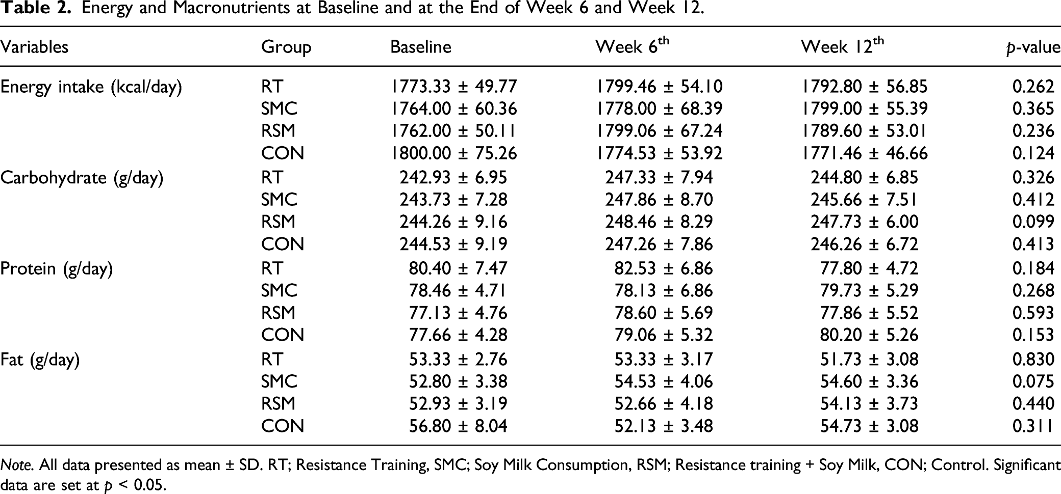
Note. All data presented as mean ± SD. RT; Resistance Training, SMC; Soy Milk Consumption, RSM; Resistance training + Soy Milk, CON; Control. Significant data are set at p < 0.05.
Blood Markers
Skeletal muscle regulatory markers are presented in Figure 2. A significant time x group interaction was observed for serum follistatin, myostatin, and GDF11 concentrations, and MFR (p < 0.001). All 3 interventions groups significantly increased serum concentrations of follistatin [RT = 25.18 pg/ml, (95% CI, 15.67 to 34.70; p < 0.001); SMC= 42.68 pg/ml, (95% CI, 15.01 to 70.34; p = 0.005); RSM = 118.31 pg/ml, (95% CI, 89.82 to 146.8; p < 0.001); Figure 2(A)], and significantly decreased serum concentrations of myostatin [RT = −30.88 pg/ml (95% CI, −49.85 to −11.92; p = 0.004);SMC -12.00 pg/ml (95% CI, −22.00 to −2.00; p = 0.017); RSM = −124.08 pg/ml (95% CI, −144.80 to −103.35; p < 0.001); Figure 2 B] over the time. The 3 interventions also decreased GDF11 [RT= −1.49 pg/ml, (95% CI, −1.93 to −1.04; p < 0.001); SMC = −0.60 pg/ml, (95% CI, −0.97 to −0.22; p = 0.004); RSM= −5.19 pg/ml, (95% CI, −6.12 to −4.26) (p < 0.001); Figure 2 D] over the time. The intervention groups also significantly decreased MFR [RT= −0.04, (95% CI, −0.06 to −0.02; p < 0.001); SMC= −0.05, (95% CI, −0.07 to −0.02; p = 0.001); RSM= −0.18, (95% CI, −0.21 to −0.15; p < 0.001); Figure 2 C] over the time. There were no alterations in these variables in the CON group (p > 0.05). Changes of these variables in the RSM group were significantly (p < 0.05) greater than those in the RT, SMC, and CON groups. Furthermore, there were significant differences between the changes in the RSM and CON group [follistatin (p = 0.001), myostatin (p < 0.001), MFR (p < 0.001), and GDF11 (p < 0.001)], RSM and RT group [follistatin (p = 0.002), myostatin (p < .001), MFR (p < 0.001), and GDF11 (p < 0.001)], and RSM and SMC group [myostatin (p < 0.001), MFR (p < 0.001), and GDF11 (p < 0.001)]. Changes in blood markers in response to 12 weeks of exercise training and soy milk consumption. RT; Resistance Training, SMC; Soy Milk Consumption, RSM; Resistance Training + Soy Milk Consumption, CON; Control group, MFR; myostatin-follistatin ratio, A; follistatin, B; myostatin, C; MFR, D; GDF11.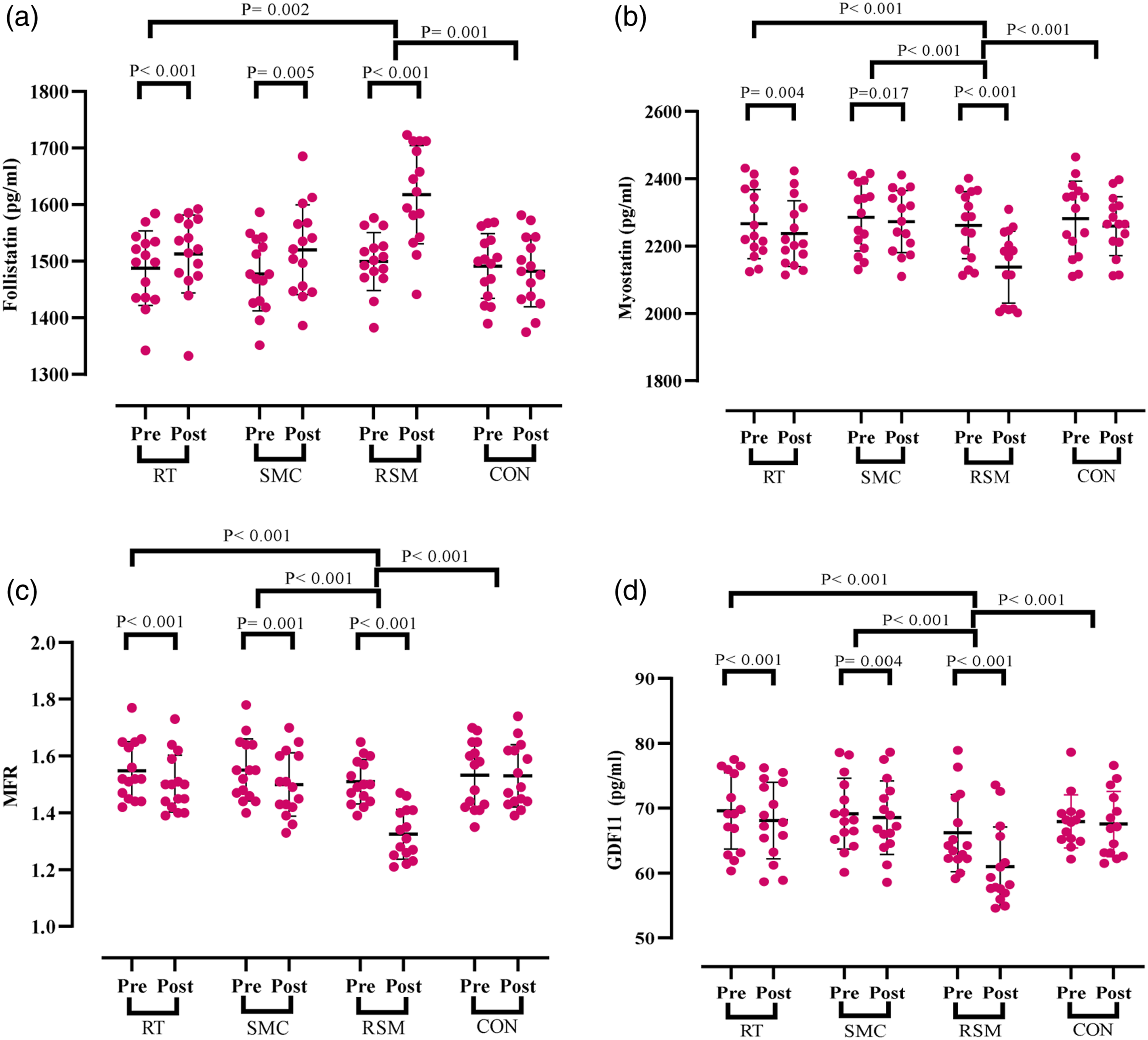
Body Composition
Anthropometric Characteristics of Participants at Baseline and Following each Intervention.
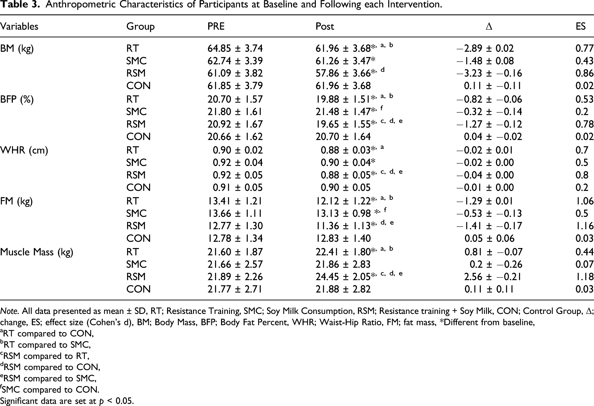
Note. All data presented as mean ± SD, RT; Resistance Training, SMC; Soy Milk Consumption, RSM; Resistance training + Soy Milk, CON; Control Group, Δ; change, ES; effect size (Cohen’s d), BM; Body Mass, BFP; Body Fat Percent, WHR; Waist-Hip Ratio, FM; fat mass, *Different from baseline,
aRT compared to CON,
bRT compared to SMC,
cRSM compared to RT,
dRSM compared to CON,
eRSM compared to SMC,
fSMC compared to CON.
Significant data are set at p < 0.05.
Physical Fitness Assessments
Physical Fitness Characteristics of Participants at Baseline and Following each Intervention.
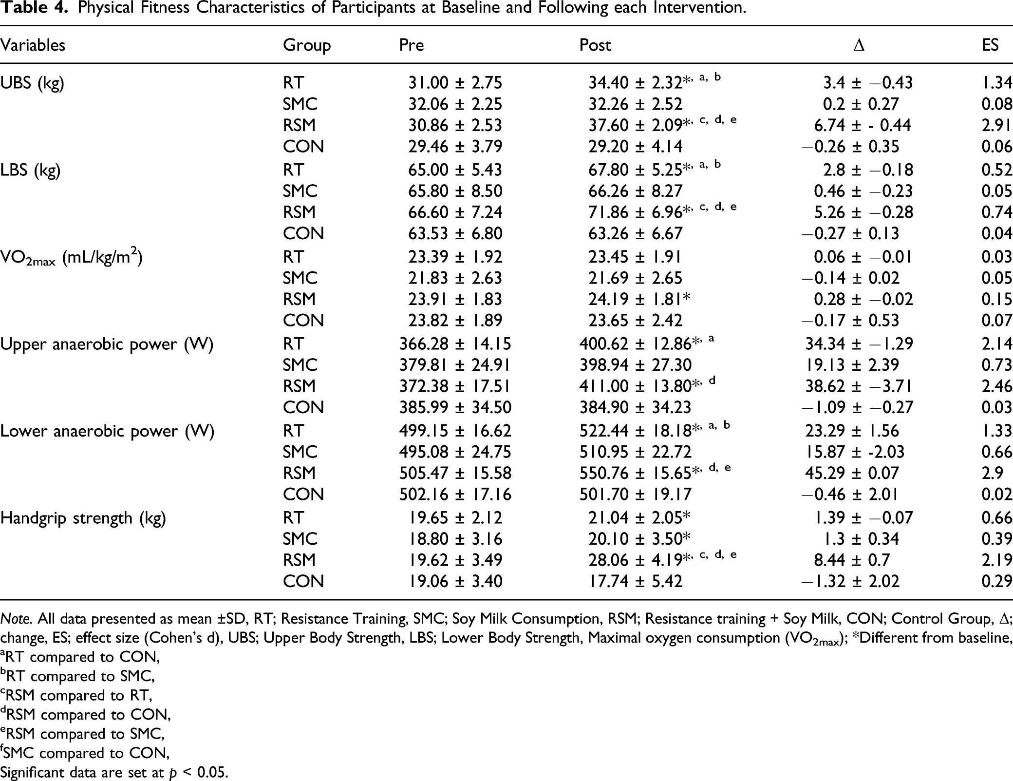
Note. All data presented as mean ±SD, RT; Resistance Training, SMC; Soy Milk Consumption, RSM; Resistance training + Soy Milk, CON; Control Group, Δ; change, ES; effect size (Cohen’s d), UBS; Upper Body Strength, LBS; Lower Body Strength, Maximal oxygen consumption (VO2max); *Different from baseline,
aRT compared to CON,
bRT compared to SMC,
cRSM compared to RT,
dRSM compared to CON,
eRSM compared to SMC,
fSMC compared to CON,
Significant data are set at p < 0.05.
Discussion
In the present study, we examined the effects of soy milk supplementation and RT on skeletal muscle regulatory markers, body composition, and physical performance in healthy older men. Our major novel finding is that the combination of a 12-week regimen of soy milk consumption and RT increased serum follistatin concentrations, decreased serum myostatin and GDF11 concentrations, reduced MFR, and increased muscle mass and performance compared to RT or soy milk consumption in healthy older men. Therefore, the hypothesis “a 12-week regimen of soy milk consumption combined with RT will improve body composition and physical performance by increasing muscle-related anabolic markers and reducing muscle-related catabolic markers in aging males” was supported.
The present study showed that all 3 interventions (RT, SMC, and RSM) significantly decreased serum concentrations of myostatin, GDF11, and MFR. Also, findings showed significant reductions in BM, BFP, WHR, and FM in all 3 intervention groups. In addition, serum concentrations of follistatin and handgrip strength significantly increased in the RT, SMC, and RSM groups. However, muscle mass, UBS, LBS, upper anaerobic power, and lower anaerobic power were increased only in RT and RSM groups. It was founded that VO2max significantly increased only in the RSM group. The delta changes in all variables were significantly greater in the RSM group compared to the other groups.
Aging displays a progressive process in which loss of muscle mass and strength is the primary cause of the progressive deterioration in the physical functional capacity in older adults (Iyer et al., 2021; Pani & Bal, 2020). Experimental studies suggested that the aging process causes an increase in the levels of catabolic factors such as myostatin which negatively regulates muscle mass and binds to muscle activin type II receptors to activate the intracellular mediator SMAD 2/3 pathway (Lee et al., 2015). In contrast, by aging, levels of follistatin, an extracellular protein, decrease which takes an important role in the regulation of muscle mass, also having paracrine and autocrine effects (Negaresh et al., 2019). It has been reported that there is a negative correlation between follistatin and myostatin expression, such that in the high expression of follistatin, myostatin is not able to bind its receptor; therefore, it leads to an increase in muscle mass (Negaresh et al., 2019). GDF11 is a myokine that is closely related to myostatin. After the circulation of GDF11 in blood, it binds to activin type II receptors and shares similar receptors and signaling pathways (Suh et al., 2020). It was demonstrated that both myostatin and GDF11 are essentially similar in suppressing muscle regeneration (Egerman et al., 2015).
Based on the results of the present study, there were significant increases in serum follistatin concentrations, and a significant reduction in serum of myostatin, GDF11 concentrations, and MFR following 12 weeks of soy milk supplementation. To the best of our knowledge, no study has been conducted to examine the effects of soy milk supplementation on the serum follistatin, myostatin, and GDF11 concentrations and MFR. While direct mechanistic actions of muscle protein synthesis and breakdown were not measured in this study, we did measure several skeletal muscle regulatory factors purported to alter muscle accretion. Numerous studies demonstrated the importance role of proteins and amino acids in various physiological and metabolic functions (Bagheri et al., 2021; Pourabbas et al., 2021). The probable mechanism of soy milk on serum concentrations of follistatin, myostatin, GDF11, and also MFR, is yet unclear. Leucine, a component of soy milk, has an important role in maintaining and increasing muscle mass during the aging process (Cruz et al., 2020). Also, it has been indicated that leucine can minimize the muscle mass in injury, cachexia, cancer, and immobilization in older individuals (Cruz et al., 2020). Leucine is able to stimulate the activation of proteins that are involved in regulating muscle protein synthesis. In this regard, Cruz et al. (2020) evaluated the effect of leucine supplementation with hindlimb immobilized for 3 and 7 days, and reported significantly upgraded serum concentrations of follistatin and decreased serum concentrations of myostatin in male Wistar rats. Verhoeven et al. (2009) found that 12 weeks of 3 gr of leucine supplementation did not significantly increase MPS in healthy elderly men. Børsheim et al. (2008) showed that 16 weeks of leucine consumption significantly increased MPS in the elderly. These disagreements in the literature may be due to different doses, different metabolites content of the administered supplements, intervention duration, and characteristics of participants. The exact mechanism explaining the effect of soy milk supplementation on skeletal muscle regulators is unknown. Soy milk contains chemical compounds including amino acids, vitamins, unsaturated fatty acids, iron, and niacin, and also has high concentrations of phosphatidylcholine and isoflavones, which can alter molecular mechanisms by affecting transcription factors, growth factors, and diverse intracellular signaling pathways (Mazumder & Begum, 2016). Previous evidence suggests that compounds of soy milk can activate mTOR, which lead to an increase in the phosphorylation of 4E-BP1, S6K1, and binding mRNAs that are involved in protein synthesis; thereby increasing the rate of MPS by improvement in levels of muscle regulator markers (Soori et al., 2017). Overall, soy milk consumption appears to be an effective and well-tolerated dietary strategy for increasing total energy and relative protein consumption leading to improvements of serum follistatin, myostatin, GDF11 concentrations, and MFR in healthy untrained older males.
In the present study, RT significantly increased serum follistatin concentrations and significantly decreased serum myostatin and GDF11 concentrations and MFR in the RT group. Numerous studies have indicated that elderly individuals maintain the capacity to increase muscle mass and strength using RT (Evans, 1996; 2002; Phillips et al., 2009). The response of skeletal muscle regulatory markers to RT is controversial. Bagheri et al. (2019) showed that 8 weeks of RT significantly increased serum concentrations of follistatin and decreased serum concentrations of myostatin in middle-aged men (Bagheri et al., 2019). In line with our results, in a study by Bagheri et al. (2020), serum concentrations of follistatin were decreased following 8 weeks of concurrent training (resistance and endurance training) in sarcopenic elderly men. Also, they found that serum concentrations of myostatin, GDF11, and MFR were significantly decreased after intervention. In a study of Negaresh et al. (2019), they evaluated the effects of 8-weeks of RT on concentrations of myostatin and follistatin in sarcopenic and healthy elderly men. Results of their study indicated that concentrations of myostatin significantly decreased in both groups after training; but in contrast, concentrations of follistatin increased in the healthy group. In contrast to ours, Willoughby (2004) found that tissue levels of myostatin did not significantly decrease after 12 weeks of RT in untrained males; however, a marked increase was observed in tissue levels of follistatin. Gaeini et al. reported that 12 weeks of RT could not significantly increase serum concentrations of GDF11 in older men (Gaeini et al., 2020). Esazadeh et al. showed non-significant changes in serum concentrations of myostatin and follistatin following eight weeks of concurrent training (resistance-aerobic) in postmenopausal women (Esazadeh et al., 2020) These disagreements in the literature may be due to the method of exercise, intensity and duration of intervention, and the gender and type of participants (age, weight, physical activity, etc.).
The main mechanisms of RT on alterations of skeletal muscle regulatory markers in elder men have not been well characterized. RT may play an important role in control and treatment of sarcopenia by increasing concentrations of follistatin and decreasing concentrations of myostatin (Wakabayashi & Sakuma, 2014). The increase in follistatin following RT may significantly contribute to activation of PI3K/AKT hypertrophy signaling pathway, satellite cell, improvement of myogenesis, and muscle regeneration (Van Kan et al., 2008; Wakabayashi & Sakuma, 2014). RT may significantly increase serum concentrations of follistatin and decreases serum concentrations of myostatin by the increase of SMAD-7 and GDF-associated serum protein-1 (GASP-1) (Santos et al., 2015). GASP-1 inhibits myostatin signaling and expression by inhibition of proteases. Also, SMAD-7 acts as an intracellular signal transducer downstream of TGF-β receptors which inhibits concentrations of myostatin in the response of mechanical stimuli such as RT (Santos et al., 2015). It was demonstrated that myostatin inhibits myoblast differentiation and blocks the activation of the PI3K/AKT hypertrophy signaling pathway (Riuzzi et al., 2018). Myostatin is responsive for 2 major functions in intracellular space (Favier et al., 2008). Myostatin increases FOXO which is responsible for increasing protein breakdown and ultimately apoptosis (Favier et al., 2008). Also, it decreases the levels of mTOR, which is the most important intracellular regulator of protein synthesis (Bagheri et al., 2019). It has been shown that by aging, myostatin functions were intensified which lead to an increase in concentrations of myostatin, followed by atrophy and cell necrosis (Sacheck et al., 2007). The levels of FOXO are higher and mTOR are lower in elder individuals (Sacheck et al., 2007). In this regard, Lundberg et al. reported that RT increased the levels of mTOR and significantly decreased tissue levels of myostatin (Lundberg et al., 2012). Therefore, it seems that the main reason for reduction in serum concentrations of myostatin is due to the increase in mTOR and the decrease in Fox1. Thus, in the present study, it seems that RT stimulus was sufficient to reduce serum concentrations of myostatin and increase serum concentrations of follistatin.
Also, the results of the present study showed a significant reduction of MFR in the RT group. Due to a significant increase in serum concentrations of follistatin and a significant reduction in serum concentrations of myostatin in the RT group, this ratio seems reasonable which indicated an anabolic environment (Motevalli et al., 2015). It has been demonstrated that serum concentrations of follistatin and myostatin are dependent on the volume and intensity of muscle involved during training (Motevalli et al., 2015). Therefore, a decrease in MFR could be due to large muscle groups during RT which would promote an anabolic state. In terms of RT on GDF11, the results of the present study indicated a significant decrease in serum concentrations of GDF11 in RT. GDF11 induces SMAD2/3 phosphorylation, inhibits myoblast differentiation, regulates signaling pathways, leads to inhibition of muscle regeneration, and decreases satellite cell expansion (Bagheri et al., 2020). It has been demonstrated that RT decreases the SMAD2/3 signaling pathway which prevents SMAD2/3, so that leads to decrease serum concentrations of GDF11. Hence, the RT may explain decrease of serum concentrations of GDF11 through the previously mentioned signaling pathways (Bagheri et al., 2020).
Other findings of the present study were significant increases in serum follistatin concentrations, significant decreases in serum myostatin and GFD11 concentrations and MFR following 12 weeks of soy milk supplementation and RT in the RMS group. Our findings showed RSM significantly improved skeletal muscle regulators compared to RT or SMC groups. To the best of our knowledge, no study has been conducted to examine the effect of soy milk consumption combined with RT on serum concentrations of follistatin, myostatin, GDF11, and MFR in elder adult men. In this regard, Soori et al. (2017) showed that 12 weeks of progressive RT and soy milk significantly decreased serum concentrations of myostatin in older adult women. Pourabbas et al. (2021) reported that 6 weeks of milk protein ingestion along with RT significantly increased serum concentrations of follistatin and follistatin-myostatin ratio, and decreased serum concentrations of myostatin in trained young males. Hulmi et al. (2009) reported that 15 g of whey protein and RT significantly decreased tissue levels of myostatin mRNA expression in young men. Also, it was found that there were increases of the phosphorylation status of mTOR (Ser 2448) and p70S6k (Thr 389) following consumption of soy protein after endurance exercise in rats (Anthony et al., 2007). Soy milk and RT can control protein synthesis by intracellular signaling pathways (Volpi et al., 2003). Protein consumption makes a positive balance which led to the transfer of amino acids into the cell (Pourabbas et al., 2021). The potential mechanisms for these changes can be the synergy and interaction of soy milk and RT. Nevertheless, the superiority of the interactive soy milk supplementation and RT elucidates the improvement of skeletal muscle regulatory markers by increasing anabolic factors (follistatin) and suppressing the catabolic factors (myostatin, MFR, and GDF11) in skeletal muscle.
Another finding of the present study was a significant increase in muscle mass in the RT and RSM groups. Hartman et al. (2007) compared effects of 12 weeks dairy milk and soy ingestion along with RT in healthy young men. They reported that dairy milk was more effective than soy in improvements of muscle mass and type I and II muscle fiber cross-sectional area. Protein consumption led to increase of MPS in young men (Elliot et al., 2006). In the present study, it may be possible that an increase of MPS led to significant enhancement of muscular accretion and strength during the intervention. However, MPS has not been evaluated in the present study, but one of the possible reasons for increase in muscle mass is MPS. Improvement of muscle mass after RT and RSM might be due to the mTORC1 signaling cascade which integrates signals from mechanical stimuli, growth factors, and nutrients to stimulate MPS. We also observed a significant decrease in BFP and FM, which might be related to a rise in resting metabolic rate via an increased muscle mass or due to the cumulative exercise energy expenditure throughout the study. In addition, it has been shown that age-related enhancement of concentrations of myostatin are associated with increase of FM (Allen et al., 2011). Hence, reduction in serum concentrations of myostatin might be partially involved in the decline in BFP. However, it must also be noted that the decrease in BFP may be driving the reduction in serum concentrations of myostatin.
UBS, LBS, upper anaerobic power, and lower anaerobic power significantly increased in RT and RMS groups. The interactive effects of soy milk consumption and RT on muscular function are less clear. The improvements in lower and upper body strength in the RT and RMS groups were significantly correlated with declines in serum concentrations of myostatin. These relationships are in line with previous evidence which indicates that the myostatin pathway might be important for strength phenotypes (Bagheri et al., 2020; Huygens et al., 2004). Exercise training improves muscle strength that is attributed to neural adaptation, such as motor unit recruitment, and increases the frequency of stimulation motor units (Ratamess et al., 2009). The nervous system is the most important factor in muscular strength; thus, this interprets gains in muscular strength (Izquierdo et al., 2005). Hence, in the present study, the intensity and repetition of the resistance program were sufficient, so that participants increased their muscle hypertrophy and strength, which may also be associated with the increase in anaerobic power. It seems that consumption of soy milk alone does not have any effect on changes in physical fitness factors, as no changes were observed in the group SMC.
Overall, based on the results of the present study, the beneficial additive effect of soy milk consumption in conjunction with RT was more effective in the improvement of follistatin, myostatin, MFR, GDF11, BFP, WHR, FM, and muscle mass in comparison to a single treatment including soy milk consumption or RT. Also, soy milk consumption along with RT had a positive effect on UBS, LBS, lower anaerobic power, upper anaerobic power, and handgrip strength. The potential mechanisms for these changes may be attributed to the synergetic interaction of soy milk ingredients and RT. However, the precise underlying mechanisms necessitates further research.
The present study also included some limitations. One of the limitations of the present study was the lack of measurement of skeletal muscle anabolism (mTORC1 signaling and MPS), which helps in the assessment of the results. Also, due to ethical considerations in human participants (elderly people), the variables were measured through blood sampling and it was not possible to measure the variables by biopsy. Also, self-reported data were used to monitor dietary intake and, therefore, dietary intake was not precisely controlled. Due to the physiological differences between men and women and the different methods, duration, and intensity of exercises, more research is needed. We expect a dose of the training program (e.g., duration, intensity, etc.) and supplementation will consider for design for older adults; however, given the uncertain influences of soy milk on the results, more investigation is needed in future work to design a more effective prescription for this elderly population.
Conclusion
In conclusion, the additive effect of soy milk consumption and RT led to a greater improvement in skeletal muscle regulatory markers (follistatin, myostatin, GDF11, and MFR), body composition (BFP, WHR, FM, and muscle mass), and physical performance (UBS, LBS, lower anaerobic power, upper anaerobic power, and handgrip strength) compared to soy milk consumption or RT alone. This strategy can be used as an effective method to improve muscle mass and physical performance in elderly men.
Footnotes
Acknowledgments
The authors thank all of the participants for their time and effort in this study.
Declaration of Conflicting Interests
The author(s) declared no potential conflicts of interest with respect to the research, authorship, and/or publication of this article.
Funding
The author(s) disclosed receipt of the following financial support for the research, authorship, and/or publication of this article: This work was supported by the Vice-Chancellor for Research Affairs, Ferdowsi University of Mashhad, Iran (grant number: 51807).
Author’s contributions
NB, BHM, and ME conceived and designed research. BHM, ME, and FG conducted the experiment. BHM, FG, and MMA analyzed data. BHM, FG, ME, MMA, and NB wrote the manuscript. All authors read and approved the manuscript.
Data Availability
The data that support the findings of this study are available from the corresponding author upon reasonable request. Some data may not be made available because of privacy or ethical restrictions.
Ethics Approval
The present study was approved by the Sport Sciences Research Institute (IR.SSRC.REC.1398.062) and registered at the Iranian Registry of Clinical Trials (IRCT20190731044398N3).
