
Abstract
Background:
To compare the vascular lesion size using optical coherence tomography angiography and indocyanine green angiography in eyes with polypoidal choroidal vasculopathy.
Methods:
Treatment-naïve cases (46 eyes of 44 patients) with polypoidal choroidal vasculopathy were retrospectively analyzed. The comparison of mean area of branching vascular network and polyp detection rate was done between indocyanine green angiography and optical coherence tomography angiography and correlated with various optical coherence tomography features.
Results:
The mean age of the study patients was 62.33 ± 10.74 years. The mean branching vascular network size was 7.47 ± 5.74 and 7.51 ± 5.69 mm² in indocyanine green angiography and optical coherence tomography angiography, respectively, with an excellent correlation (r = 0.997). Optical coherence tomography angiography overestimated (mean ± SD: 0.28 ± 0.19 mm²) and underestimated branching vascular network area (0.36 ± 0.33 mm²) in 23 eyes each as compared to indocyanine green angiography. However, the difference in branching vascular network size was not statistically significant (p = 0.53). Indocyanine green angiography and optical coherence tomography angiography could identify polyps in 43 of 46 (93.48%) and 32 of 46 (69.57%) patients, respectively.
Conclusion:
Branching vascular network size measurements with indocyanine green angiography and optical coherence tomography angiography were comparable and showed significant correlation, albeit the polyp identification rate was lower with optical coherence tomography angiography. Optical coherence tomography angiography may serve as a useful substitute to indocyanine green angiography in measurements of branching vascular network for photodynamic therapy and follow-up of polypoidal choroidal vasculopathy eyes.
Keywords
Introduction
Polypoidal choroidal vasculopathy (PCV), described by Yannuzzi et al. in 1990, is now considered a variant of age-related macular degeneration (AMD).1,2 The reported incidence of PCV is higher in Asian population and comprises up to 25%–50% of neovascular AMD cases compared to the Caucasian population (<10%).3–6 This disease is characterized by the presence of both exudative and hemorrhagic complications, which formed the basis of previous classification systems. 7 PCV has been classified as type 1 and type 2 based on the basis of location of polyps, branching vascular network (BVN), and the presence of feeder vessel in sub–retinal pigment epithelium (RPE) space and choroidal space, respectively. 8
Optical coherence tomography (OCT) in eyes with PCV may show the presence of intraretinal fluid (IRF), subretinal fluid (SRF), and hemorrhagic pigment epithelial detachment (PED), along with the double-layer sign. 4 Although structural OCT B-scan has shown a high sensitivity and specificity in diagnosing PCV,9,10 indocyanine green angiography (ICGA) still remains the mainstay investigative modality for the diagnosis of PCV and has the ability to demonstrate polyps and BVN.3,11,12 The detection rates of BVN and polyp in ICGA are high and range from 71% to 100% and 69% to 100%, respectively.13–17 However, the technique is invasive, requires intravenous dye administration, and rarely can have systemic adverse effects. Thus, performing ICGA at every follow-up visit becomes challenging to evaluate disease activity.
Recently, the advent of optical coherence tomography angiography (OCTA) provided an acceptable, non-invasive alternative in the follow-up of PCV patients. However, the detection rate of polyps has been lower in OCTA compared to ICGA, whereas BVN detection rates are comparable between the two modalities.14,18 Greatest linear dimension (GLD), an ICGA-based quantitative parameter, is used to guide photodynamic therapy (PDT) for the treatment of BVN and polyps. Whether OCTA can substitute ICGA for GLD measurements at the baseline and follow-up visits remains unanswered at present.
There have been few publications in the literature comparing the angiographic features of PCV based on ICGA and OCTA.14,18–20 Few authors have described the location and area of BVN and/or polyps on ICGA and OCTA in a smaller number of patients.14,18,19 Our study aimed to compare imaging characteristics, specifically size of BVN and polyp detection rate, between ICGA and OCTA in eyes with treatment-naïve PCV.
Methods
This was a retrospective, cross-sectional study which included treatment-naïve cases with PCV visiting the retina department at a tertiary eye care setup in Southern India between March 2016 and June 2018. The study was approved by the institutional review board. A written informed consent was obtained from the patients, and the study conformed to the tenets of the Declaration of Helsinki.
PCV was defined as the presence of orangish nodule(s) and subretinal hemorrhages and/or hemorrhagic PED by fundus examination. The cases were included only when BVN was identified at the posterior pole on both ICGA and OCTA. The cases with peripheral PCV (extramacular location of BVN and polyps) or significant subretinal hemorrhage leading to difficulty in identifying the size of BVN were excluded. Patients with other ocular pathologies such as dense cataract and advanced glaucoma or with other chorioretinal pathologies such as diabetic retinopathy, venous occlusion, central serous chorioretinopathy (CSCR), or any history of previous laser photocoagulation, anti–vascular endothelial growth factor (VEGF) injections, or PDT were also excluded.
Multimodal imaging including fluorescein angiography and ICGA (using HRA-II; Heidelberg Engineering, Dossenheim, Germany), along with OCT and OCTA (swept-source OCT (SS-OCT); Topcon DRI OCT Triton® plus, Japan), was performed for detailed evaluation of PCV eyes. The area of BVN was calculated in the mid- or late phase of ICGA after at least 60 s of dye injection as per the criteria set by EVEREST study. 4 Topcon SS-OCT employs OCTA Ratio Analyses (OCTARA) and is based on intensity ratio analysis. It uses a wavelength of 1050 nm with an A-scan rate of 100 kHz. 21 We used 6 mm × 6 mm OCTA scans of the macula to evaluate BVN and polyps in each eye. Manually segmented OCTA en face scans in outer retina or choriocapillaris were used to define the BVN and polyps in cases in which autosegmentation was unable to provide the topographical details.
The polyps were localized, and their number was calculated and compared in both ICGA and OCTA. Based on their reflectivity pattern, polyps were described on OCTA as hyperreflective, hyporeflective with hyperreflective border, or absent. Two retinal physicians (P.G., J.C.) performed the image analysis independently, and in cases of conflict of diagnosis, confirmation was made on mutual consensus.
The results were expressed as mean ± SD. The comparison of parameters between ICGA and OCTA was done using paired t-test (parametric) or Wilcoxon signed-rank test (non-parametric) based on the normality of the data. Statistical analysis was done using SPSS V.23 (IBM, Chicago, Illinois, USA). Values of p ⩽ 0.05 were considered statistically significant.
Results
A total of 46 eyes of 44 patients were evaluated using OCTA and ICGA. Bilateral presentation was seen in two patients. The mean age of the study patients was 62.33 ± 10.74 years, with 23 males and 21 females. Mean best-corrected visual acuity (BCVA) of the study eyes was 0.48 ± 0.31 logarithm of minimum angle of resolution (logMAR; Snellen’s equivalent: 20/60). Baseline structural OCT characteristics included central macular thickness (CMT) (mean ± SD: 260.11 ± 204.5 µm) and PED height (359.63 ± 326.17 µm). The mean (± SD) subfoveal choroidal thickness was 270.02 ± 87.28 µm in the study cohort. A total of 19 and 31 eyes had the presence of IRF and SRF, respectively. The mean height of neurosensory detachment (NSD) was 175.29 ± 114.09 µm in patients with the presence of SRF (Figures 1 and 2).
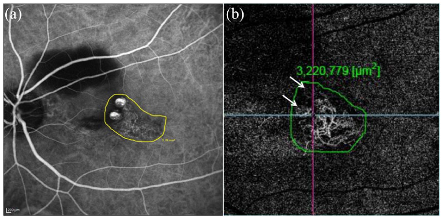
A 47-year-old male presented with diminution of vision in the left eye with a best-corrected visual acuity of 20/200. (a) Late phase of indocyanine angiography (ICGA) showed the presence of branching vascular network (BVN) of area 3.19 mm2 and two polyps on the superonasal aspect of the BVN. (b) Optical coherence tomography angiography (OCTA) also showed BVN correlating with ICGA with an area of 3.22 mm2. However, superior polyp was not localized on OCTA, while inferior polyp was seen as hyperreflective lesion (white arrows).
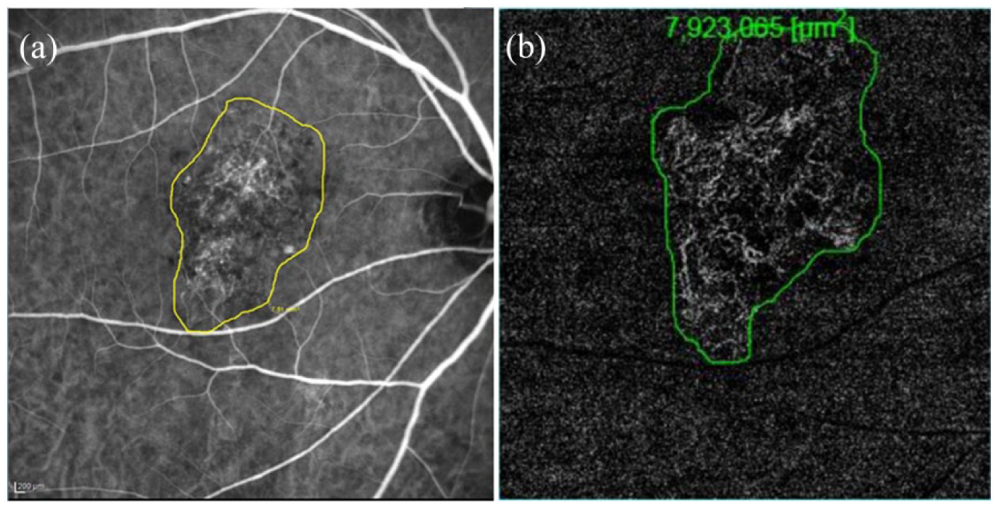
Indocyanine angiography (ICGA) image of a 66-year-old male with best-corrected visual acuity of 20/100 (a) showing branching vascular network of area of 7.51 mm2 with polyp at nasal aspect of branching vascular network (BVN). (b) Optical coherence tomography angiography (OCTA) showed a BVN area of 7.92 mm2 with the presence of polyp seen as hyperreflective structure nasal to BVN.
The measured BVN area in both ICGA and OCTA was 7.47 ± 5.74 and 7.51 ± 5.69 mm², respectively, and the difference was not statistically significant (p = 0.53). The measured area of BVN on ICGA and OCTA showed excellent correlation (r = 0.997). Compared to ICGA, OCTA underestimated the BVN area (mean ± SD: 0.36 ± 0.33 mm²) in 23 eyes. Conversely, overestimation of BVN area (0.28 ± 0.19 mm²) on OCTA was seen in 23 eyes compared to ICGA. The two groups where OCTA underestimated and overestimated BVN size compared to ICGA had a mean PED height of 471.04 and 248.22 µm, respectively, which was significantly different (p = 0.01). The correlation between BVN area (measured in ICGA) and PED height was 0.47, which was significant (p = 0.001).
Polyps were identified in 43 (93.48%) and 32 (69.57%) of the total 46 eyes using ICGA and OCTA, respectively. In the three eyes in which polyps were not detected on ICGA, polyps were not identified on OCTA as well. OCTA was unable to identify polyps in another 11 eyes (23.91%) where ICGA could identify polyps. The mean BVN size in these 11 eyes was 6.67 mm² (ICGA) and 6.68 mm² (OCTA). We also compared the PED height in these 11 eyes (247.45 ± 153.27 µm) with the rest of the cohort (394.89 ± 358.53 µm). PED height was not significantly different in the two groups (p = 0.19).
The imaging characteristics of the remaining 32 eyes in which polyps were identified on both ICGA and OCTA were analyzed separately. The mean number of polyps was 1.89 ± 1.53 and 0.85 ± 0.77 on ICGA and OCTA, respectively, and the difference between the two groups was statistically significant (p = 0.002). The differential reflectivity pattern of polyps on OCTA revealed that 45.1% and 39.2% of polyps were hyperreflective and hyporeflective with a hyperreflective border, respectively. We could not detect 15.7% of polyps on OCTA compared to ICGA.
Discussion
ICGA is the gold standard test for the diagnosis of PCV. 22 However, it is not commonly performed especially in Western countries due to the lower incidence of PCV in their population and the invasive nature of ICGA. Still, ICGA remains one of the commonly used imaging modalities in the diagnosis and management of PCV. OCTA, a recently described technique, uses low-coherence interferometry and motion contrast to provide three-dimensional en face reconstruction of the chorioretinal vasculature. 23 It has an additional advantage of being non-invasive with a lower scan acquisition time. However, major limitations include motion artifacts and no detection of leak or blood flow below a certain threshold. These lead to difficulty in the identification of polyps in PCV compared to ICGA.14,17,18,20,24
In this study, the angiographic characteristics of treatment-naïve PCV eyes were evaluated by means of ICGA and OCTA. The mean area of BVN was comparable between the two imaging diagnostic tools (p = 0.53). OCTA could better delineate the margins of BVN when the BVN size was small.
The polyps were not seen in 6.52% (3/46) of patients on ICGA. However, OCTA had a significantly lower polyp detection rate and polyps were not identified in 30.43% (14/46) of patients. Previous publications have consistently shown lower polyp detection rate with OCTA compared to ICGA, ranging from 47% to 85%.18,19,24,25 Wang et al. 20 and Kim et al. 25 have reported a higher identification rate of polyps (50%–92.3%) in the outer retina slab compared to the choriocapillaris. The anatomical correlation for this observation relies on the finding that polyps are generally located on the top of PED and BVN lies between RPE and Bruch’s membrane. Therefore, a single en face plane may not be sufficient to identify both the vascular components. In addition, the manual segmentation plays an important role in detecting the vascular lesions, as suggested by previous authors. 19 Moreover, Kim et al. 25 have reported a differential reflectivity of polypoidal lesion based on the reference plane. The slow blood flow on OCTA beyond a detection threshold may also lead to certain polyps being hyperreflective and others being hyporeflective. 25 Wang et al. 20 have used multiple descriptors based on reflectivity to define the pattern of polyps on OCTA, such as nodular, ring, clustered, or dot. Takayama et al. 14 have reported lower size of polyps on OCTA compared to ICGA with a lower rate of polyp detection. However, OCTA had a higher detection rate of BVN in their study. Peiretti et al. 17 have reported 90% and 100% detection rate of polyps and BVN on OCTA, thereby highlighting the utility of OCTA in cases with chronic central serous chorioretinopathy with progression to PCV.
We noted that among the 11 eyes in which OCTA could not identify polyps, there was no significant variation in BVN size (ICGA: 6.67 mm²; OCTA: 6.68 mm²), and the polyps were located at the end of BVN in 9 out of 11 eyes. This suggests that apart from BVN size, other factors such as sluggish blood flow below the detection threshold, signal attenuation due to the presence of subretinal hemorrhage, and hemorrhagic PED also may play a role in limiting the role of OCTA in the identification of polyps. The similarity of BVN size values between the two diagnostic tools suggests that OCTA can be considered as a reliable method with which to analyze BVN area. Therefore, GLD measurements based on OCTA may be used to plan treatment with PDT as well.
Various studies have shown high polyp regression rates with anti-VEGF monotherapy or combination of anti-VEGF and PDT with higher polyp regression associated with better visual outcomes.4,15,26 However, the PLANET study showed lower polyp regression rates with aflibercept monotherapy and combination therapy (38.9% and 44.8%, respectively) with acceptable visual outcomes. 27 This suggests that successful visual outcomes may not be directly related to polyp regression. With this background, the role of combination of OCTA and structural B-scan in the follow-up of PCV eyes seems more feasible because other relevant information regarding BVN and the presence of disease activity (SRF, IRF) can be obtained from a combination of structural OCT B-scan and OCTA.
Compared to ICGA, OCTA overestimated and underestimated BVN size in 23 eyes each, although the difference was statistically insignificant. The eyes in which ICGA measured higher BVN area had a significantly higher PED height compared to the other subgroup. PED is known to cause hypofluorescence in ICGA and may therefore mask or lead to underestimation of BVN size. 11 Higher PEDs also lead to signal attenuation on OCTA, which can lead to difficulty in identifying borders of BVN. 28
The main strength of the study is the inclusion of treatment-naïve eyes with PCV. This is helpful as polyps are known to regress following treatment with anti-VEGF agents or PDT. The axial resolution of OCTA is 5–7 µm, so polyps smaller than 5 µm in size may not be detected on OCTA. 21 OCTA has certain advantages compared to ICGA. OCTA images can be depth encoded to identify the level of BVN and polyps and may be helpful to delineate the type 1 membrane. Moreover, unlike ICGA, OCTA can be performed at each follow-up visits.
The limitations of the study include its retrospective nature and its cross -sectional design. Thus, the changes in BVN and the polyp area with treatment could not be quantified. Moreover, video ICGA was not performed in the study eyes. We were therefore unable to comment on the presence of feeder vessels. The inherent disadvantages of OCTA in the form of segmentation errors also pose challenge in delineating polyps. The boundaries of large-sized BVN could have been missed in few of the eyes because the view of OCTA was limited (6 mm × 6 mm).
The possibility of OCTA replacing ICGA for measurement of GLD and characterizing BVN and polyps is difficult at present. However, OCTA in conjunction with structural OCT B scans can provide more information on the disease activity. The future advancements in the imaging techniques especially OCTA may provide more information about polyps and BVN and improve our disease management.
Footnotes
Declaration of conflicting interests
The author(s) declared no potential conflicts of interest with respect to the research, authorship, and/or publication of this article.
Funding
The author(s) received no financial support for the research, authorship, and/or publication of this article.