
Abstract

Dear Editor,
Detection of an intraocular calcified cyst to the extent seen here is exceedingly rare.1–3 This case provides histologic evidence of extensive intraocular osseous metaplasia with bone marrow formation in a malformed eye several decades from birth.
A 35-year-old man with type 1 diabetes mellitus, blind from birth in his left eye, was admitted to our Unit because of an open globe injury in the same eye. The injury occurred at work 6 h earlier, while he was repairing an iron gate. The patient was unable to provide any further information about his blindness. Best corrected visual acuity was 20/20 in the right eye and no light perception in the left one. Slit-lamp examination of the right eye was unremarkable. The left eye showed a full-thickness scleral injury close to the upper limbus, with prolapse of the lens and vitreous fluid. Moderate corneal pannus was also present. Computed tomography disclosed an atrophic left globe with intraocular calcifications in the posterior segment (Figure 1A). The patient was offered to save the eyeball but opted for its removal. Evisceration was performed and, during surgery, a red grey “stone” of the size of a hazelnut was removed (Figure 1B). A 14-mm spherical hydroxyapatite orbital implant was inserted.
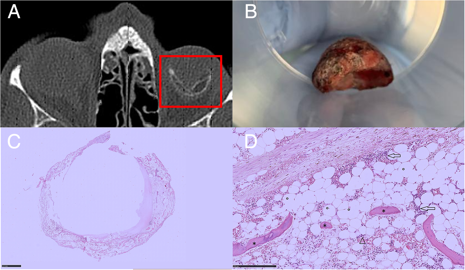
Histopathologically, gross examination of the surgical specimen revealed a solid, oval lesion with a 1.5 cm diameter. This was decalcified by chemical solution, fixed in formalin, and routinely processed with representative tissue section embedded in paraffin and stained with hematoxylin and eosin. Microscopic findings are described in Figures 1C and D.
Intraocular osseous metaplasia with formation of hematopoietic bone marrow is very rare.1,2 The incidence of heterotrophic bone formation in enucleated eyes has been reported to range from 4 to 18%. 3 It is commonly associated with phthisis bulbi and may occur in eyes with history of ocular trauma, chronic inflammation, long-standing retinal detachment, intraocular tumours (e.g., choroidal hemangioma, osteoma, and melanoma), as well as congenital abnormalities, including hyperplastic primary vitreous, microphthalmos, and buphthalmos. Intraocular osseous metaplasia may be subretinal, preretinal or both. 3 The subretinal form is believed to be caused by metaplasia of the pluripotent retinal pigment epithelium (RPE) cells into osteoblasts secondary to vascular delivery of osteoblasts through the choroidal circulation. It is more common in phthisis bulbi and usually occurs years after a retinal detachment. In the preretinal form, usually associated with proliferative vitreoretinopathy and Eales’ disease, the breakdown of the internal limiting membrane may be responsible for RPE cell migration and consequent formation of osseous metaplasia.
Detection of an intraocular calcified cyst to the extent seen here is exceedingly rare, and this case provides histologic evidence of extensive intraocular osseous metaplasia with bone marrow formation in a malformed eye several decades from birth.
Footnotes
Patient consent
Consent to publish this case report has been obtained from the patient in writing.
Author contributions
All authors attest that they meet the current ICMJE criteria for Authorship.
Declaration of conflicting interests
The author(s) declared no potential conflicts of interest with respect to the research, authorship, and/or publication of this article.
Funding
The author(s) received no financial support for the research, authorship, and/or publication of this article