






Abstract
Purpose
To evaluate outcomes of fornix deepening with retractor recession and repositioning for conjunctivochalasis (CCh) on improvement of conjunctival folds and ocular surface symptoms, particularly epiphora.
Methods
Retrospective, single-centre, observational case series of patients with refractory CCh who underwent fornix deepening and retractor recession. CCh was graded using the Hoh classification (grades 0 to 3 depending on the number and height of folds). Epiphora, reflex tearing, and dry eye symptoms were assessed using the validated 'TEAR' score pre- and post-CCh correction.
Results
18 eyes of 11 patients with CCh (average age 68, range 46–82 years) were treated with fornix deepening and retractor recession. All had shallow fornices pre-operatively with a mean CCh grade of 1.7 (typically lower than the tear meniscus). Locations of the folds were variable: diffuse/middle (n = 10), nasal (n = 4), and temporal (n = 4). At 15-month mean follow-up, conjunctival redundancy was absent in 17 of 18 eyes postoperatively, resulting in a restored tear meniscus and reservoir. 91% saw a reduction in tearing frequency (T), with 73% gaining ≥ 2-point improvement. Improvements in clinical effects (E) and activity limitation (A) were seen in 82% and 91% of patients, respectively, with 36% and 64% gaining ≥ 2-point improvement. R scores (related to reflex tearing) improved in 73%, with 64% seeing ≥ 2-point gains. (P < 0.05 for all).
Conclusion
Restoration of the tear reservoir by inferior fornix deepening with retractor recession and repositioning can result in improvement of CCh and epiphora.
Introduction
The tear film is critical for ocular protection, lubrication, and refraction. It comprises lipid, aqueous, and mucin, which all play a significant role in its homeostasis and stability. 1 Tears are distributed in three ocular compartments. 2 The main lacrimal gland secretes aqueous into the conjunctival sac and fornices (third compartment), spreads to the tear meniscus (second compartment), and finally, extends over the corneal surface (first compartment). 3
Conjunctivochalasis (CCh) is characterised by redundant, loose, and non-oedematous bulbar conjunctival folds resulting in tear film instability.4–7 It is hypothesised that the repeated friction of the eyelids against the conjunctiva contributes to the breakdown of the elastic fibres8,9 and the development of the characteristic lid-parallel conjunctival folds (LIPCOF). These folds obstruct the tear drainage and obliterate the meniscus and forniceal compartment. 4 CCh can cause an array of symptoms, including ocular irritation, disturbance of tear outflow, and exposure problems. 10
Management can be challenging, ranging from lubricants in mild cases to surgical intervention in refractory cases. Several surgical techniques have been described for treating CCh; however, there has yet to be a consensus on the optimum approach. Most of these surgical procedures involve excising or resecting the conjunctiva and may be effective in some cases, as they relieve the mechanical obstruction.
Under normal circumstances, the tear reservoir rapidly refills the meniscus. Huang et al 6 examined the original meniscus height recovery rate following depletion with a capillary tube and found it significantly slower in symptomatic CCh patients. In a study by Cheng et al, 2 the tear volume was significantly increased when the fornix tear reservoir was restored with conjunctival recession and amniotic membrane transplant (AMT) in 18 eyes with CCh. The authors also reported a significant resolution of corneal staining, conjunctival inflammation, and reduction of topical medications. We feel similar results can be achieved using similar principles of fornix deepening but without AMTs, which can be costly and require storage facilities. This study evaluates outcomes of fornix deepening with retractor recession and repositioning for CCh on improvement of conjunctival folds and ocular surface symptoms, particularly epiphora.
Materials and methods
Patients
This is a retrospective, non-comparative, single-centre study of consecutive patients with refractory CCh who consented to and underwent fornix deepening with retractor recession and repositioning by a single surgeon (RM) between 2017 and 2022 at the Queen Victoria Hospital NHS Trust. The patient's medical records were assessed, including demographics, ocular co-morbidity, previous therapies, clinical presentations, and concomitant medications at presentation. The study was conducted in accordance with the declaration of Helsinki and was approved by the local institutional review board.
The height and location of the folds were graded by a blinded observer (CSL) using pre- and post-operative photographs. The Hoh classification (grade 0 - no persistent fold; grade 1 - a single, small fold; grade 2 - two or more folds but not higher than the tear meniscus; grade 3 - multiple folds and higher than the tear meniscus) was utilised to document the height. 11 In addition, the location of folds was specified as T, M, and N, similar to Meller and Tseng's system 5 if they were predominantly found in the temporal, the middle (or inferior to the limbus), and the nasal aspect of the lower lid, respectively.
All cases were refractory to conservative treatment, such as topical preservative-free lubricants, short courses of corticosteroid drops (≤ 4 weeks), and/or bandage contact lenses. Exclusion criteria included a history of lower lid surgery or trauma, lid malposition disorders (e.g., ectropion, entropion, floppy eyelid syndrome), lagophthalmos on gentle closure, trichiasis, punctal stenosis, and nasolacrimal obstruction.
Tear Score
Epiphora was assessed for each patient using a validated ‘TEAR’ score pre- and post-CCh correction 12 (Figure 1). The TEAR score is a scale of clinical and patient-reported severity in patients with epiphora, comprising four subscales: Times wiping (T), clinical Effects (E), Activity limitation (A), and symptoms of Reflex epiphora (relating to symptoms suggestive of reflex tearing) (R). A fifth subscale is used during follow-up review to record patient-reported success (S) as a percentage.
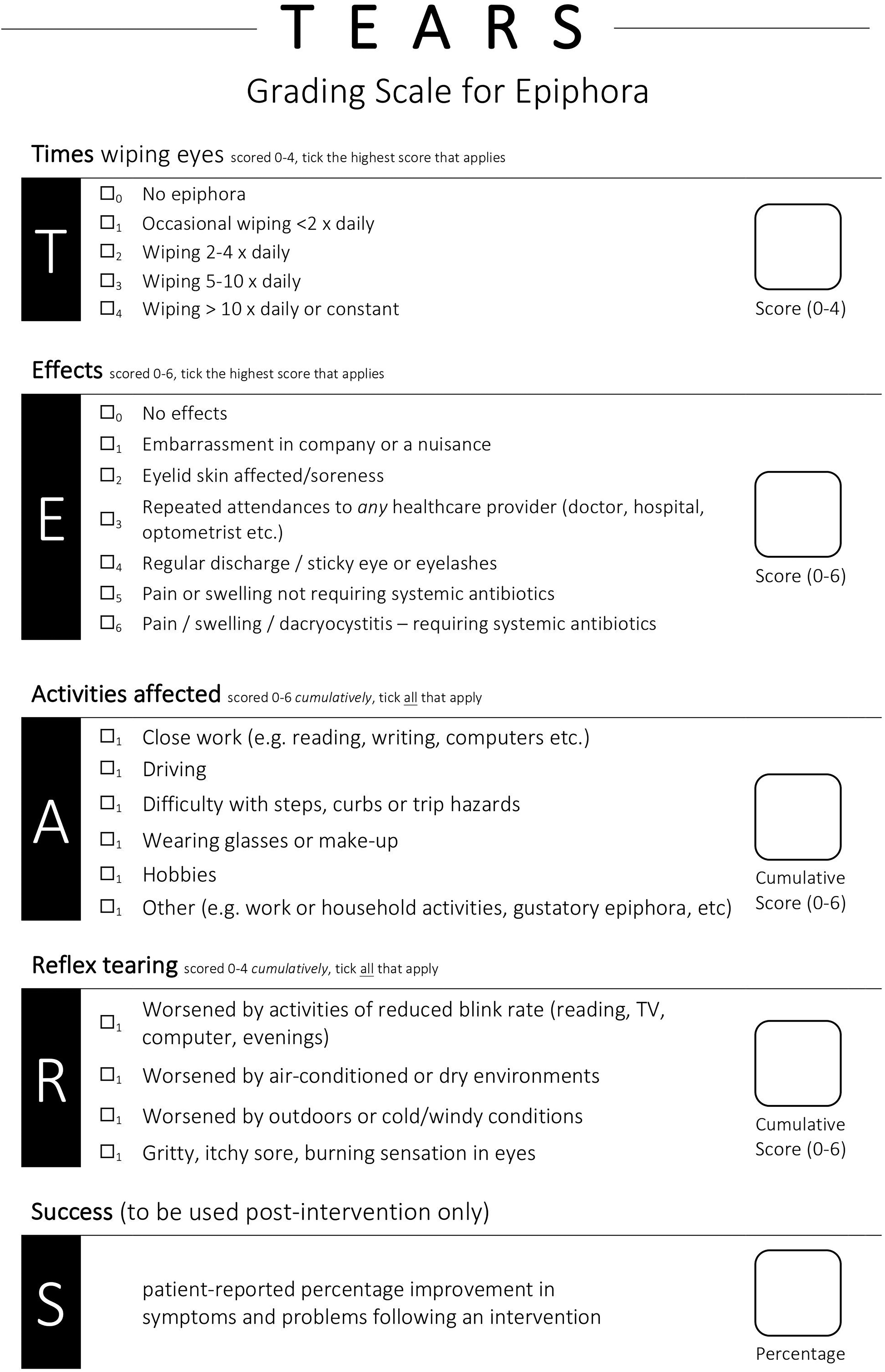
Tear score, comprising 4 subscales: Times wiping (T), clinical Effects (E), Activity limitation (A), and symptoms of Reflex epiphora (R).
Subjective Evaporative Dry Eye Scores
The patients scored the frequency of dry eye symptoms (e.g., discomfort, grittiness, foreign-body sensation, dryness, and light sensitivity) from 0 to 4. The grading system was based on the Ocular Surface Disease Index (OSDI) questionnaire13,14 where 0 indicates none of the time; 1, some of the time; 2, half of the time; 3, most of the time; and 4, all of the time.
Surgical Technique (Figure 2)
The inferior conjunctival fornix, lower eyelid skin, and lateral canthus were infiltrated with anesthetic (Bupivacaine 0.5% with 1:200,000 adrenaline, 27-gauge needle). A corneal shield was inserted. A lateral canthotomy/cantholysis incision was made with a 15-blade. The lower eyelid retractors were visualised via a swinging eyelid approach (Figure 2a), then dissected from the conjunctiva posteriorly and the septum/inferior fat pads anteriorly with Westcott scissors (Figure 2b). The inferior retractors’ horns were lysed until the inferior retractors were freely mobile. The location of loose conjunctival folds was identified, and its posterior conjunctival surface located (Figure 2c). Inferior retractors were then recessed and repositioned to the inferior fornix and, in particular, to the loose conjunctiva, with interrupted 6-0 vicryl sutures (typically 3 or 4 sutures) taking partial thickness bites of the conjunctiva to ensure the vicryl remained buried and not exposed onto the epithelial surface of the fornix (Figures 2d and 2e). The conjunctival edges were closed with interrupted 6-0 vicryl sutures. The lateral canthus was de-epithelialised and repositioned to the periosteum at the lateral orbital wall, without resecting the tarsus nor shortening the lower lid. Postoperatively, all patients received chloramphenicol (1%) ointment three times a day for one week.

Photographs of retractors recession and fornix deepening. A conjunctival–retractor flap is created (a). The retractors are dissected off the conjunctiva posteriorly (b). The locations of loose conjunctival folds are identified and held from their posterior conjunctival surface with St Martins forceps (c). The distal edges of the retractors are then sutured to the posterior surface of the loose conjunctival folds (d and e). Three interrupted 6-0 vicryl sutures are placed partial thickness to recess the retractors and deepen the fornix.
Results
Eighteen eyes of 11 patients with CCh (average age 68, range 46–82 years) were treated with fornix deepening, retractor recession and repositioning. All had shallow fornices pre-operatively with a mean CCh grade of 1.7 (typically lower than the tear meniscus). The locations of the folds were variable: diffuse/middle (n = 10), nasal (n = 4), and temporal (n = 4). There were no patients with upper bulbar conjunctival folds. Ten patients (16 out of 18 eyes) had signs of lower lid laxity with a delayed snap-back test but no malposition. In addition to fornix deepening with retractor recession and repositioning, they also underwent lateral tarsal repositioning, with the attachment of the de-epithelialised lateral tarsal edge to the lateral orbital rim at or above the level of Whitnall's tubercle.
Tearing frequency (T) using the TEAR grading scale 12 improved from 3.7 ± 0.9 (range 1–4) to 1.3 ± 1.3 (range 0–4) (P < 0.05), clinical effects (E) improved from 1.5 ± 0.9 (range 0–3) to 0.5 ± 1.0 (range 0–3) (P < 0.05), activity limitation (A) improved from 2.0 ± 1.0 (range 0–3) to 0.2 ± 0.4 (range 0–1) (P < 0.05) and R scores (related to symptoms suggestive of reflex tearing) improved from 2.3 ± 0.6 (range 1–3) to 1.0 ± 1.3 (range 0–4) (P < 0.05). Ten patients (91%) saw a reduction in tearing frequency (T), with 8/11 (73%) gaining ≥ 2-grade improvement. Improvements in clinical effects (E) and activity limitation (A) were seen in 9/11 (82%) and 10/11 (91%) of patients, respectively, with 4/11 (36%) and 7/11 (64%) gaining ≥ 2-grade improvement. R scores improved in 8/11 (73%), with 7/11 (64%) seeing ≥ 2-grade improvement. All patients improved symptomatically (Figure 3), with an average improvement “S” score of 78% (range 30%-100%). The subjective dry eye grading score improved from 2.3 ± 1.4 (range 0–4) to 1.1 ± 0.8 (range 0–2) (P < 0.05).
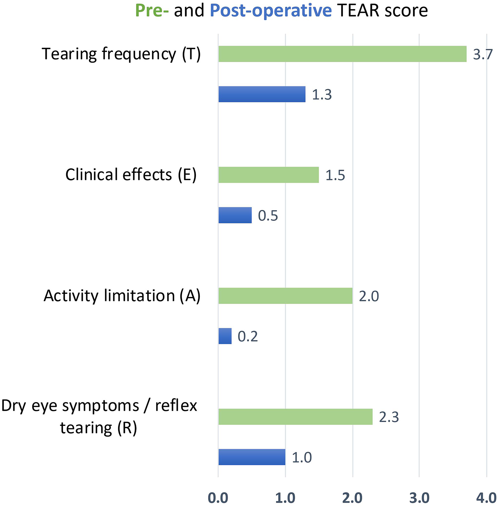
Bar chart showing mean pre-operative and post-operative TEAR scores. See Table 1 for definitions of sub-scale T, E, A, and R scores. Microsoft® Excel was used to create the artwork.
Surgical procedures used to correct CCh with respective advantages and disadvantages and literature review. Adapted from Yvon et al. 47

Abbreviations: AMT indicates amniotic membrane transplant; CCh, conjunctivochalasis; LIPCOF, lid-parallel conjunctival folds; OSDI, Ocular Surface Disease Index; SI, symptomatic improvement; TBUT, tear break-up time; TMA, tear meniscus area.
At a mean follow-up of 15 months, conjunctival redundancy was absent in 17 out of 18 eyes postoperatively, resulting in a restored tear meniscus and reservoir (Figure 4). The distance from the corneal light reflex to the lower eyelid (MRD2) was used to measure lower eyelid height. In our case series, retractor recession +/- lateral tarsal repositioning achieved 0.7 mm (range 0–2mm) in lid elevation. Complications included one case of temporary focal conjunctival inflammation due to a persistent suture and one conjunctival cyst that required deroofing. No cases of ocular motility restrictions nor fornix shortening were observed.
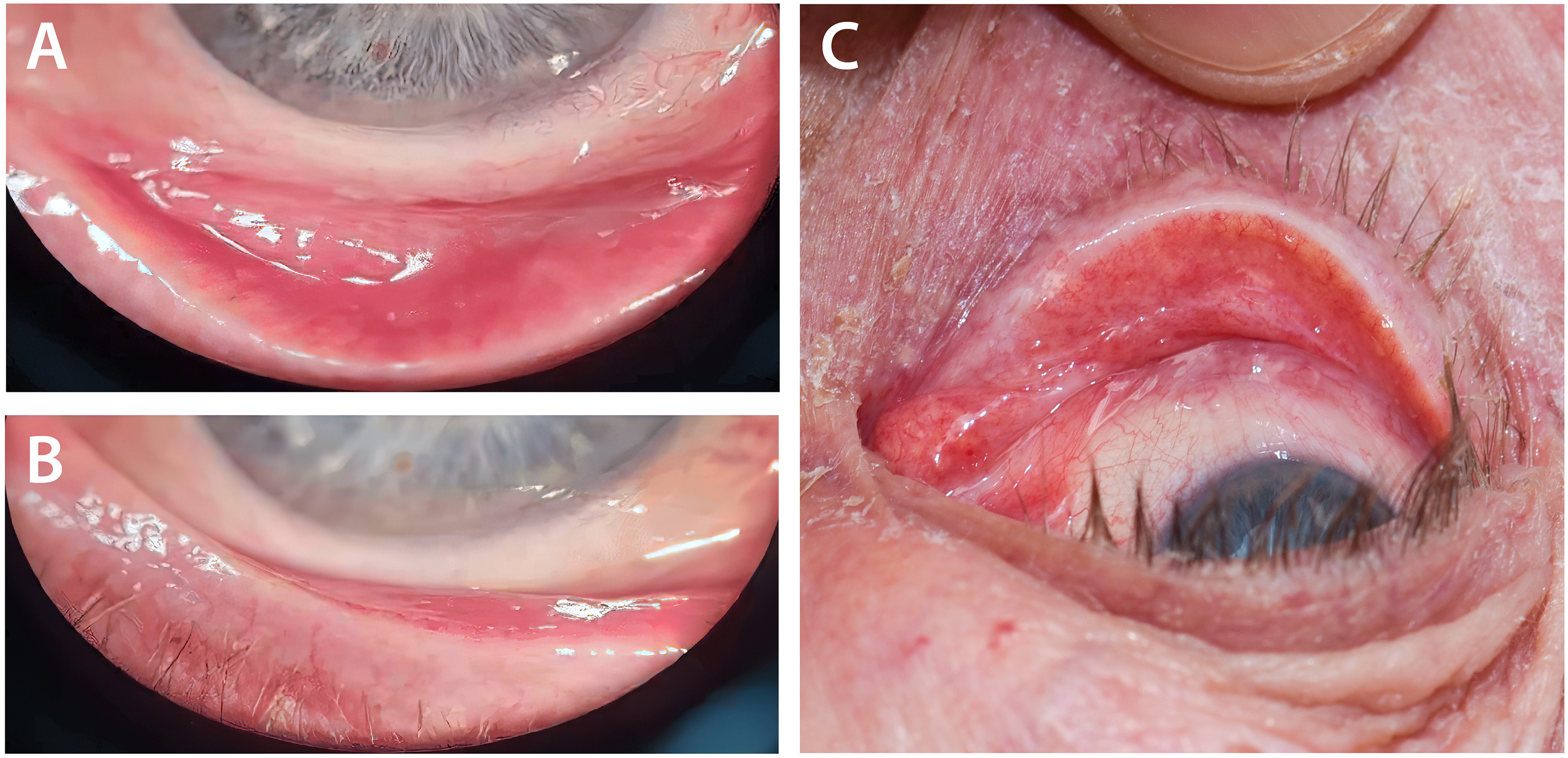
Pre-operative photograph showing a grade 2 (two or more folds but not higher than the tear meniscus) middle CCh (a); 4 months post-operative showing resolution of conjunctival folds (grade 0 - no persistent fold) (b); and 6 months post-operative showing a deepened fornix (c).
Discussion
In CCh, the LIPCOF shortens the fornices and obliterates the tear meniscus and reservoir. 6 This may lead to the loss of the second and third tear film compartments and subsequently exacerbates ocular surface symptoms. In our study, we showed that restoration of the tear reservoir by fornix deepening and retractor recession can result in improvement of CCh, epiphora, and evaporative dry eye symptoms. This ensures a continuous supply of tears from the reservoir to the tear meniscus and the corneal surface. To our knowledge, this is the first study to assess an improvement in epiphora using a validated score.
CCh is often associated with dry eye symptoms and epiphora.7,15 It is likely that CCh exacerbates an unstable tear film by disrupting the tear meniscus. 16 It is, however, unclear if the unstable tear film precedes or is a consequence of the conjunctival folds. Examination shows a discrete punctate or linear staining on the mucosal side of the lid margin near the LIPCOF, which is different from the exposure pattern in patients with dry eye syndrome. It is thought that CCh is caused by lipid tear film deficiency, not an aqueous deficiency. 5 The lipid and mucin layers of the tear film are mainly secreted from meibomian glands and conjunctival epithelial cells, respectively. We postulate that the obliteration of the tear meniscus and reservoir affects this secretion, leading to a deficient lipid tear film layer and rapid tear evaporation.
Ocular inflammation plays a vital role in the pathophysiology of CCh. Studies have reported an increased expression of matrix metalloproteinase in cultured conjunctival fibroblasts, as well as higher levels of inflammatory markers in the tear profiles of CCh patients.17–20 This, in turn, may lead to a hyperosmolar tear film. This is in keeping with our significant R score improvement, indicative of dry eyes and reflex tearing. Studies have shown that dry eye symptoms and abnormal tear parameters were more significant if the folds were nasal. 21 In addition, inflammation was found to be more pronounced in eyes with nasal CCh, likely secondary to the delayed tear clearance. 18
Management can be challenging; hence, multiple surgical approaches to tighten the redundant conjunctiva have been trialed with varying success rates (Table 1). These include conjunctival excision,22,23 cauterisation,24–28 scleral fixation of the conjunctiva, 29 conjunctival ligation, 30 laser conjunctivoplasty,31–33 radio-wave electrosurgery,34,35 tissue grafting (e.g., AMT)36–39 and more recently, plasma-based conjunctivoplasty. 40
Numerous studies have shown an improvement in epiphora following redundant conjunctival excision.41–43 This was mainly evident in a subgroup of patients where the LIPCOF was mechanically obstructing the punctum. A study by Liu 16 noted redundant conjunctiva over the punctum in 15 patients with epiphora, who all improved with simple conjunctival excision. However, most surgical procedures do not tackle fornix reconstruction and can lead to scarring or relapses. Conjunctival recession is thought to be more effective because it rebuilds the fornix tear reservoir. 2
Three studies examined CCh outcomes following conjunctival recession and AMT.2,36,44 Cheng et al demonstrated an improvement in symptoms, corneal staining, basal wetting, and a reduction of topical medications in 18 eyes, 2 in addition to an increased fornix depth in 7 eyes. 44 Kheirkhah et al 36 showed a smooth conjunctival surface with complete or significant improvement of symptoms in 44% and 56%, respectively. In all three studies, the abnormal Tenon's capsule was dissected from the overlying conjunctival, and AMT was applied to the bare sclera. AMT is known to have anti-inflammatory, anti-fibrotic, and anti-angiogenic properties but does carry disadvantages, including the risk of transmission of communicable diseases, early disintegration, increased costs, and requires complex storage facilities. 45 Our study shows the resolution of conjunctival folds and improvement of epiphora and dry eye symptoms without using an amniotic membrane.
Fornix deepening with retractor recession and repositioning is more time-consuming than other techniques, such as simple excision. However, fornix shortening and ocular motility restrictions would be unusual with this technique as no conjunctiva is resected. Complications are typically related to secondary focal conjunctival inflammation (e.g., granuloma, papillary conjunctivitis). Kheirkhah and colleagues 36 used fibrin glue to secure the amniotic membrane; however, they had similar rates of conjunctival inflammation after the procedure. We postulate the inflammation may be related to the manipulation of the conjunctiva and Tenon's capsule. AMT does not appear to negate those effects. In our case series, topical corticosteroids were only added post-operatively if the patients had signs of inflammation, but have been used routinely in the other studies. Given the small number of patients, it is difficult to determine whether their use reduces complications.
Our study's limitations are the retrospective nature, small patient number, and lack of control. A blinded observer determined the presence, location, and height of folds; however, the fornix depth and tear meniscus height were not measured. Furthermore, most of our patients with CCh were elderly and had signs of mild lateral canthal or lower lid laxity but no eyelid malposition. It is difficult to ascertain if the repositioning of the lateral tarsus also improved the epiphora. However, it is unlikely that this would have led to such a significant improvement in both epiphora, evaporative dry eye symptoms, and reflex tearing in the absence of obvious eyelid malposition. Furthermore, there is a paucity of literature performing standard lateral tarsal strips or repositioning for CCh. Given what has been published already on fornix reconstruction for CCh, we could not ethically do such procedures to compare as a control.
We believe this study sheds new light on the clinical management of patients presenting with both CCh and epiphora and provides proof of concept to the role of retractor recession and repositioning to achieve fornix deepening and improved symptoms. It also provides evidence to support the need for a study evaluating the role of transconjunctival approach fornix deepening by retractor recession and repositioning in the absence of a lateral canthotomy.
Once the cause of epiphora is resolved, dry eye disease can be accurately assessed and diagnosed. In effect, an improvement in dry eye symptoms was observed in the study; however, the clinical signs, such as osmolarity, were not formally evaluated. Further studies examining pre- and post-operative osmolarity following retractor recession and repositioning may also be valuable. 46
Conclusion
Our study highlights the importance of fornix deepening during CCh surgery as a logical step in the treatment algorithm for epiphora. CCh impacts the preocular tear film by delaying tear clearance and disrupting tear flow to the meniscus and fornix reservoir. As shown by the improved TEAR scores, lower eyelid retractor recession and repositioning directed to restoring and deepening the fornix is effective in treating CCh. Future prospective studies are needed to compare the outcomes of the modifications to this technique.
Footnotes
Acknowledgment
Miss Christina S Lim, FRCOphth;
Queen Victoria Hospital, Holtye Rd, East Grinstead RH19 3DZ
Author contributions
The study conception and design were performed by Raman Malhotra. Material preparation, data collection and analysis were completed by Camille Yvon. The first draft of the manuscript was written by Camille Yvon and all authors commented on subsequent versions of the manuscript. All authors read and approved the final manuscript.
Consent
Informed consent was obtained from all individual participants included in the study.
Declaration of conflicting interests
The authors declared no potential conflicts of interest with respect to the research, authorship, and/or publication of this article.
Ethics approval
The study was conducted in compliance with the Declaration of Helsinki and was approved by the local ethics review committee (Queen Victoria Hospital NHS Foundation Trust, East Grinstead, UK)
Funding
The authors received no financial support for the research, authorship, and/or publication of this article.
Meeting presentation
Presented at 41st ESOPRS Annual Meeting in Naples (Italy), 14–16 September 2023.
