
Abstract
Purpose
To compare the clinical outcomes of conventional photorefractive keratectomy (PRK) and transepithelial PRK (TransPRK) for the correction of myopic refractive errors.
Methods
This prospective, paired-eye comparative study included 48 eyes of 24 patients. Each patient underwent conventional PRK in the right eye and TransPRK in the left eye. Both patient and outcome assessors were blinded to treatment allocation. Preoperative and postoperative parameters including uncorrected distance visual acuity (UDVA), spherical equivalent (SE), postoperative pain, epithelial healing time, and complications were evaluated. Paired statistical analyses were used to compare outcomes between the two procedures.
Results
There were no significant differences in baseline characteristics between the groups. Postoperatively, there were no statistically significant differences in UDVA (p = 0.084) or final SE (p = 0.512). Epithelial healing was faster in the TransPRK group but failed to show statistical significance (p = 0.371). However, postoperative pain was significantly higher in the TransPRK group, with a median pain score of 8 (IQR 8–10) compared to 6 (IQR 6–8) in the PRK group (p = 0.014). Clinically significant postoperative haze was observed in 3 (12.5%) of patients, with an identical pattern in both eyes of every affected patient.
Conclusion
Both conventional PRK and TransPRK are safe and effective procedures for myopic correction, providing comparable visual and refractive outcomes. In this cohort, TransPRK was associated with significantly greater early postoperative pain, a finding that contrasts with much of the existing literature and warrants further investigation. The contralateral study design highlights the consistency of other outcomes while isolating this key difference.
Introduction
Laser corneal refractive surgery is a well-established and widely utilized alternative to glasses or contact lenses for correcting refractive errors, including myopia, hyperopia, and astigmatism. 1 Following the introduction of the excimer laser, multiple surgical techniques have been developed to reshape the cornea, with photorefractive keratectomy (PRK) recognized as one of the earliest and most enduring procedures. 2 Conventional PRK entails mechanical or alcohol-assisted removal of the corneal epithelium, followed by excimer laser ablation of the anterior corneal stroma to achieve the desired refractive correction. 3, 4 Although effective, the manual de-epithelialization step introduces variability, extends surgical time, and is frequently associated with significant postoperative pain, delayed visual recovery, and an increased risk of haze formation5,6.
Transepithelial PRK (TransPRK) was developed to address these limitations. This single-step, no-touch technique employs the excimer laser to ablate both the epithelium and the underlying stroma in a single, continuous procedure. The proposed advantages of TransPRK include greater precision, reduced surgical time, decreased risk of stromal dehydration, and the potential for smoother postoperative recovery with less pain and more rapid epithelial healing5,6.
Multiple studies have compared the outcomes of TransPRK and conventional PRK, however, the results remain inconsistent. Several meta-analyses and prospective studies indicate that TransPRK is associated with reduced postoperative pain, faster visual recovery, and more rapid epithelial healing compared to conventional methods5,7,8. In contrast, other reports have found similar outcomes or have suggested that TransPRK may result in greater discomfort and longer recovery times in certain cases9,10. These conflicting findings underscore the necessity for controlled studies to clarify the true differences between the two techniques.
Contralateral eye studies, where each patient serves as their own control by receiving a different treatment in each eye, represent a powerful study design in ophthalmology. This approach minimizes the influence of inter-subject variability in factors such as genetics, systemic conditions, and healing responses, thereby enabling a more precise comparison of the procedures themselves11,12.
The primary outcome was postoperative pain during the first three postoperative days, assessed using a numerical rating scale (NRS). Secondary outcomes included epithelial healing time, uncorrected distance visual acuity (UDVA), and spherical equivalent (SE) at six months postoperatively.
Methodology
Study design and patient population
This prospective, paired-eye, comparative study was conducted at Hamad Medical Corporation (HMC), Ophthalmology Department. A total of 24 consecutive patients (48 eyes) with myopia or myopic astigmatism were enrolled. The study protocol was reviewed and approved by the Medical Research Council (MRC) (approval No. MRC-01-25-1282). We adhered to the tenets of the Declaration of Helsinki, and all participants provided informed consent after a thorough explanation of the procedures and potential risks. Each patient underwent conventional PRK in the right eye (OD) and TransPRK in the left eye (OS), making each subject their own control. Both eyes were treated during the same operative session, with the right eye (conventional PRK) operated first, followed by the left eye (TransPRK). The assignment of procedures to specific eyes was fixed by protocol rather than determined by random allocation; this was done to standardize the surgical sequence and ensure procedural consistency across patients.
Inclusion criteria were age over 18 years, stable refraction for at least one year, a corrected distance visual acuity of 20/20 or better and eligibility for refractive surgery after excluding any contraindications on corneal tomography (Pentacam) and slit lamp examination. Exclusion criteria included a history of previous ocular surgery, corneal pathologies, autoimmune diseases, and pregnancy or lactation.
Surgical technique
All surgical procedures were performed by the same experienced surgeon. For the conventional PRK group, the corneal epithelium was removed mechanically after application of a diluted 70% alcohol solution followed by ablation of the anterior stroma with the WaveLight EX500 by Alcon Laboratories, Inc. For the TransPRK group, a single-step transepithelial ablation was performed using the StreamLight protocol in the same excimer laser platform. The optical zone (OZ) was set to 7 mm for all eyes. Mitomycin C (MMC) 0.02% was applied to the stromal surface for 1 minute using a sponge followed by ocular surface wash with copious amounts of chilled balanced salt solution (BSS). After instilling one drop of moxifloxacin 0.5%, a bandage contact lens was placed on the cornea. MMC was applied systematically to all eyes regardless of ablation depth, as a uniform prophylactic measure.
Postoperative care and follow-up
Following surgery, all eyes received a standardized postoperative regimen comprising topical moxifloxacin 0.5% four times daily for one week, Dexamethasone 0.1% four times daily tapered over six weeks, and preservative-free artificial tears. Oral analgesics (Ibuoprofen 400 mg as needed, up to three times daily) were prescribed uniformly to all patients for the first three postoperative days; no topical anesthetic eye drops were used and no inter-eye differences in analgesic use were documented. The applied bandage contact lens was removed upon complete re-epithelialization. Patients were examined on day 3, day 8, and at 1, 3 and 6 months postoperatively.
At each follow-up visit, uncorrected distance visual acuity (UDVA), corrected distance visual acuity (CDVA), and manifest refraction were recorded. Postoperative pain was rated by patients using a numerical rating scale (NRS) from 0 (no pain) to 10 (worst imaginable pain) on each of the first three postoperative days (day 1, day 2, and day 3); the peak (highest) NRS score recorded across these three days was used as the pain outcome for analysis. The area of the epithelial defect (ED) was estimated at the slit lamp on days 3 and 8 using slit-beam measurement in two perpendicular meridians to approximate an elliptical area, performed by the same masked observer at each visit. Corneal haze was graded at the slit lamp during the 6 months follow up visit with an ordinal scale described by Fantes et al (Supplementary Table 1).13,14
Patients were blinded to the treatment allocation for each eye. Follow-up slit lamp and refractive assessments were conducted by an experienced refractive surgeon and an optometrist, respectively, both independent of the operating surgeon. These assessors were likewise blinded to the treatment administered to each eye.
Statistical analysis
All statistical analyses were performed using R software (version 4.5.0; R Foundation for Statistical Computing, Vienna, Austria). Given the paired-eye design, all comparisons were conducted using paired statistical tests. The Shapiro-Wilk test was used to assess the normality of data distribution. Normally distributed continuous variables were compared using the paired t-test and are presented as mean ± standard deviation (SD). Non-normally distributed variables were compared using the Wilcoxon signed-rank test and are presented as median and interquartile range (IQR). Categorical variables were compared using the McNemar test and are presented as counts and percentages. A p-value of < 0.05 was considered statistically significant. No correction for multiple comparisons was applied given the exploratory, hypothesis-generating nature of this pilot study.
Results
Baseline characteristics
A total of 24 patients (48 eyes) were included in the study, with each patient contributing one eye treated by PRK (right eye) and the fellow eye treated by TransPRK (left eye).
The median age of participants was 27 years (IQR: 25–32), with 54.2% females (n = 13). There were no significant differences between PRK and TransPRK eyes regarding baseline parameters including age, gender distribution, unaided and aided visual acuity, or preoperative spherical equivalent (SE) (all p > 0.05) (Table 1). The mean preoperative SE was −2.69 ± 0.97 D overall, with no difference between the two procedures (p = 0.448). Similarly, the median preoperative unaided LogMAR visual acuity was 0.69 [0.46–0.78], showing no significant intergroup variation (p = 0.490). The mean ablation depth was 53.4 ± 18.6 µm for PRK eyes and 56.4 ± 17 µm for TransPRK eyes, also with no statistically significant difference between groups. The overall preoperative central corneal thickness was comparable between groups, with a median of 557.5 µm (IQR: 535.8–575.5 µm).
Baseline characteristics.
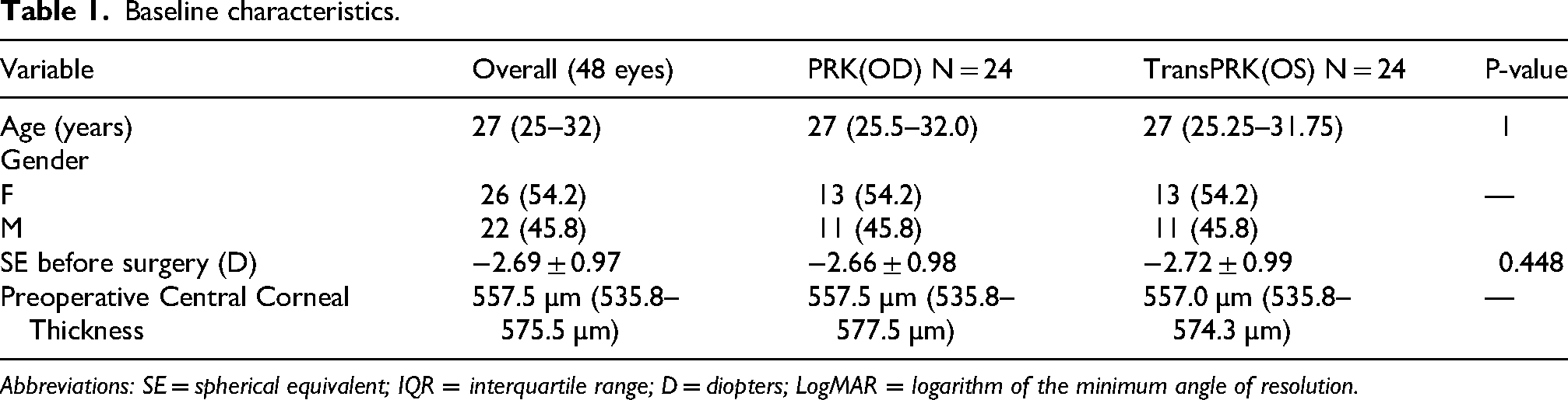
Abbreviations: SE = spherical equivalent; IQR = interquartile range; D = diopters; LogMAR = logarithm of the minimum angle of resolution.
Postoperative outcomes
The primary postoperative outcomes are summarized in Table 2. At six months postoperatively, there were no significant differences between the PRK and TransPRK groups in terms of postoperative UDVA (p = 0.084) or final SE (p = 0.512). Both procedures demonstrated excellent efficacy and predictability.
Postoperative outcomes.
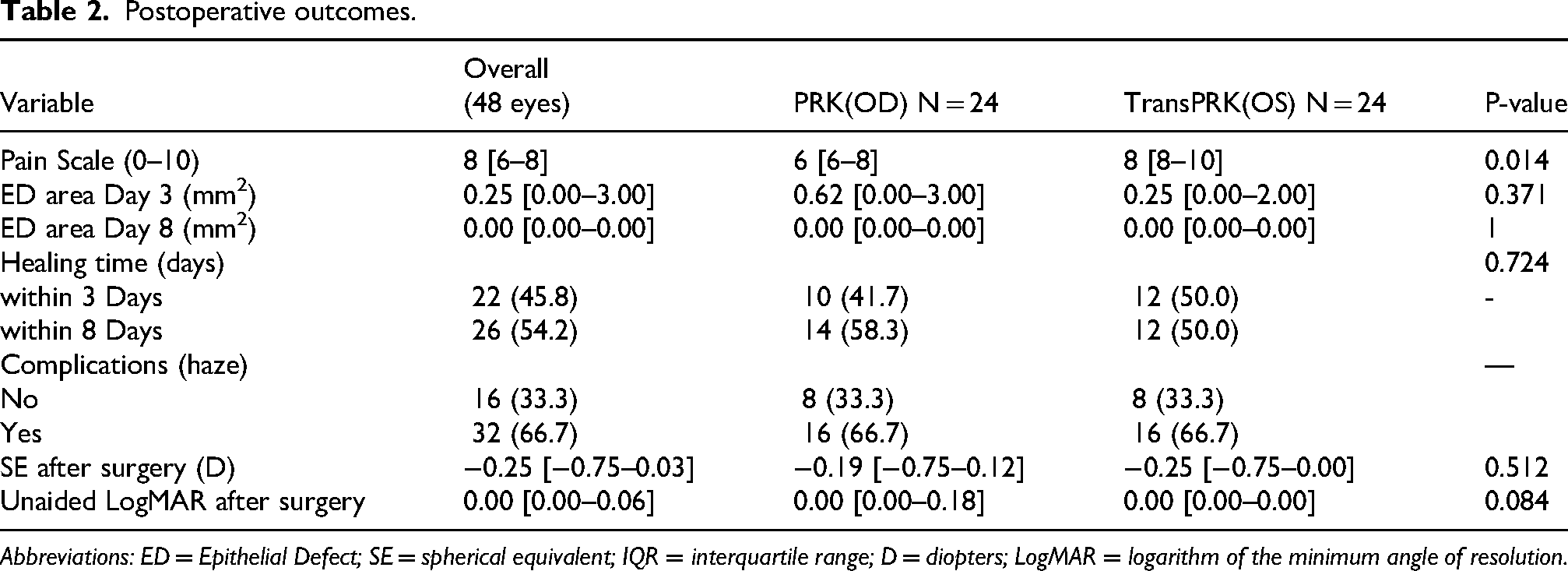
Abbreviations: ED = Epithelial Defect; SE = spherical equivalent; IQR = interquartile range; D = diopters; LogMAR = logarithm of the minimum angle of resolution.
Postoperative pain
A statistically significant difference was observed in postoperative pain scores. Patients reported significantly higher pain in the eye that underwent TransPRK (median 8, IQR 8–10) compared to the eye that underwent conventional PRK (median 6, IQR 6–8) (p = 0.014). This finding is illustrated in Figure 1. Median pain was 6 (IQR 6–8) following PRK and 8 (IQR 8–10) following TransPRK. The difference was statistically significant (p = 0.014, Wilcoxon signed-rank test), indicating greater postoperative discomfort after TransPRK.
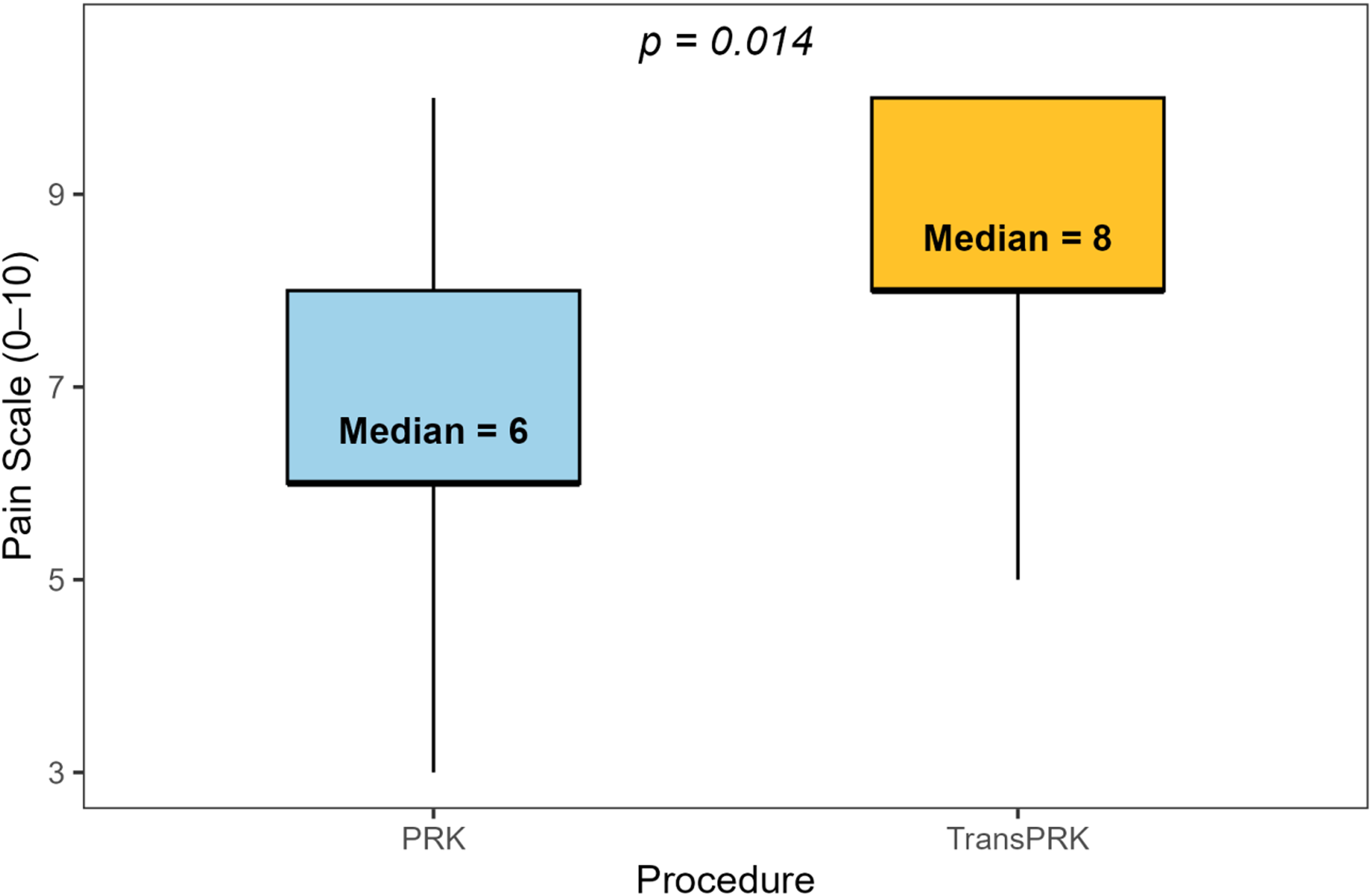
Paired boxplot showing pain score difference per patient between PRK and TransPRK eyes.
Epithelial healing and complications
There was a trend toward a difference that was not statistically significant in the rate of epithelial healing between the two groups. By day 3, the median epithelial defect area was 0.62 mm2 in the PRK group and 0.25 mm2 in the TransPRK group (p = 0.371). All eyes in both groups had achieved complete re-epithelialization by day 8. The healing time distribution was comparable between techniques: 41.7% of PRK and 50.0% of TransPRK eyes healed within 3 days, while 58.3% and 50.0%, respectively, required up to 8 days (p = 0.724).
Postoperative corneal haze was observed in 16 of the 24 patients (66.7%). Out of these patients, only 3 (12.5%) had grade 2 haze (mild haze easily visible with direct focal slit lamp illumination), while the other 13 (54.2%) only showed grade 0.5 haze (trace haze seen only by indirect, broad tangential illumination). Notably, the presence and severity of haze were identical in both eyes of every affected patient, suggesting a patient-specific predisposition rather than a technique-dependent complication.
Discussion
This prospective, contralateral eye study was conducted to enable a direct, intra-patient comparison of conventional PRK and single-step TransPRK. The results indicate that both procedures are highly effective and predictable for myopia correction, producing similar uncorrected distance visual acuity and final spherical equivalent outcomes. These findings align with the broader literature, including a meta-analysis by Alasbali et al., which reported comparable efficacy, predictability, and safety for both techniques. 8 Although some contralateral studies, such as Rymer et al., have suggested a slight advantage for mechanical PRK in visual outcomes at six months 12 , our data support the general consensus that both methods achieve excellent and equivalent refractive results.
A notable and unexpected finding of our study was the statistically higher level of postoperative pain reported in eyes treated with TransPRK. This outcome contrasts with a substantial body of evidence. Multiple studies and meta-analyses have concluded that TransPRK is associated with less postoperative pain than conventional PRK, attributing this to the elimination of manual or alcohol-assisted epithelial debridement, which can cause inflammation and irregular epithelial defects5,7,8,15. For example, Gaeckle et al. reported a median pain score of 5.0 for TransPRK compared to 11.0 for conventional PRK (on a 15-point scale) 7 , and Fadlallah et al. found a mean pain score of 2.0 for TransPRK versus 4.1 for conventional PRK (on a 10-point scale) 5 .
This divergent finding warrants careful consideration. Although the methodology for pain assessment (a 0-10 numerical rating scale) is standard, the result may have been influenced by the specific laser platform or ablation profile. It is possible that the thermal effect of the laser during the epithelial ablation phase of TransPRK could induce a greater inflammatory response in certain settings, although this remains speculative in the absence of objective inflammatory markers. Notably, a review by Chang et al. described TransPRK as potentially being the “most uncomfortable” postoperative experience among modern refractive procedures, which lends some support to our observation 16 . The power of our contralateral design, which eliminates patient-specific differences in pain tolerance, suggests that this is a genuine, technique-related effect within the context of our specific treatment parameters used.
Regarding epithelial healing, although no statistically significant difference was observed between the two groups, a numerically notable trend toward faster healing was noted in the TransPRK group. By day 3, the median epithelial defect area measured 0.62 mm2 in the PRK group and 0.25 mm2 in the TransPRK group (p = 0.371), suggesting a faster healing process in the TransPRK group. The small sample size may have contributed to the lack of statistical significance; however, this finding aligns with most studies reporting faster re-epithelialization with TransPRK5,7,8,17 with the exception of one study that reported slower re-epithelialization with TransPRK 9 .
Another notable finding was the high incidence of corneal haze (66.7%) in this cohort. Although this rate appears much higher than the 1-10% typically reported in recent literature18,19, only 3 patients (12.5%) exhibited grade 2 haze, defined as mild haze easily visible with direct focal slit lamp illumination. The remaining 13 patients (54.2%) demonstrated only grade 0.5 haze, characterized as trace haze seen only by indirect, broad tangential illumination, which is comparable to other studies. In all affected patients, the haze was symmetrical and identical in both the PRK and TransPRK eyes. This observation suggests that haze development was more likely related to the individual patient's healing response rather than to the specific surgical technique used for epithelial removal. Multiple risk factors for haze have been identified, including ablation depth, ultraviolet exposure, vitamin D deficiency, and ethnic predisposition 20 . The high prevalence of vitamin D deficiency in Qatar and other Gulf Cooperation Council (GCC) countries 21 , combined with increased ultraviolet exposure and darker iris color, may contribute to the higher rate of haze formation observed. These factors are noted as plausible contributors in the context of our cohort; however, we did not measure vitamin D levels in this study, and this association remains speculative.
Strengths and limitations
A key strength of this study is its prospective, contralateral-eye design, widely regarded as a gold standard for comparing two treatments because it minimizes confounding due to inter-patient variability. Nevertheless, certain limitations should be acknowledged. First, we did not randomize which eye received which treatment; instead, the protocol assigned PRK to the right eye and TransPRK to the left. While we did this to keep the procedure consistent across patients, it's not true randomization and could theoretically introduce a laterality effect. Second, we didn't run a formal power calculation beforehand. This was designed as an exploratory pilot study, so our sample size of 24 patients might have been too small to pick up on subtle differences in secondary outcomes like uncorrected distance visual acuity (UDVA) or how fast the epithelium healed. Because of this, results should be considered hypothesis-generating rather than confirmatory, and non-significant findings should not be interpreted as equivalence.
Third, we relied on slit-lamp measurements rather than objective imaging to estimate epithelial defect size, and we didn't use corneal densitometry to quantify haze, both of which introduce some subjectivity and are worth addressing in future work. Furthermore, having only 24 patients and the fact that only patients with low to moderate myopia were included in this study make it harder to generalize these findings to a broader population. Also, postoperative factors such as sun exposure, which may influence outcomes like haze formation, were not systematically addressed or controlled for. Finally, the Mitomycin C (MMC) protocol used in this study (0.02% for 1 minute) appears longer than commonly used in other settings for low to moderate myopia, which may further limit the external validity of our findings.
Future studies should replicate these findings in larger, multicenter contralateral trials to determine whether increased pain associated with TransPRK is consistently observed with specific platforms or represents a random anomaly. Further research into the relationships among laser ablation profiles, thermal effects, and postoperative inflammatory responses may provide valuable insights. Additionally, investigations into genetic and molecular markers that predispose patients to haze formation could facilitate personalized treatment strategies and improve the management of patient expectations. An especially promising research direction is to examine whether vitamin D deficiency constitutes a significant risk factor, as it is readily treatable and may substantially reduce patient suffering related to haze formation.
Conclusion
This contralateral eye study demonstrates that both conventional PRK and TransPRK are safe, effective, and predictable procedures for the correction of myopia, providing comparable and excellent visual and refractive outcomes. The contralateral design supports the conclusion that haze development is primarily patient-dependent rather than technique-dependent. TransPRK was associated with significantly greater early postoperative pain. These results suggest that although the “no-touch” technique of TransPRK offers theoretical benefits, its influence on patient experience is multifaceted and may be affected by specific procedural parameters.
Supplemental Material
sj-docx-1-ejo-10.1177_11206721261461914 - Supplemental material for Clinical outcomes of conventional Vs. transepithelial photorefractive keratectomy: A prospective contralateral eye study
Supplemental material, sj-docx-1-ejo-10.1177_11206721261461914 for Clinical outcomes of conventional Vs. transepithelial photorefractive keratectomy: A prospective contralateral eye study by M. Basil Marchi, Adeya Al Harami, Ahmed Maher, Omar Al Qahtani, Abdulaziz Al Qahtani, Joenie Anilao, Shanudheen Chekkumpadykunnu and Hashem Abu Serhan in European Journal of Ophthalmology
Footnotes
Author contributions
Conceptualization: M.B.M., A.A.H., H.A.S.; Methodology: M.B.M., A.A.H., A.M., H.A.S.; Data curation: O.A.Q., A.A.Q., J.A., S.C.; Formal analysis: M.B.M., A.M., H.A.S.; Investigation: O.A.Q., A.A.Q., J.A., S.C.; Writing—original draft: M.B.M., A.A.H.; Writing—review & editing: A.M., O.A.Q., A.A.Q., J.A., S.C., H.A.S.; Supervision: H.A.S.; Project administration: H.A.S. All authors reviewed and approved the final manuscript.
Funding
The authors received no financial support for the research, authorship, and/or publication of this article.
Declaration of conflicting interests
No conflicting relationship exists for any author.
Supplemental Material
Supplemental material for this article is available online.
References
Supplementary Material
Please find the following supplemental material available below.
For Open Access articles published under a Creative Commons License, all supplemental material carries the same license as the article it is associated with.
For non-Open Access articles published, all supplemental material carries a non-exclusive license, and permission requests for re-use of supplemental material or any part of supplemental material shall be sent directly to the copyright owner as specified in the copyright notice associated with the article.