






Abstract
Introduction:
The aim of this study was to compare the accuracy of preoperative templating in total hip arthroplasty (THA) using conventional 2-dimensional (2D) and computed tomography (CT)-based 3-dimensional (3D) measures.
Methods:
One hundred and sixteen consecutive primary THAs were analysed. The preoperative diagnosis was primary osteoarthritis in all cases. The 2D templating and the 3D templating were performed by two different residents. All templating results were available for the orthopaedic surgeon performing the procedure. Accuracies with regard to the predicted and actual implant sizes were determined for each procedure. Implantation of the size as planned was defined as “exact”, whereas the use of components within one size larger or smaller (±1) as planned were defined as “accurate.”
Results:
The 3D templating was significantly more accurate in predicting implant sizing compared to 2D templating for primary total hip arthroplasty (THA). The difference was statistically significant for the cup templating (‘‘exact’’ p = 0.02; ‘‘accurate’’ p = 0.01) and for the stem templating (‘‘exact’’ p = 0.04; ‘‘accurate’’ p = 0.01).
Conclusion:
Our results support the superiority of 3D templating over 2D templating in predicting implant size.
Introduction
Appropriate preoperative planning in total hip arthroplasty (THA) helps to reduce complications and its importance for implant selection as well as orientation of components has been documented previously.1–8 Conventional templating is based on plain radiographs. However, individual patient anatomy is not always illustrated reliably on conventional radiographs, and crucial parameters such as the anticipated diameter of the femoral canal may differ significantly with rotation of the hip. 9 Preoperative planning using computed tomography (CT) for 3-dimensional (3D) templating has been developed to improve the placement of components and promising applications have been reported.4,5 It aims to select the best fitting implant and to improve canal fill to maximise metaphyseal engagement. Ideal fitting is achieved if the size of the implanted stem optimises the proximal femoral fit-and-fill, thus maximising the stem–bone contact area. 10 For best initial stability the prosthesis requires a precise fit in the local bone stock of the proximal femur to restore the individual hip geometry. 11 However, 2-dimensional (2D) templating still remains the gold standard technique in clinical practice and current literature.1,3,12
The purpose of this study was to assess and compare the accuracy of preoperative templating for 5 different stem designs and 1 cup using 2D and 3D techniques. The hypothesis of this study was that 3D planning using the software ZedHip (Lexi, Japan) is more accurate than the conventional 2D templating technique regarding the prediction of implant size.
Methods
In this retrospective study 116 patients (40 men and 76 women) who underwent primary cementless THA between July 2014 and July 2016 were included. The mean age of the patients was 69.2 ± 9.9 years (range 50–91 years). The appropriate ethics committee approved this study. The preoperative diagnosis was primary osteoarthritis in a non-dysplastic hip for all cases. There were 39 patients with a flexion contracture of the hip. THA was performed using 5 different femoral stem components with different caput-collum-diaphyseal (CCD) angles: the Fitmore short stem (Zimmer, Neu-Ulm, Germany), the SPP II anatomical stem (LINK, Hamburg, Germany), the MIA stem (Smith & Nephew, London, UK), the Alloclassic stem (Zimmer, Neu-Ulm, Germany) and the Avenir stem (Zimmer, Neu-Ulm, Germany). In all hips the Allofit pressfit cup (Zimmer, Neu-Ulm, Germany) was used. For optimal press-fitting, the cavity was under-reamed by 2 mm in relation to the peripheral dimension of the cup. A load-bearing, stable acetabular floor and solid lateral bony tissue were desired, as an extensively preserved osseous circumference of the acetabulum is a precondition for primary stability of the cup. The aim is to create an anatomically shaped acetabular implant bed so that the cup is gripped by bone on all sides and anchored in well-vascularised bone. This establishes the requirements for primary and secondary stability.13,14
70 procedures were performed by AK (n > 1000 THA), 18 procedures by TK (n = 500–1000 THA) and PK (n < 500 THA) completed 28 procedures. An anterolateral approach (Watson-Jones) for total hip arthroplasty was used in all cases. In all patients Rippstein I (anteroposterior [AP] view of the hip with the patient supine and knees flexed at 90° over the X-ray table in a defined position) views of the pelvis, lateral view of the hip (magnification marker 32 mm) and low-dose CT scans. CT scans had a field of view from the femoral head to the ankle and were saved in DICOM format. The primary slice direction was axial with a slice pitch of 2 mm and a 0° tilt of the gantry. All radiographs and CT scans were performed using the same calibrated equipment.
All patients consented to participate in this study knowing the main goal of this study being to compare templating methods. The intraoperative selection of implant size was based on the surgeon’s judgment, joint stability, leg length, and intraoperative X-ray examination aiming to reconstruct patient’s individual anatomy.
A day before surgery resident A (post-graduate year [PGY] 4, n < 500 THA) planned the arthroplasty using the 2D software (HECTEC, Altdorf, Germany) based on the radiographs. Based on the CT-images another resident B (PGY 4, n < 500 THA) performed 3D planning using the 3D ZedHip software (Lexi, Japan) in all patients. All templating results were available for the surgeon performing the procedure.
The 3D software enables the surgeon to navigate the prosthetic components into the desired position based on the 3D images (Figure 1). 8 reference points (femoral head, piriformis fossa, the most posterior point of the proximal femur, bilateral femoral condyles, bilateral posterior condyles and knee centre) were used at the femoral side. At first, stem anteversion and valgus angle were adjusted to match anatomical femoral neck anteversion and shaft axis. Moreover, the size of the femoral stem was determined to fill both the medial calcar and the lateral flare of the proximal femur as much as possible. Intraoperatively the neck osteotomy plane was selected in order to reconstruct the correct leg length and femoral offset based on radiographs. Then, the level from top of the greater trochanter was measured to ensure accurate positioning during surgery. The acetabular component size was chosen to meet the anterior and posterior acetabular walls in the orientation determined by 3D templating.
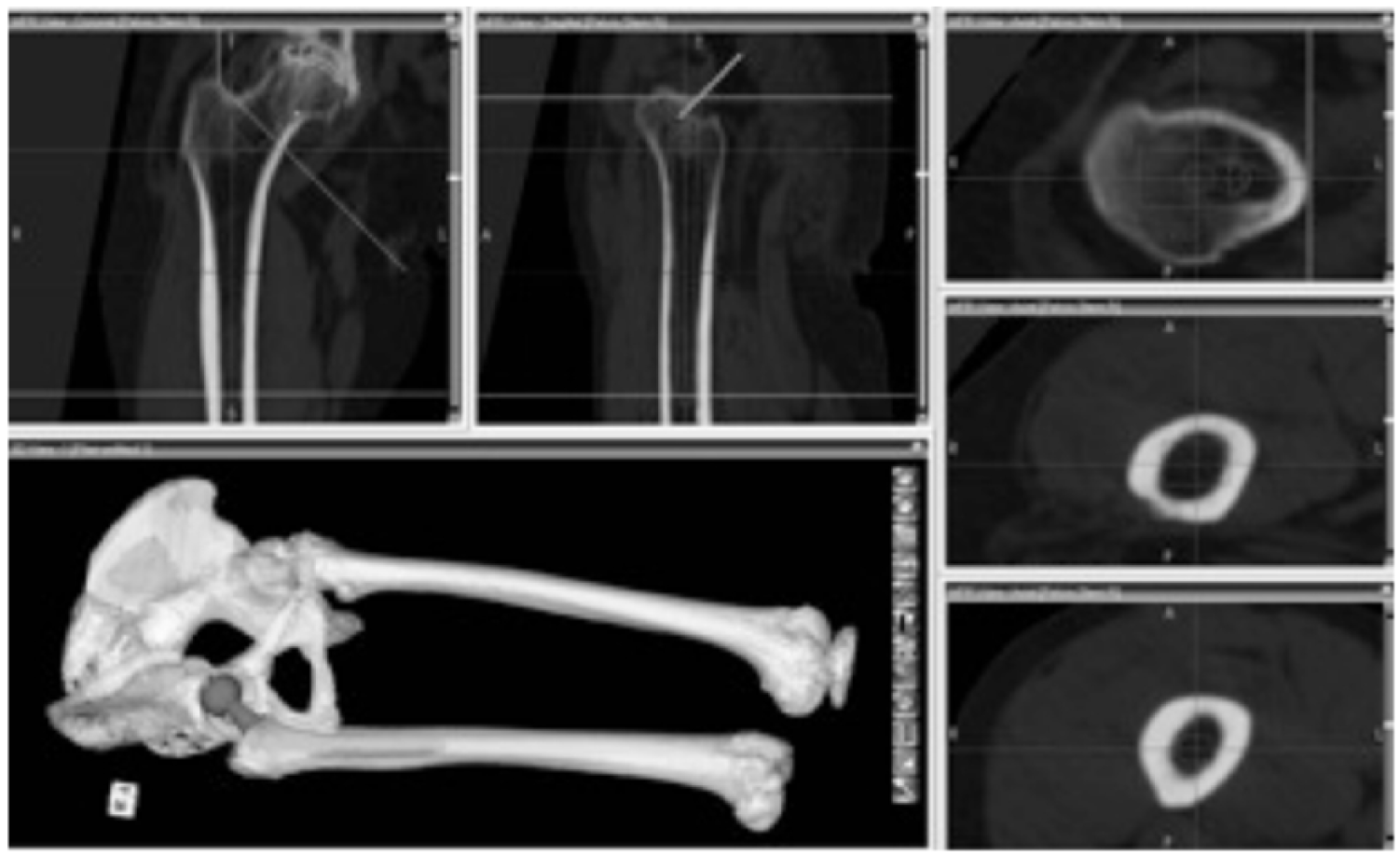
A Photograph of 3D templating ZedHip software (Lexi, Japan).
The mean effective dose for CT scanning was found to be about 11.6 mSv (9.8–14.0), which is equivalent to 5 plain radiographs each with an effective dose of 2.3 mSv. The additional cost created by the CT scan was €53–116 (German Scale of Medical fees) for each patient.
For postoperative evaluation a Rippstein I view was performed about 2 weeks after surgery. We checked that the stem fills or nearly fills the femoral canal on AP radiographs and has cortical contact in specifically determined areas. Final stem size and the final cup size were evaluated from records. Implantation of the size as planned was defined as “exact” (fit and fill the determined area), whereas the use of components within ±1 size as planned were defined as “accurate”.6,14,15
Statistics
For statistical analysis Kruskal-Wallis non-parametric test followed by the post-hoc Dunn’s test using SPSS software pack (version 23, IBM, New York, USA) was used. P value < 0.05 was considered for statistic significance.
Results
Surgical procedures
There was no difference in selection of implant type between 2D and 3D templating. In all cases the type of implant was used as planned. No intraoperative or postoperative complications were observed. AK implanted in 55 of 70 cases (85.7%), TK in 15 of 18 cases (83.3%), and PK in 21 of 28 cases (78.5%) the final stem within ±1 size as preoperatively determined by 2D templating.
In 61 of 70 cases (92.9 %) AK, in 17 of 18 cases (94.4%) TK, and in 27 of 28 cases (96.4%) PK implanted the final stem within ±1 size as preoperatively determined by 3D templating.
2D planning
The size of the femoral stem matched the planning exactly in 45.7% and was accurate in 83.6% of cases. The cup sizes matched the planned size in 44.8% and were accurate in 80.17% (Figures 2 and 3).

Comparison templating cup size (*significant), ± 1 SD.

Comparison templating stem size (*significant), ± 1 SD.
3D planning
The stem size corresponded exactly to the planned size in 58.6 % of the cases. 94 % of the femoral stem size estimates were accurate. In 56.9 % of all cases the final acetabular cup size corresponded exactly to the preoperatively planned size. In total, 86.2% of the cup size estimates were accurate ± 1 size. (Figures 2 and 3)
Overall, 3D templating was significantly more accurate in predicting component size compared to 2D templating for primary total hip arthroplasty (THA). The difference was statistically significant for the results of cup templating (‘‘exact’’ p = 0.02; ‘‘accurate’’ p = 0.01) and for the results of stem templating (‘‘exact’’ p = 0.04; ‘‘accurate’’ p = 0.01).
The mean time for 2D templating was 12 minutes (range 8–23 minutes) using the 3D templating method the mean time was 17 minutes (range 10–25 minutes, p < 0.05).
Further analysis of the data demonstrated a tendency in 2D and 3D templating to overestimate the cup sizing (Figure 4). For the stem size there was no tendency.
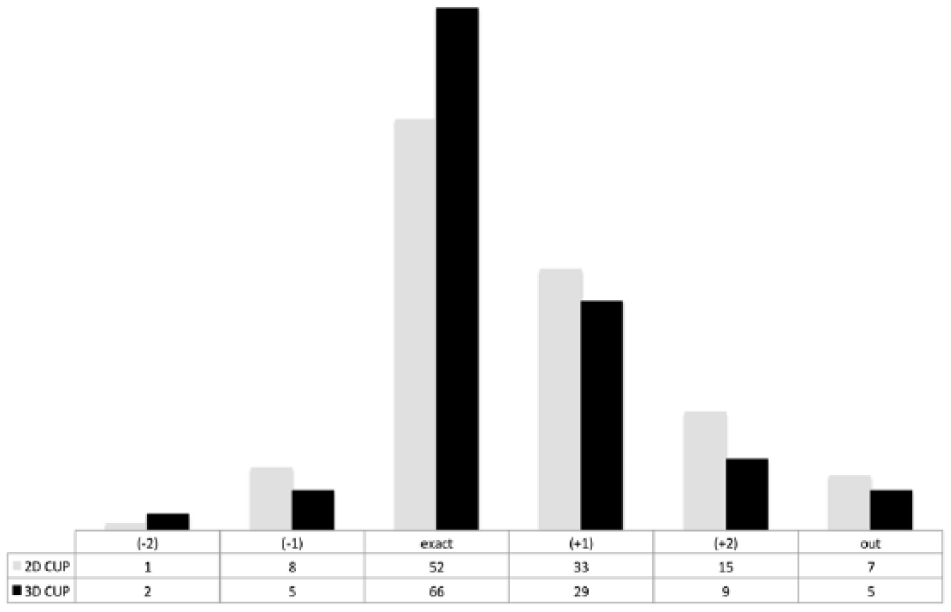
Error from implanted cup size (positive values indicate larger component selection).
Discussion
Preoperative planning is an essential part of hip joint reconstruction. It reduces operative time and minimises surgery related complications.8,16–19 Previous studies have evaluated the accuracy of templating using the 2D planning methodology.1,4,6,20,21 However, 3D planning has also been used in primary THA with superior accuracy compared to 2D templates.4,22 Eckrich et al. 9 reported that the prediction rate of templating is related to the intrafemoral anatomy and the final cranio-caudal blockage level of the stem cannot be accurately predicted without a 3D volume of the femur.
The main finding of this study was that by using 3D templating software ZedHip (Lexi, Japan) the accurate prediction of stem and cup size is improved significantly compared to 2D surgical planning (86.2% vs. 80.1%, p = 0.01 for the cup and 94% vs. 83.6%, p = 0.01 for the stem). In this study there were no differences between the selected type of implant between 2D templating and 3D templating. Both planning methods are equally effective in selecting the type of stem and cup. The current results for 2D templating are in agreement with other studies.6,23–25 However, variability of femoral neck anteversion and rotational contracture of the hip may make the 2D templating method inaccurate. 7 The lack of accuracy for the 2D templating may be due to inaccurate appreciation of that the hip anatomy on plain radiographs, especially for the femur. The preoperative prediction of implant size of the cup and the femoral component was improved significantly using 3D CT templating compared to 2D templating in our study and compared well with the results reported in literature.2,4,5,26 Hassani et al. 2 evaluated the accuracy of 3D preoperative planning in primary cementless THA. They reported that the implant size was correctly predicted in 100% of the stems and 94% of the cups. In a prospective randomised study, Sariali et al. 4 compared the accuracy of 3D preoperative templating using CT scanning to 2D templating. Patients were divided in 2 groups: in 1 group, planning was made on calibrated X-rays using 2D templates (n = 30). In the other group, a CT-scan based 3D computerised planning (n = 30) was performed with dedicated software. A relatively inexperienced surgeon performed all the surgical procedures. In the 3D group, accuracy in predicting the final sizing was twice as high; 96% for the cup and 100% for the stem. As a limiting factor for this study, the implanted stem type was different between the 2D templating and the 3D templating group.
Inoue et al. 15 reported that preoperative 3D templating was able to predict the stem size exactly in 65% of cases and ranged within ±1 size in 98% of cases. The final cup size corresponded exactly to the preoperatively planned size in 92% of cases. 100% of the cup size estimates were within ±1 size. Our results are in accordance with these findings and we include more patients.
Despite the promising results for the accuracy of 3D templating there may still be potential for improvements regarding the prediction of stem and cup sizes. Still, preoperative planning relies on subjective decision of the examiner, which may lead to inaccuracy. Hsu et al. 27 evaluated the effects of training level on the accuracy of digital templating for primary THA and total knee arthroplasty (TKA). An implant sales representative, physician assistant, medical student, resident, and fellowship-trained arthroplasty surgeon templated all cases independently after a standardised orientation and were blinded to the actual component sizes used for surgery. The medical student, resident, and arthroplasty surgeon re-templated the same 97 cases 1 month later to determine intra-observer reliability. There was no difference in quality of templating between different levels of training. However, contradictory results regarding the influence of experience, familiarity with the templating software and reproducibility of preoperative 3D templating in primary THA were reported by other authors.27–29 In this study the 2 planning examiners were residents (PGY 4, n < 500 THA) and were experienced with the 2D and the 3D software used. In this study none of the examiners planning the templating performed the hip arthroplasty.
To restore normal hip biomechanics, it is essential that the cup used to fit the acetabulum is of the correct size and also placed in the correct position. 14 In this study a tendency to overestimate the cup size was observed. A reason to select a larger cup is to prevent acetabular loosening due to poor bone quality.15,30
Even though the applied radiation is 5 times higher for 3D planning compared to 2D planning, the dose of 11–12 mSv is far beneath the reported dose related to an increase in cancer risk (200–5000 mSv). These results are comparable to other studies.5,31 In clinical use, only low-dose CT should be performed for the preoperative 3D planning. Henckel et al. 32 reported that specific protocols combining filters and image post-processing on multiple detector helical CT scan may reduce the radiation dose to a level comparable to standard radiographs.
We found that 2D templating was a faster templating method when compared to 3D templating. On average, 2D templating was 5 minutes quicker. To our knowledge, this is the first study to evaluate time as an outcome measure for 2D and 3D templating.
3D templating increases the overall costs. However, Huppertz et al. 31 reported that preoperative CT for THA is associated with slight increases in costs per-patient. This is justifiable because the CT reveals a higher accuracy and may improve patient outcome and reduce revision rates.
A further strength of our study is the large and homogenous population of patients. One limitation of this study is its retrospective nature, and the surgeons not being blinded to the results of the preoperative templating. However, for practical and ethical reasons the surgeon were aware of all available information on patient characteristics and anatomy preoperatively. Sizing of components is only one part of preoperative planning especially in difficult cases so another limitation of this study is no measurement of femoral offset, cranio-caudal position of the stem, femoral head centre, and cup position, centre of rotation and leg length using 3D imaging methods were not performed in current study. However, this was not scope of this study and may be subject of further evaluations.
Conclusion
This study demonstrates that 3D preoperative templating with the software ZedHip (Lexi, Japan) is an accurate and reproducible process for the orthopaedic surgeon planning primary THA. It allows a significantly higher accuracy in predicting implant sizing compared with conventional 2D templating for THA in primary osteoarthritis. However, the higher accuracy may not be associated with better clinical outcomes. Further studies are needed to analyse the clinical relevance.
Footnotes
Declaration of conflicting interests
The author(s) declared no potential conflicts of interest with respect to the research, authorship, and/or publication of this article.
Funding
The author(s) received no financial support for the research, authorship, and/or publication of this article.