






Abstract
Eponyms, while inherently flawed, remain a constant in medical vernacular, especially in orthopaedic surgery. It is essential to understand how these eponyms came to be named and for whom they were named after in order to understand their correct usage and definitions. In this second part of a 2-part review, we describe the history of eponym usage in the radiography of the adult hip; who, when, what, where, and how. We hope to provide a historical perspective of interest, resolve any controversies in semantic definitions, and create a comprehensive library of eponymous terms related to adult hip radiography.
Keywords
Background and introduction
The definition of an eponym is as follows; “(1) one for whom or which something is or is believed to be named”, or “(2) a name (as of a drug or a disease) based on or derived from an eponym”, based on Merriam-Webster. 1 Throughout its history, medicine, particularly orthopaedic surgery, has used eponymous terms to describe common presentations, often attributed to the first person to describe said pathology. Eponyms continue to be frequently used in orthopaedic surgery,2,3 including but not limited to describing wrist to pelvic fractures, approaches for hip surgery, and even orthopaedic instruments.4–7 While the accuracy and validity concerning the usage of such terms have often been questioned,8,9 eponyms remain a prominent part of daily practice. Thus, it is essential to remember from where, when, and whom we obtained these terms.
This review highlights eponymous terms of hip and pelvis radiography, with this second part focusing on adult pathologies, such as dysplasia, femoroacetabular impingement, and osteoarthritis radiography. An excellent review by Clohisy et al. 10 describes technical points on several of these radiographic views, but who were these techniques named after?
Adult hip radiographic eponyms
Shenton’s line
Edward Warren Hine Shenton (12 July 1872–12 October 1955) was an English radiologist beginning his medical studies at the famed Guy’s Hospital in London in 1893. 11 While he was at medical school, William Roentgen invented roentgenographs on 08 November 1895, and Shenton and a fellow medical student, AHB Kirkman, having been appointed radiographers at Guy’s Hospital in 1899, immediately adopted their use.11,12 They both presented the practical application of Roentgen rays in medicine, published in Guy’s Hospital Gazette in 1899. Notably, Shenton did not qualify for his medical license until 1901. Hence, as a medical student, he developed the radiology department at Guy’s Hospital and published a radiography system in his Day Book in 1900. 13
Perhaps most famously, Shenton11,14 published his textbook entitled “Disease in Bone: And Its Detection by the X-Rays” in 1911. In this book, to which another famed English contemporary radiologist, Robert Bayley Osgood (who was born and passed 1 year after Shenton14–16) was a contributor, along with several orthopaedic surgeons, Shenton described a projected arc from the inferior border of the femoral neck to the superior border of the obturator foramen (Figure 1). Shenton,12,14 who described himself as a “Senior Surgical Radiographer,” actually described a break in said line to be helpful in the early detection of tuberculosis affecting the hip joint. He said, “. . .I found a line, the broken continuity of which infallibly indicates displacement or trouble in the hip region”.12,14 Shenton’s line, or arc, has held up in diagnostic validity even 100 years later and remains a critical radiographic marker for paediatric and adult hip pathology. 17 Shenton resigned from Guy’s Hospital in 1919, not for retirement, but due to his work for the Royal Air Force Central Hospital, for which he received the Red Cross gold medal, and in his later career worked in his practice in Harley Street and as an honorary radiologist to both St. Peter’s Hospital in London and St. Bartholomew’s Hospital in Rochester.11,12 Outside of the medical field, Shenton held many hobbies that aligned with that of the surgeons he closely worked with, including violin making (which he accompanied with composing several songs) and boat building, handcrafting a 10-ton yacht in his home of Golders Green.11,12 Nevertheless, he did not retire, due to his passion for work, until age 78, passing at age 83.
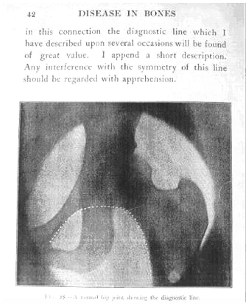
Edward Shenton’s original roentgenograph of his line from the inferior border of the femoral neck to the superior border of the obturator foramen.
Dunn and modified Dunn view
Denis Michael Dunn (25 February 1916–08 March 2001) was an English orthopaedic surgeon interested in the hip, not to be confused with another English orthopaedic surgeon Naughton Dunn (1884–1939). 18 He was born in Newcastle-upon-Tyne, attended Durham University, and then undertook his medical studies at the medical school of Newcastle-upon-Tyne (now Newcastle University Medical School) in 1938. 19 As a pacifist, he did not serve in the English Armed Forces in WWII, instead training with several renowned orthopaedic hip surgeons such as Joseph Trueta at Oxford, and Sir Reginald Watson-Jones at the London Hospital, eventually settling into his role at the Colchester and District Hospital in Colchester, England. 19
It was in Colchester when his interest in hip pathology and deformity, in particular, that of slipped upper femoral epiphysis (SUFE), led to his development of a new radiographic view to better assess the anteversion of the proximal femur and more accurately identify proximal deformity, which he published in the Journal of Bone and Joint Surgery, British Volume in 1952. 20 In this paper, he identified the difficulty of accurately assessing proximal femur anteversion using standard frog-leg or cross-table laterals, and, using a model of a 3-year-old child’s femur, determined the most accurate limb positioning to be 90° of hip flexion, 15° of hip abduction, with the roentgenograph beam positioned midline over the pubic symphysis. 20 To be most precise, Dunn reported 0° of abduction, but for patient positioning and decrease of soft tissue overlap determined up to 20° abduction to be accurate. He found that increasing abduction angles underestimated femoral anteversion (45° of hip abduction made a 90° anteversion appear to be 55°) and hip flexion more significant than 90° underestimated anteversion. In contrast, hip flexion <90° overestimated anteversion (Figure 2). Of note, Dunn made no mention of head-neck offset or the morphology of the head-neck junction in this seminal publication.
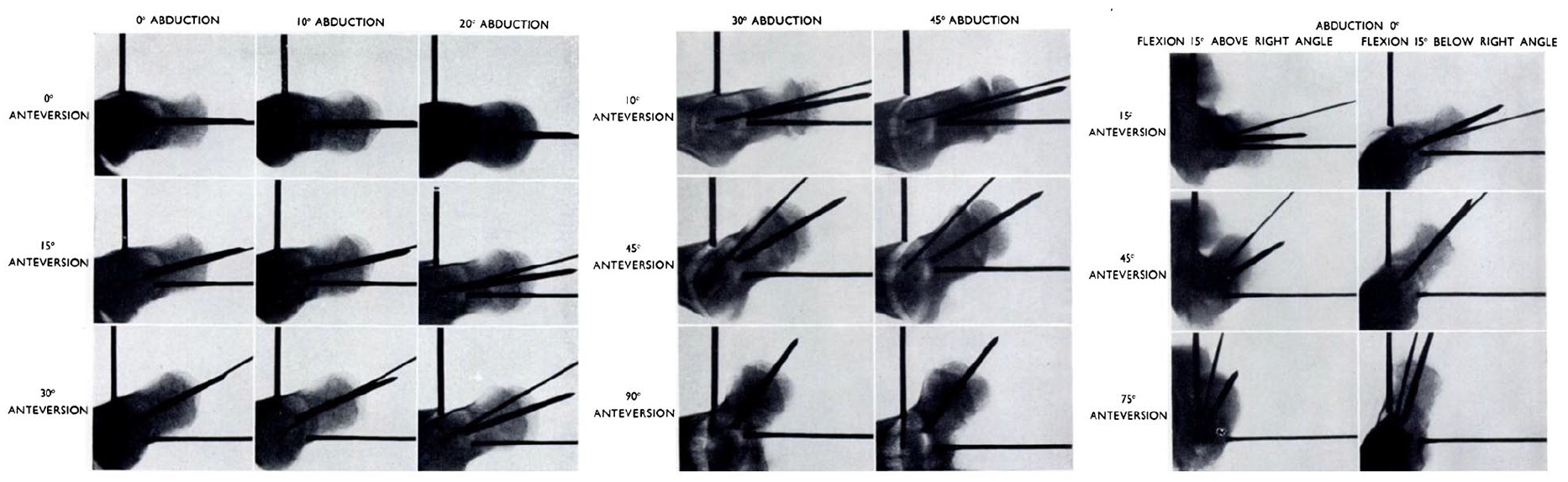
Denis Dunn’s radiographs of a model 3-year-old proximal femur in various positions of hip flexion and abduction to determine the most accurate measurement of anteversion.
More recent studies, in particular by Eijer et al., 21 have modified Dunn’s view to 45° of hip flexion, and have additionally identified the ability to assess proximal femoral head-neck offset, and with an increasing background knowledge of femoroacetabular impingement (FAI) and proximal femoral morphology, used these views to assess cam deformities of the proximal femur.10,21,22 1 small note in Clohisy et al.’s 10 review, and a common misconception, is that the Dunn view should be performed at 20° of hip abduction. In contrast, Dunn’s original publication describes hip abduction at 15°, although the clinical significance of this discrepancy is likely minor.
Dunn’s contributions to orthopaedics spanned beyond radiography, describing an open treatment for SUFE of severe chronic slips with open epiphysis, in which the retinacular vessels have already shortened. In his 1964 publication, he described his cervical proximal femoral osteotomy, removing a “trapezoidal rather than cuneiform [segment of bone] and includes the posterior bony beak” (Figure 3).23,24
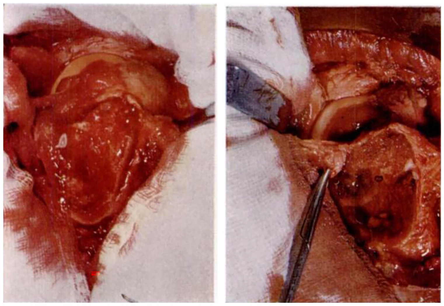
The Dunn procedure for the treatment of severe chronic slips from his original 1964 publication, with the left figure demonstrating a severe chronic slip and the right figure demonstrating an acute on chronic slip.
Lateral centre-edge angle of Wiberg
Gunnar Wiberg (1902–08 September 1988) was a Swedish orthopaedic surgeon born in Gothenburg, Sweden. He studied medicine at the Karolinska Institute in Stockholm, rejoining the faculty after his surgical residencies in 1937. 25 During his time there, he published 2 theses on hip dysplasia in 1939 and patellofemoral chondromalacia in 1941 that led to international renown.26,27 This led to his recruitment to the department of orthopaedics at Lund University as an associate professor in 1944. He became chairman within the year, a post he held until his retirement in 1969. 25
Wiberg published his thesis on the dysplastic acetabulum and congenital subluxation of the hip joint in 1939 in Acta Chirurgica Scandinavica from the Karolinska Institute, with perhaps its most famous takeaway being the now eponymously named lateral centre-edge angle (CEA) of Wiberg. 26 In his original description, he took anteroposterior (AP) roentgenographs of a pelvis and, using 2 concentric circles around the femoral head and acetabulum, quantified the lateral coverage of the acetabulum roof (Figure 4). Wiberg’s 27 thesis on the patellofemoral joint utilised anatomical dissections and many roentgenographs to study 158 “femoropatellar” joints in 101 patients to investigate both patellar dislocations and chondromalacia (Figure 5). He grouped the patellas into 3 groups, which has come to be known as the eponymous Wiberg Classification and been cited numerous times.28–30
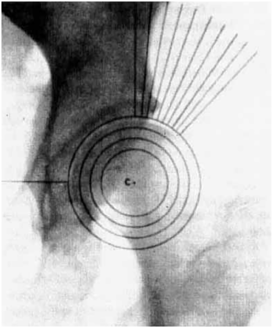
Gunnar Wiberg’s original roentgenograph in his 1939 thesis demonstrating his technique for measuring lateral center edge angle using concentric circles around the femoral head and acetabulum.
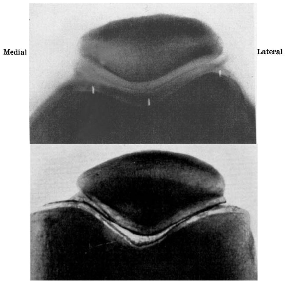
Wiberg’s roentgenographs of the patellofemoral joint with arthrogram (top panel) and with red lead preparation (bottom panel) demonstrating his type I patella.
Wiberg’s contributions were vast, and he was a board member of the Scandinavian Orthopaedic Association and President of the Swedish Orthopaedic Society from 1960 to 1966. However, perhaps none of his accomplishments spoke to his character as much as his campaign to transfer the care of fractures from general surgeons to orthopaedists. Bauer, 25 as secretary of the Swedish Orthopaedic Society at that time, witnessed the signing between Wiberg and Stig Lindgren, a general surgeon, in the Nobel Foundation building in Stockholm of this momentous shift in fracture care, but Wiberg’s background in general surgery training also led to his semi-ironic quote on handling the poly-traumatised patient as an orthopaedist; “We should handle the abdomen without any help from the [general] surgeons”.
2 present day titans and a question
False profile view and anterior centre-edge angle of Lequesne
Michel G Lequesne (20 December 1924–current) is a French rheumatologist born in the Neuilly-sur-Seine district of Paris, France. After his medical training in the Hôpitaux de Paris, he obtained his medical degree in 1954 and his board certification in Rheumatology in 1961. 31 It did not take long for him to make his mark in hip arthritis. His original publication in 1961 on a new false profile view of the pelvis to better evaluate hip dysplasia and osteoarthritis was widely adopted in France, followed by the remainder of Europe and the United States.32,33 Lequesne’s original publication described the “faux profil” technique as the ipsilateral foot axis parallel to the radiographic table with the hip in contact with the plate. The pelvis and body are then rotated 25° backward to profile the antero-superomedial edge of the acetabulum, leaving the patient’s back at an angle of 65° to the radiographic plate, with the beam 110 cm away (Figure 6).32,33 A few years later, Lequesne34–36 published 2 articles in 1963 and 1964 detailing his technique of measuring all radiographic parameters of hips, including a new vertical-centre-anterior (VCA) angle on his false profile view of the hip, which is now referred to as the anterior CEA.
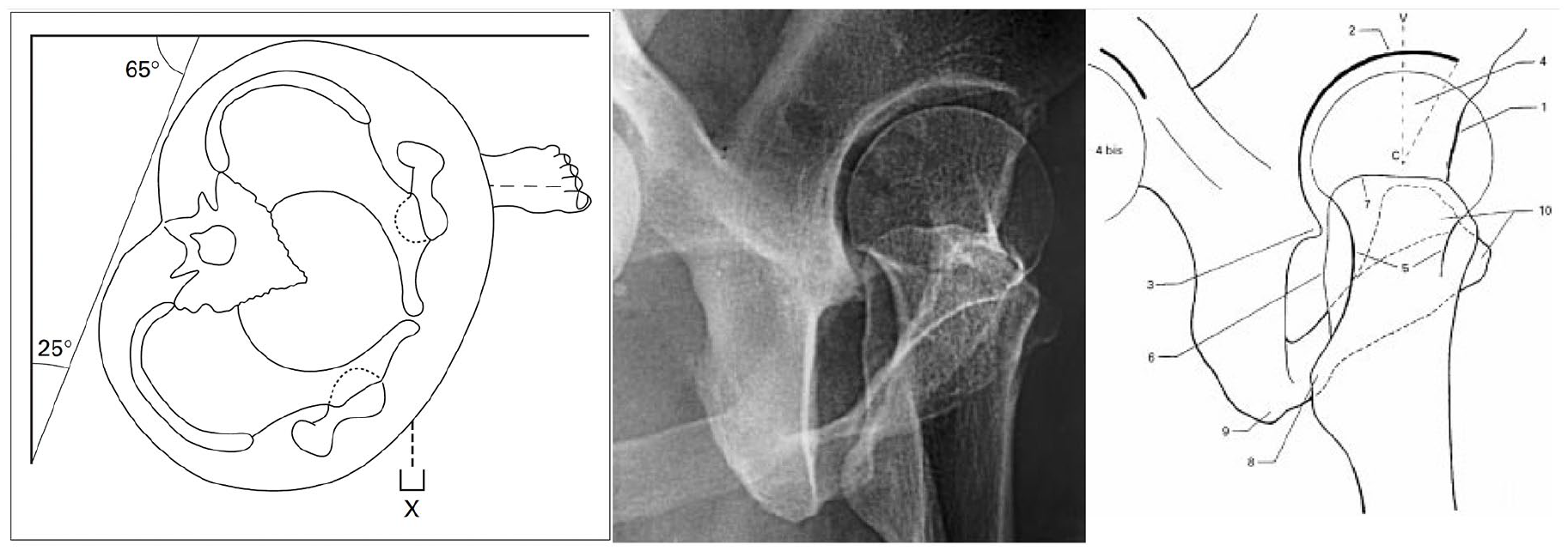
Michel Lequesne’s original diagram and updated radiograph demonstrating: (left panel) cross-sectional diagram of the pelvis demonstrating the 25-degree posterior rotation of the pelvis with the ipsilateral foot parallel to the radiograph plate, (middle panel) a normal radiograph of Lequesne’s “faux profil” view, and (right panel) Lequesne’s schematic labeling anatomic landmarks of the false profile view.
Lequesne has continued his prolific work in hip arthritis and rheumatology, publishing over 200 peer-reviewed papers, multiple textbooks, and over 50 lectureships in his career. His other contributions included developing the Lequesne37,38 algofunctional indices for hip and knee osteoarthritis in 1987 which he modified himself in 1991 to include morning stiffness, as well as serving on several large recent clinical trial groups validating the use of diacerein, an interleukin-1beta inhibitor, and Piascledine, an avocado-soybean unsaponifiable-Expanscience, in the treatment of osteoarthritis.39,40 His many accomplishments earned him the President of the European League against Rheumatism (EULAR) from 1987 to 1989. 31 He served as the chief of service of Rheumatology at the Hôpital Léopold Bellan in Paris until his retirement.
Alpha angle of Nötzli
Hubert Nötzli is a current Swiss orthopaedic surgeon who is the chief of orthopaedics at Sonnenhof Hospital in Bern, Switzerland, although little is known of his private life. He is a disciple of Ganz et al.41–43 and has published over 40 peer-reviewed articles, specialising in the hip, and in particular FAI. He is best known for describing the alpha angle, a method for quantifying an aspherical femoral head with cam deformities in FAI.
Nötzli et al. 44 published his method for quantifying the femoral head-neck junction using axial cuts oriented along the femoral neck axis of a 1T MR arthrography unit in The Journal of Bone and Joint Surgery-British Volume. He found the apex of the concavity of the femoral neck to be a point where the distance from the bone to the femoral head center first exceeds the radius of the femoral head. The angle between a line from this point to the head center and a line along the axis of the femoral neck was defined as the alpha angle (Figure 7). 44
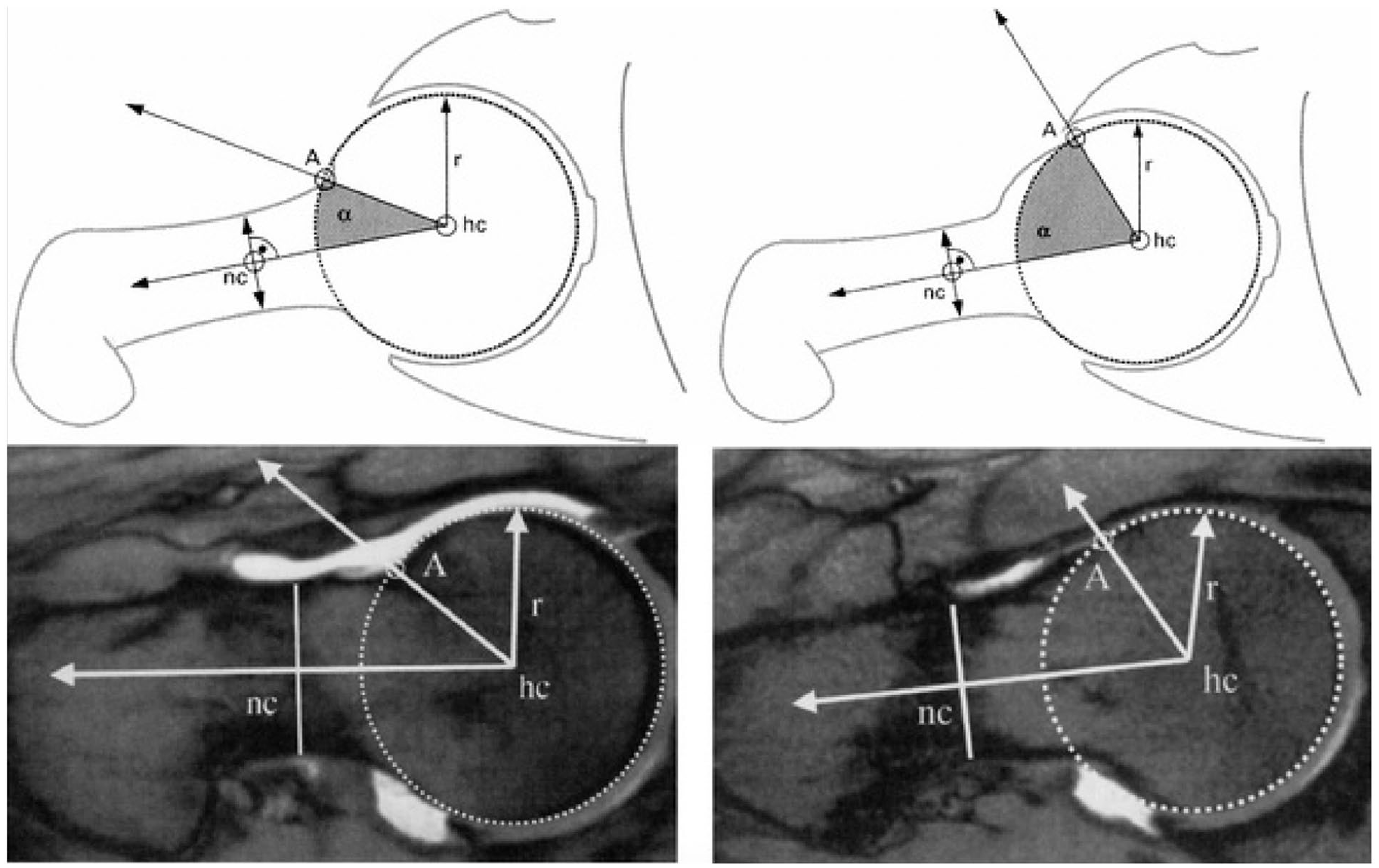
Hubert Nötzli’s method of calculating the alpha angle to quantify the asphericity of the anterior femoral head-neck junction (top panel), with the measurement demonstrated on 1T MRI (bottom panel).
Who coined Köhler’s line?
Köhler’s line is an eponymous term for the ilioschial line, representing the posterior column of the acetabulum and a confluence of cortical densities on the AP pelvis radiograph. Disruption of this radiographic line indicates posterior column disruption, and coxa profunda and acetabuli protrusion are defined by the medial wall of the acetabulum and femoral head being medial to Köhler’s, respectively. With such a ubiquitous eponym used in arthroplasty, trauma, hip preservation, and paediatrics, one would expect a well-described attribution of this eponym, yet there are no discernible reports as to where this line received its name.
There was a prominent German radiologist named Alban Köhler (01 March 1874–26 February 1947) whose career aligned with the rise of roentgenographs and who quickly became a large proponent. 45 Within the first 15 years of Roentgen’s invention, Köhler had already published 3 textbooks on the subject, on the diseases of bones in 1901, the hip joint and femur in 1905, the diagnosis of pulmonary tuberculosis in children in 1906, and his mea culpa, the “Radiologist’s Bible,” “The Borderlands of the Normal and the Early Pathological Findings in Skeletal Radiography” in 1915, which continues to be published to this day.45–47 His publication of a proximal calcified protrusion of the medial femoral condyle in 1905 led to Pellegrini and Stieda’s popularisation of the Köhler-Pellegrini-Stieda lesion. 48 His discovery of a new disease in children of avascular necrosis of the navicular in 1908 led to the eponym Köhler’s disease. However, newer literature has shown his initial roentgenograph to be incorrect in its diagnosis (Figure 8).49,50
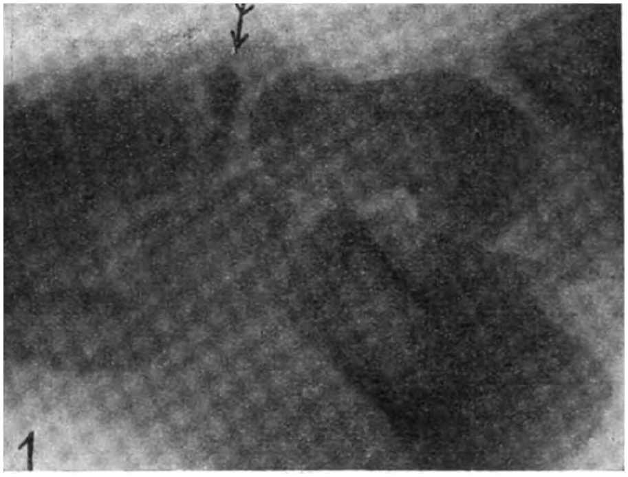
Alban Köhler’s original roentgenograph of his discovery of avascular necrosis of the navicular in a child “Erkranktes Os naviculare”.
Moreover and lastly, in his 1915 publication of the “Typical Disease of the Second Metatarsophalangeal Joint,” in which he reported on “a most peculiar disease. . . at the 2nd metatarsophalangeal joint. . . an infraction- or fracture-like intermediary space. . .” and called out Freiberg for his incomplete description has led to the eponym Freiberg-Köhler disease or Köhler disease II.45,51 Despite these notable accomplishments, there is no mention of his description of the ilioischial line. The authors thus theorise that this eponym arose from 1 of his earlier publications in 1901 or 1905.
Conclusion
Along with the eponyms of radiography of the paediatric hip, which can be found in part 1 of this 2-part review, the purpose of this review was threefold. First, with the common usage of eponyms in orthopaedic surgery on the wards and in conversation, it is imperative to standardise the definitions and the context in which we use them. The second purpose was to compile the history of these pioneers in orthopaedic surgery and radiology who have expanded our knowledge of radiography of the adult hip into 1 manuscript. Thirdly, it is essential to acknowledge and understand where we as orthopaedic surgeons came from and whose shoulders we stand on to advance the field further.
Footnotes
Declaration of conflicting interests
The author(s) declared no potential conflicts of interest with respect to the research, authorship, and/or publication of this article.
Funding
The author(s) received no financial support for the research, authorship, and/or publication of this article.
