






Abstract
Introduction:
As a countermeasure against the organized thrombi frequently observed in vascular access occlusion, we devised a percutaneous excision method for organized thrombi under superficial ultrasound guidance using biopsy forceps. The Radial JawTM 3 (2 mm) biopsy forceps of Boston Scientific Corporation was used. The usefulness of this method was investigated.
Method:
The following protocol, which is suitable for endovascular treatment, was prepared and used: (1) all procedures were performed under ultrasound guidance and (2) use of the forceps was assisted via a long sheath in cases involving a complicated bloodstream.
Subjects:
Among 564 cases of arterio-venous access occlusion that received treatment at this hospital between May 2014 and April 2017, the subjects of this study were 468 cases who could be followed up. Thrombectomy of an organized thrombus using the biopsy forceps was performed 194 times in 138 cases. Biopsy forceps were used in the initial treatment in a total of 115 cases, including 71 cases of acute occlusion of an autologous vein, 3 cases of chronic occlusion of an autologous vein, and 41 cases of acute occlusion of a prosthetic vessel.
Results:
The initial success rate for all cases of occlusion was 97.2%. The initial success rate for cases in which biopsy forceps were used was 98.2%. For cases using biopsy forceps, the primary patency rates were 47.1% at 3 months, and the secondary patency rates (until occlusion) were 89.1% at 3 months.
Conclusion:
This method was considered to be a valuable choice as a countermeasure against organized thrombi.
Background/Objective
Organized thrombi are often observed in vascular access (VA) occlusion and may represent a major cause of difficulty in endovascular treatments. While the primary conventional countermeasure has been surgical excision, it is often difficult to resect mural thrombi with the Fogarty catheter, which is likely to be highly invasive, and new countermeasures have been desired. In this study, we devised a percutaneous excision method under superficial ultrasound guidance using the forceps employed in gastrointestinal biopsy, and herein report the superior results obtained using the method.
Method
In this study, the Radial JawTM 3 (2 mm) biopsy forceps of Boston Scientific Corporation was used (Figure 1).1–3 A device originally used for extravascular tests was applied for endovascular treatment, and ingenuity in manipulation was required. The following protocol, which is suitable for endovascular treatment, was prepared and performed (Figure 2(a)–(c)):
All procedures were performed under ultrasound guidance; 4
Protective devices (bandage, tourniquet) were used during the procedure to prevent micro-thrombi from flowing into the center of a vessel;
The blood around the thrombi was aspirated through a sheath or by an aspiration device after resection;
The forceps were inserted through a 5 or 6-Fr sheath toward a target site. Use of the forceps was assisted with a long sheath in cases involving a complicated bloodstream (Figure 3).
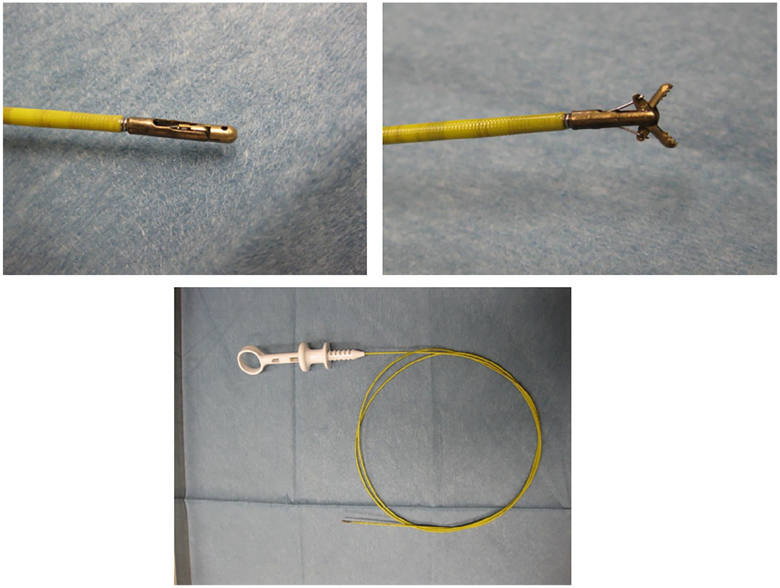
Biopsy forceps. These consist of a tip, catheter, and handle. The cup at the tip can be opened or closed by maneuvering the handle.

Cases in which biopsy forceps were used for occlusion of a prosthetic vessel. (1) 6-Fr sheaths are inserted facing each other. (2) Aspiration using the Thrombuster II. (3) Thrombectomy with a biopsy forceps is performed for rigid thrombi which cannot be aspirated. Tourniquet were used during the procedure All procedures were performed under ultrasound guidance (Figure 2(a)). The forceps were inserted through a 6Fr sheath (Figure 2(b)). (4) Fragmented thrombi and residual thrombi are aspirated. (5) Balloon dilatation for the stenosis site after reopening (Figure 2(c)). After confirmation that no thrombus remains, the procedure is terminated.

Guidance of forceps using a long sheath: (a) a case of autologous venous shunt occlusion, where thrombus was adjacent to the grafted section; (b) place a long sheath in the target site and move the forceps inside the sheath; and (c) when the forceps reach the target site, slide the sheath frontward. It is possible to guide the forceps to the target site safely using these procedures.
For ultrasound equipment, the Logiq S7 or S8 11-MHz linear probe of GE Healthcare was mainly used. In procedure, the physician performed probe manipulations and guided the tip of the forceps, while the assistant opened and closed the tip under the direction of the physician. Blood pressure, pulse, and oxygen saturation were monitored during the procedure.
The initial success rates, complication rates were obtained for all cases of occlusion. The patency rates were obtained for cases using forceps and for the cases in which biopsy forceps were not used (Figure 4).

Patency rates for the cases (a) in which biopsy forceps were used and (b) in which biopsy forceps were not used.
Subjects
A total of 468 patients were included (313 male, 155 female; mean age, 68.8 ± 12.2 years; dialysis history, 5.7 ± 6.0 years) of the 564 patients with VA occlusion who received treatment at our clinic during the period from May 2014 to April 2017 and who could be followed up. A breakdown of these cases (cases with multiple treatments counted once) is as follows: 366 cases of acute occlusion of an autologous vein, 49 cases of acute occlusion of a prosthetic vessel, 51 cases of chronic occlusion of an autologous vein, and 2 cases of chronic occlusion of a prosthetic vessel. The treatments included surgical revascularization in 252 cases, endovascular treatments in 207 cases, and hybrid treatments with both performed simultaneously in 9 cases. In one case, treatment was changed to a cuffed catheter. Thrombectomy of an organized thrombus using biopsy forceps was performed 194 times in 138 cases. Biopsy forceps were used in the initial treatment in a total of 115 cases, including 71 cases of acute occlusion of an autologous vein, 3 cases of chronic occlusion of an autologous vein, and 41 cases of acute occlusion of a prosthetic vessel. The procedural techniques used were hybrid treatment in three cases, and endovascular treatment only in the other cases.
Results
The initial success rates among 467 cases, excluding one case in which treatment was changed a cuffed catheter, among the 468 cases of VA occlusion are presented here. The initial success rate was 251 of the 252 cases (99.6%) with surgical revascularization, 195 of the 207 cases (94.2%) with endovascular treatments, and 9 of the 9 cases (100%) with hybrid treatment. Organized thrombi and intimae could be excised and resected using forceps in 113 of the 115 cases (98.2%) in which biopsy forceps were used. The two cases where such excision was impossible required surgical excision and revascularization. There was one case in which massive bleeding occurred at the sheath extraction site on the night following the operation, which resulted in multiple organ failure due to circulatory failure. No serious complications such as extravascular deviation or pulmonary embolism were observed. No case required intraoperative fluoroscopy or contrast radiography. A thrombus-aspiration device was concomitantly used in 109 cases. The procedural time in the cases in which forceps were used is 65.3 ± 10.3 min, and the blooding volume was small in all cases.
For cases using biopsy forceps, the primary patency rates by the Kaplan–Meier method were 85.0% at 1.5 months and 47.1% at 3 months, the secondary patency rates (until occlusion) were 93.3% at 1.5 months and 89.1% at 3 months, and the tertiary patency rates (until requiring surgical revascularization) were 95.8% at 1.5 months and 95.8% at 3 months. For all cases of occlusion, the primary patency rates were 86.1% at 1.5 months and 55.6% at 3 months, the secondary patency rates were 93.0% at 1.5 months and 89.2% at 3 months, and the tertiary patency rates were 94.7% at 1.5 months and 93.8% at 3 months.
Discussion
There are treatment methods of thrombotic occlusion in VA currently in the main stream such as surgical thrombectomy, thrombolysis device, thrombus fragmentation device or thrombus suction device, and so on. These methods show a good result for a long period as in the previous works,5–10 but, except for surgical thrombectomy, they require medical treatment during the hyperacute phase after obstruction. Fragmentation will be impossible when time passed after obstruction as the thrombus degenerates and stiffens. This time, we employed biopsy forceps for three cases of chronic obstruction, in which significantly organized thrombus could be removed for each case. This method, capable of treating significantly organized thrombus, can be considered to be extremely useful.
The main countermeasure against organized thrombi has been surgical thrombectomy. However, this technique is highly invasive, some thrombi are difficult to excise by surgical measures, and surgical methods have practical limitations. Furthermore, in the occlusion of a prosthetic vessel, the infection of an incision wound may occur, or it may be difficult to maintain the site of skin incision given frequent occlusions. Accordingly, conventional methods present various problems, and new countermeasures have been desired. We devised a method which employs the forceps originally used in biopsy, under the guidance of gastrointestinal endoscopy for the resection of thrombi and intimae in endovascular treatments. Rigid thrombi can be excised and resected by using the forceps.
While compatible sheaths are 5 Fr or larger, the volume of resectable thrombus per time becomes smaller when a sheath with a smaller inner diameter is used. Accordingly, use of a sheath with a large inner diameter should be considered.
Since a device originally used for extravascular tests is applied for endovascular treatments in this method, ingenuity in manipulation is required. The length of the shaft reaches 120 cm, which presents some manipulative problems. A long sheath is necessary to safely pass forceps into severely curved, tortuous blood vessels. By inserting the tip of a sheath into a target site in advance, feeding the forceps through the lumen to the target site, and pulling the sheath forward, the forceps can be safely and securely advanced to the target site.
Flow of a fragmented thrombus into the center of a vessel presents the risk of arterial embolism in cases involving a pulmonary embolism or right-to-left shunt, and countermeasures against the flow of such fragmented thrombi are important.11,12 Protective devices, including a tourniquet and manchette, were used to reliably collect fragmented, dislodged thrombi. Thrombus-aspiration catheters have been used in many cases using biopsy forceps and are considered to be useful in treatments for fragmented thrombi as well as unstable thrombi around organized thrombi.
One of the problems accompanying use of a thrombus-aspiration catheter is that the surrounding blood is simultaneously aspirated in thrombus aspiration. As a result, the bleeding volume tends to be larger with frequent aspiration. However, the intra-procedural bleeding volumes were small in all of the cases in which forceps were used. It is considered that the concomitant use of an aspiration catheter and forceps reduced the frequency of aspiration.
Although post-procedural bleeding from the site of sheath extraction, which was observed as a complication, was not attributed to the intra-procedural manipulation of the forceps, it cannot be ruled out that frequent movements of the sheath position due to the use of the forceps and a prolonged procedural time might have affected hemostasis after extraction of the sheath. Securing hemostasis at the site of sheath extraction is one future challenge.
Post-procedural pseudoaneurysm was attributed to balloon dilatation at the stenotic site. Performing the procedures under ultrasound guidance and use of a long sheath, as appropriate, could prevent serious complications such as extravascular deviation and blood vessel perforation.
For countermeasures against occlusion cases, surgical revascularization, endovascular treatment, or concomitant use of both may be selected. Procedural techniques should be determined by comprehensively judging many factors, including the properties of the blood vessel and thrombus. In particular, countermeasures against thrombi will be a factor that is heavily involved in the selection of a procedural technique.
Endovascular treatments could be performed in 40% or more of all cases of occlusion. This suggests that biopsy forceps enables the resection of thrombi which are conventionally difficult to resect by endovascular treatment, so that the indication of endovascular treatment has been extended. In addition, the patency rates of all cases of occlusion treatment as well as those of cases using biopsy forceps were superior.13–17 It is considered that the appropriate selection of treatment improved the overall patency rate of the occlusion cases. It is important to determine which cases of occlusion endovascular treatment or surgical revascularization will be suitable for the appropriate use of biopsy forceps.
We will work to obtain approval for this device for use in endovascular treatments in Japan and improve the product to yield a more suitable structure for endovascular treatments.
Conclusion
We performed the percutaneous thrombectomy of organized thrombi under superficial ultrasound guidance, using the forceps that are used in gastrointestinal biopsy. This method was considered to be a valuable choice as a countermeasure against organized thrombi.
Footnotes
Acknowledgements
We are deeply grateful to the staff of the catheter laboratory in Bousei Daiichi Clinic for their excellent work and cooperation.
Declaration of conflicting interests
The author(s) declared no potential conflicts of interest with respect to the research, authorship, and/or publication of this article.
Funding
The author(s) received no financial support for the research, authorship, and/or publication of this article.
Research ethics and informed consent
Approval from the Ethics Committee (approval no. 2014-01) and consent from the patient for use of biopsy forceps in endovascular treatment were obtained for this study.