






Abstract
Purpose:
To evaluate the use of a dual-chambered venous access port for extracorporeal apheresis therapy.
Methods:
This was a single-center retrospective analysis of all patients who received a dual-chambered venous access port for apheresis therapy over a 36-month period. Clinical success was defined as successful completion of at least one round of apheresis via the venous access port. Major complications were defined as any event requiring elevation of patient care and/or venous access port removal or repositioning. Minor complications were defined as venous access port issues resolved with clinical intervention.
Results:
Forty-four patients had a venous access port placed at the time of this study. Patients underwent red cell exchange (n = 33), therapeutic plasma exchange (n = 6) or extracorporeal photopheresis (n = 5). Forty (90%) patients had autoimmune diseases and four (10%) had neoplastic processes. Clinical success was achieved in 42 (95.5%) patients. Average venous access port dwell time was 632 days (range = 42–1191 days). All therapies through the venous access ports were well tolerated and no patients reported pain or discomfort. Major complications were seen in nine (20.5%) patients–the majority (n = 7) of which were due to venous access port malfunction–and resolved with catheter revision. One (2.27%) major complication involved an infected venous access port, and one involved a large hematoma at the venous access port site. Minor complications were seen in eight (18.2%) patients, where simple flushing of the catheter with saline or tissue plasminogen activator resolved the issue.
Conclusion:
The dual-chambered venous access port was successfully used for sustained blood flow in apheresis therapy with a moderate, yet correctable complication rate.
Keywords
Introduction
Apheresis plays a crucial role in treatment of several conditions such as hypercholesterolemia, post-transplantation disorders, sickle cell disease, and certain neurological syndromes such as myasthenia gravis and Guillain–Barre syndrome. 1 In its original form, a large-bore needle is percutaneously inserted into the venous system and draws out blood, returning it to the body via a second large-bore needle following separation of target particles. More improved methods (i.e. tunneled and non-tunneled catheters) have since evolved that allow for continuous venous access required in patients with prolonged illnesses, while obviating the need for repeated venous puncture. 2 Although the catheter method allows for decreased incidences of vein obliteration or thrombosis associated with repeated venous puncture, it is not devoid of its own adverse effects. 3 Notable complications include infection (especially in pediatric patients) and, with prolonged catheter dwell times, thrombosis.4–8
One of the current standards of care for repeated venous access is placement of a central venous access port (VAP), devised with the intent of bypassing issues with limited peripheral access. 9 As VAPs are generally safe and carry very low complication rates, they can be used for repeated venous access with a significantly decreased incidence of the aforementioned risks.9,10 A fairly novel approach to apheresis therapy combines the standard VAP method—via the use of a dual-chambered VAP—with apheresis in an attempt to maximize care and comfort, while minimizing the risks of infection, bleeding, venous stenosis and obliteration associated with the previously mentioned methods. The aim of the present retrospective study is to report on the use of the dual-chambered VAP in carrying out apheresis therapy.
Materials and methods
Study design
This was a single center retrospective analysis of all patients whom underwent placement of a dual-chambered VAP over a 36-month period. Following approval by the institutional review board and informed consent, the electronic medical records were searched in order to identify all patients having undergone placement of a dual-chambered VAP for apheresis therapy. Only authors on this publication—all of whom had the proper and necessary authorization—accessed the patient records for this study.
Study population
All patients were confirmed alive at time of data collection. Thirty-two (72.7%) patients were female and 12 (27.3%) were male, all with an average age of 41 years (median = 32 years, range = 20–68 years). All patients in the study had previous conditions requiring red cell exchange (n = 33), therapeutic plasma exchange (n = 6), or extracorporeal photopheresis (n = 5). Thirty-one (70.5%) patients had sickle cell disease, three (6.82%) had myasthenia gravis, two (4.5%) had mycosis fungoides, two had leukemia, one (4.35%) had beta-thalassemia, one had cryoglobulinemia, one had acute lymphoblastic leukemia with acute graft-versus-host disease post stem-cell transplantation, one had multiple sclerosis, one had bronchiolitis obliterans, and one had T-cell lymphoma. Thirty (68.2%) patients were already undergoing apheresis therapy prior to VAP placement; the remaining 14 (31.8%) began apheresis therapy following VAP placement. The five patients who underwent extracorporeal photopheresis had completed treatment at time of data analysis and had their VAPs removed thereafter.
Procedural and apheresis technique
Following informed consent, patients were taken to the interventional radiology (IR) suite where they underwent VAP placement. All patients received the Vortex LP Dual Titanium Port System (Angiodynamics, Latham, NY), which consisted of a titanium dual-chambered VAP (0.9 mL internal volume per chamber), an 11.4 French silicone catheter (76 cm length), and a 12 French introducer. The right internal jugular vein was accessed with a 0.018 inch guidewire, which was then exchanged for a 0.035 inch guidewire. The right chest was then anesthetized over the third rib and a 1.5 inch transverse incision was made. Through the incision, a pocket was bluntly dissected in the subcutaneous soft tissues to fit the VAP. The VAP catheter was tunneled up through the subcutaneous soft tissues of the right chest and lateral neck to the venotomy site, where the 0.035 inch guidewire was exchanged for a peel-away sheath. Through the sheath, the VAP catheter was advanced down to the level of the superior vena cava (SVC)–atrial junction. Confirmation of the catheter tip at the SVC–atrial junction was confirmed on fluoroscopic imaging. The back end of the catheter was trimmed and attached to the VAP, which was, in turn, placed in the subcutaneous pocket. The VAP was aspirated and flushed, and if normal blood return was present, the pocket was sutured closed. Following successful placement, the VAP was ready for immediate use. For apheresis, one chamber was accessed with an access needle and used for the inflow channel and the other chamber with a second access needle and used for the outflow channel.
Clinical and technical success
Clinical success was defined as the successful completion of at least one full round of apheresis therapy via the VAP. Clinical failure was defined as the inability to successfully begin and/or complete at least one full round of apheresis following two separate attempts. Complications were defined as major if they required any elevation in patient care, including further VAP revision by IR for issues such as infection, repositioning or replacement occurring any time after successful apheresis began. Complications were defined as minor if any clinical interventions were required during apheresis (e.g. line occlusions requiring flushing with tissue plasminogen activator [t-PA] or sterile saline) for continued successful VAP usage.
Statistical analysis
Demographic and clinical parameters were expressed as mean, median, range, and interquartile range (IQR) as appropriate. Analyses were performed using SPSS, version 20, software package (IBM Corp, Armonk, NY, USA).
Results
Forty-six patients underwent placement of a dual-chambered VAP. Of the 46 patients, only 44 began apheresis therapy at the time of this study, and as such were the only ones included in the final data analysis. The average dwell time for all VAPs was 632 days (median = 678 days, range = 42–1191 days), with 38 (86.4%) patients still with VAPs in place at time of data analysis.
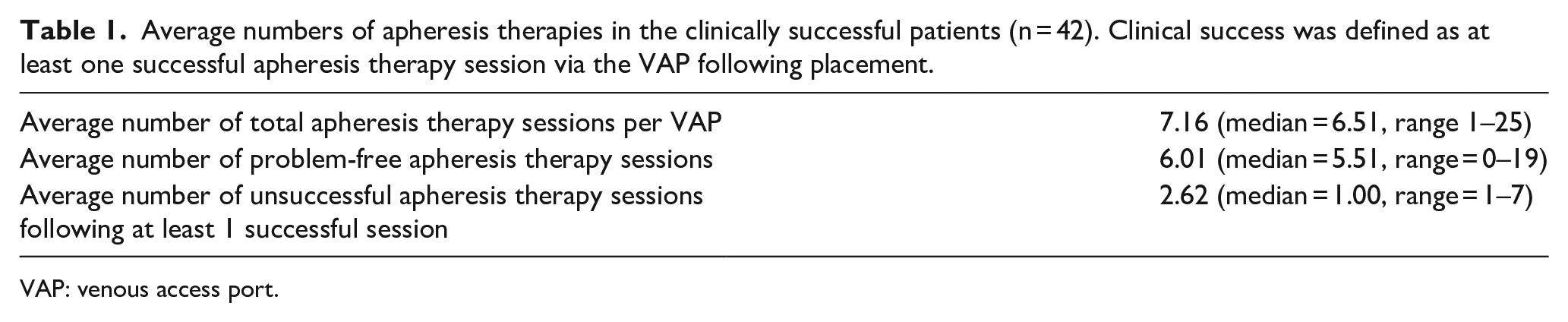
Clinical success (defined as at least one successful apheresis session via the VAP) was achieved in 42 (95.5%) patients (95% confidence interval (CI) 88.6%–101%). However, following initial clinical success, all 42 of these patients underwent repeated subsequent apheresis therapy sessions, each. Some of these subsequent apheresis sessions were unsuccessful and are further detailed below. One (2.27%) patient experienced clinical failure of the VAP, and one patient had no data available, as their treatment took place at an outside institution following VAP placement. In the 42 patients achieving clinical success, the average number of total apheresis therapies per VAP was 7.16 (median = 6.51, range 1–25), the average number of problem-free apheresis therapies per VAP was 6.01 (median = 5.51, range = 0–19), and the average number of unsuccessful apheresis sessions was 2.62 (median = 1.00, range = 1–7; Table 1). No patients reported pain or discomfort at any time during usage of the VAP, and all therapies through the VAPs were well-tolerated.
Average numbers of apheresis therapies in the clinically successful patients (n = 42). Clinical success was defined as at least one successful apheresis therapy session via the VAP following placement.
VAP: venous access port.
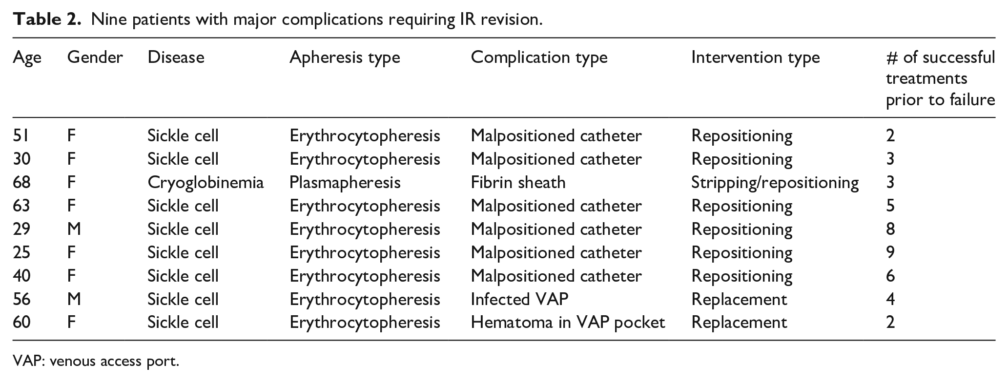
Major complications were seen in nine (20.5%) patients. All nine of these patients were initially a clinical success, in that they all underwent at least one round of successful apheresis therapy via the VAP. The major complications mentioned below occurred following initial clinical success. One (2.27%) of these nine patients required VAP removal (and eventual replacement) due to VAP-related infection (frank purulent fluid was found in the VAP and VAP pocket at time of removal). One patient required VAP removal due to massive hematoma at the VAP site. The remaining seven (15.9%) patients required IR revision due to VAP malfunction not responsive to clinical interventions (Table 2). All nine patients were taken back to IR for a VAP check that consisted of a flush/aspiration attempt, as well as evaluation of the catheter integrity and appropriate positioning by radiographic imaging. In the seven cases of malfunction, the catheter appeared intact; however, all catheters had migrated superiorly by an average of 8 mm (median 11 mm; range 6–18 mm). None of the seven VAPs were able to be flushed or aspirated. All seven VAPs required catheter repositioning, which was successfully achieved via a forceful hand injection of saline, or using a snare device when hand injection was unsuccessful.
Nine patients with major complications requiring IR revision.
VAP: venous access port.
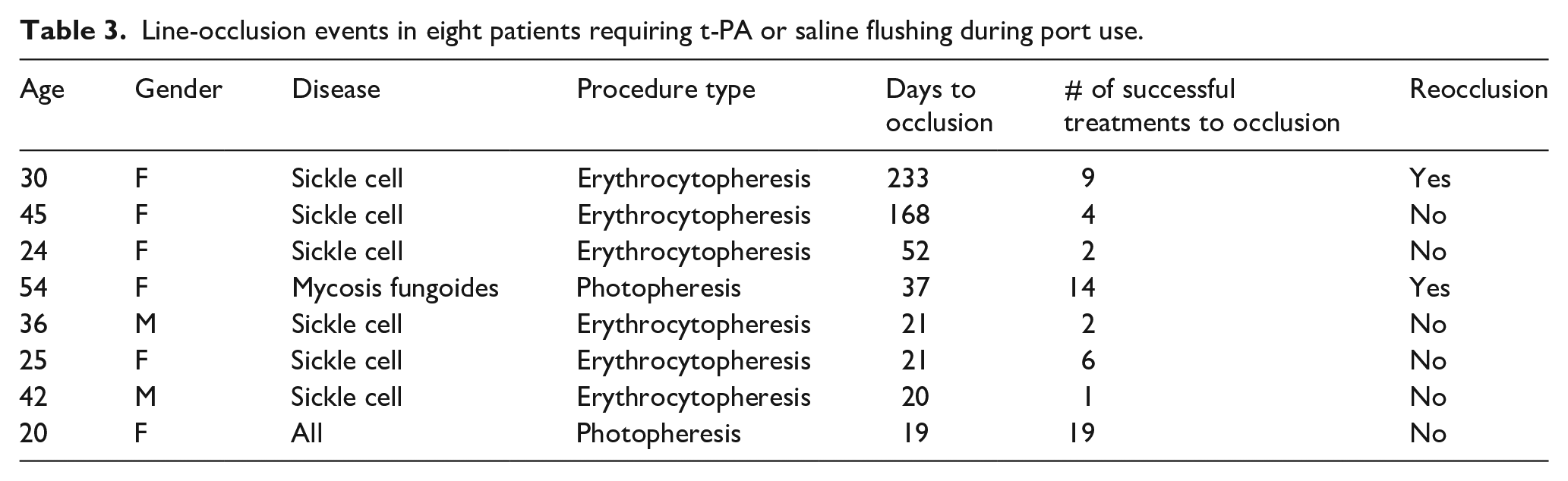
Minor complications were seen in eight (18.2%) patients. As was the case with the major complications, all minor complications occurred following initial clinical success. All minor complications involved catheters that were obstructed during or before apheresis (Table 3). Full VAP function resumed immediately following a few flushes with sterile saline, or a 30- to 60-min incubation with 2 mg of t-PA. Two (4.6%) patients had repeat catheter obstruction at follow-up apheresis sessions, both of which were similarly resolved.
Line-occlusion events in eight patients requiring t-PA or saline flushing during port use.
Flow rates through the VAP averaged at approximately 47 (range: 30–55) mL/min for all apheresis types. These rates remained generally consistent.
Discussion
Our study demonstrated the dual-chambered VAP’s ability to sustain prolonged blood flow required for apheresis therapy, as evidenced by the clinical success rate of 95.5%. Although the major complication rate was 20%, the overwhelming majority (n = 7) of these nine complications were due to functional issues with the VAP. The issues were all resolved with a VAP revision or replacement by IR. As such, we surmise that the major complications in this study were likely more related to initial VAP placement—specifically, the final position of the VAP catheter tip—rather than to VAP usage for apheresis. For example, though studies have demonstrated that VAP catheter tips placed in the SVC carry a greater risk of malfunction than when placed in the right atrium, many physicians who place VAPs still use the SVC-atrial junction as the final location for catheter tip placement on radiographic imaging. 11 Studies have also demonstrated that once the patient is no longer in the supine position following VAP placement, the catheter invariably retracts cephalad by 1–2 cm. 11 In our study, final catheter tips were also placed at the SVC-atrial junction. It very well may be that the longer usage time, faster flow rates, and increased throughput during apheresis versus that encountered with chemotherapy hastens the time to malfunction with a tip in the SVC. In our study, the VAP demonstrated efficacy and a good safety profile when used for apheresis therapy. However, the major complication rate, though moderate, was still too high (20%). As such, if VAPs are placed for apheresis, a final catheter tip in the right atrium should strongly be considered to diminish the number complications encountered in our study.
Minor complications were seen in 18.2% of the patients and were all associated with inability to flush/aspirate through the VAP catheter. This issue was solved with a saline or t-PA flush. The underlying cause for the catheter obstruction or whether this could have been obviated with catheter tip placement into the right atrium as opposed to at the SVC-atrial junction are both unknown at this time.
Another advantage to the usage of a VAP for apheresis is the decreased infection rates associated with VAPs for repeated venous access. 9 This was also demonstrated in our study with a low infection rate of 2.27%. Given that the clear majority of patients in this study required apheresis due to underlying autoimmune disease (90.1%) and that individuals with autoimmune diseases have a higher rate of infection due to underlying immune compromise, VAP usage for apheresis may confer an added benefit in this patient population.
Although the results of this study are encouraging, the study is limited by its retrospective nature and leaves some questions unanswered. For example, and as alluded to earlier in the discussion, it is not known whether the inability to flush/aspirate the VAP catheter experienced in all minor complications was due to catheter occlusion secondary to thrombus, or due to mechanical obstruction. As these devices were not originally intended to have blood continuously cycled through the VAP or VAP catheter as was done with apheresis, the off-label usage of the devices further confounds any hypotheses. It was noticed that t-PA injection into the VAP resolved most of the occlusions; however, it is not known whether a simple saline flush could have accomplished the same results. Because approximately 40% of overall real-world catheter occlusions do not occur as a result of thrombi, but as a result of mechanical obstructions (e.g. catheter migration, kinking), it is possible that the use of saline would have been just as effective as t-PA. 12 Also, complications related to VAP access can at times be operator dependent, as VAPs are made to be accessed in a particular manner and with a specialized non-coring needle. The level of experience that our apheresis nurses had with VAP access and whether this played a role in the minor complication rate is not known.
Finally, and again due to its retrospective nature, this study was unable to fully evaluate flow rates, information that is essential in apheresis therapy. It was mentioned earlier that the average flow rate was 47 (range: 30–55) mL/min across all apheresis types. However, more comprehensive and quantitative information such as maximum allowable flow rate and flow rates for various types of apheresis (e.g. photopheresis vs plasmapheresis) were not attainable due to the study’s design.
In conclusion, apheresis via a dual-chambered VAP is efficacious, well-tolerated and maintains a good safety profile. However, with a moderate complication rate of 20%, which appeared to be due to final catheter tip positioning at the SVC-atrial junction, VAPs placed for apheresis are likely to suffer a much lower complication rate with a final catheter tip position in the right atrium. However, a prospective large-scale study would be needed to further elaborate on the device’s capabilities and limitations with respect to apheresis therapy.
Footnotes
Declaration of conflicting interests
The author(s) declared no potential conflicts of interest with respect to the research, authorship, and/or publication of this article.
Funding
The author(s) received no financial support for the research, authorship, and/or publication of this article.
