
Abstract
Clinicians commonly place ultrasound-guided intravenous catheters in peripheral veins for the diagnostic and therapeutic treatments of patients. This procedural skill requires practice and static phantom models are a commonly used education tool. Several commercial models that simulate blood vessels within tissue are available; however, they can be expensive. There are many examples of “Do-It-Yourself” models proposed; however, many of these require time to create the model. Mixing water and gelatin to make a gelatinous material, and the time necessary to set and store the phantom may deter people from pursuing these options. We propose Konnyaku jelly, or “yam cake,” found in many Asian grocery stores, as the substrate to create a phantom model. When imaging with ultrasound, this model is similar to commercially available models, however the cost is less than $3.00 and preparation is about 5 min. We believe that Konnyaku jelly should be a more generally accepted homemade static model for phantom preparation.
Keywords
Introduction
Clinicians commonly place ultrasound-guided intravenous (IV) catheters in peripheral veins for the diagnostic and therapeutic treatment of patients. 1 The ultrasound-guided insertion of peripheral IVs is helpful when blind insertion of an IV catheter is unsuccessful or difficult. 2 This procedural skill requires practice and static ultrasound phantoms are a commonly used education tool. Commercially available ultrasound models, that allow clinicians to practice placing ultrasound-guided IVs, can be cost prohibitive. 3 Less expensive, Do-It-Yourself (DIY) ultrasound models have been described; however, most require ingredients including water and gelatin tubing or balloons to simulate a blood vessel and a multi-step preparation process.3–7 This may take hours and is thus time intensive. Ultrasound phantoms using food, such as chicken, tofu and spam are described.8–10 Selame et al. 11 compared several non-commercial phantoms including models made from tofu, spam, gelatin, ballistic and gel. The cost of production of various models ranged from $5.00 to $30.00 and production time ranged from 10 to 120 min. 11
We present a superior static phantom model using Konnyaku jelly. Konnyaku jelly, made from the konjac plant, is a food primarily consumed in Japan and Korea and commonly used in hot pot dishes. It is gelatinous in structure and texture, packaged in individual blocks, and in the United States, found primarily in Asian grocery stores. Ingredients listed on the packaging are water, yam flour, and calcium hydroxide. When imaging with ultrasound, Konnyaku jelly appears similar to soft tissue. The material is inexpensive, requires minimal additional materials, and can be set up for use in less than 5 min.
In this paper, we describe the process of creating an ultrasound IV phantom out of Konnyaku jelly. We further demonstrate its appearance on ultrasound and perform basic durability and degradation tests.
Methods
Materials
Konnyaku jelly
Plastic tub
Plastic straw
Water
Preparation time: 5 min
Cost: Approximately $3
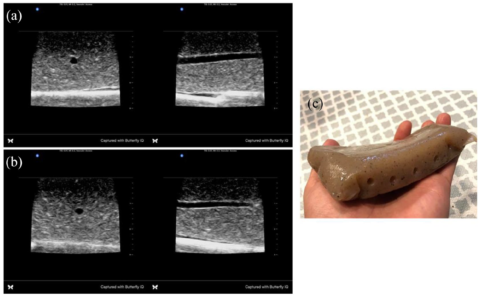
Konnyaku jelly was removed from its packaging. The representative sample (Image 1) measured approximately 15 cm × 9 cm × 2 cm and weighs 255 g. A hole was bored through the jelly using a straw. The straw was taken out completely through the other end, which took the luminal content along with it. The model now contained a cylindrical hole extending through its entire length, simulating a vessel (Image 2(a) and (b)). The model was placed in a tub of water such that the water level was adequate to fill the hole created by the straw but low enough to not cover the Konnyaku jelly surface (Image 2(c)). This water simulated blood within a vessel. The model was compressed perpendicular to the surface of the container to expel any air bubbles that remained within the lumen of the vessel to avoid artifacts. Multiple vessels were created in one block of Konnyaku jelly (Image 3).
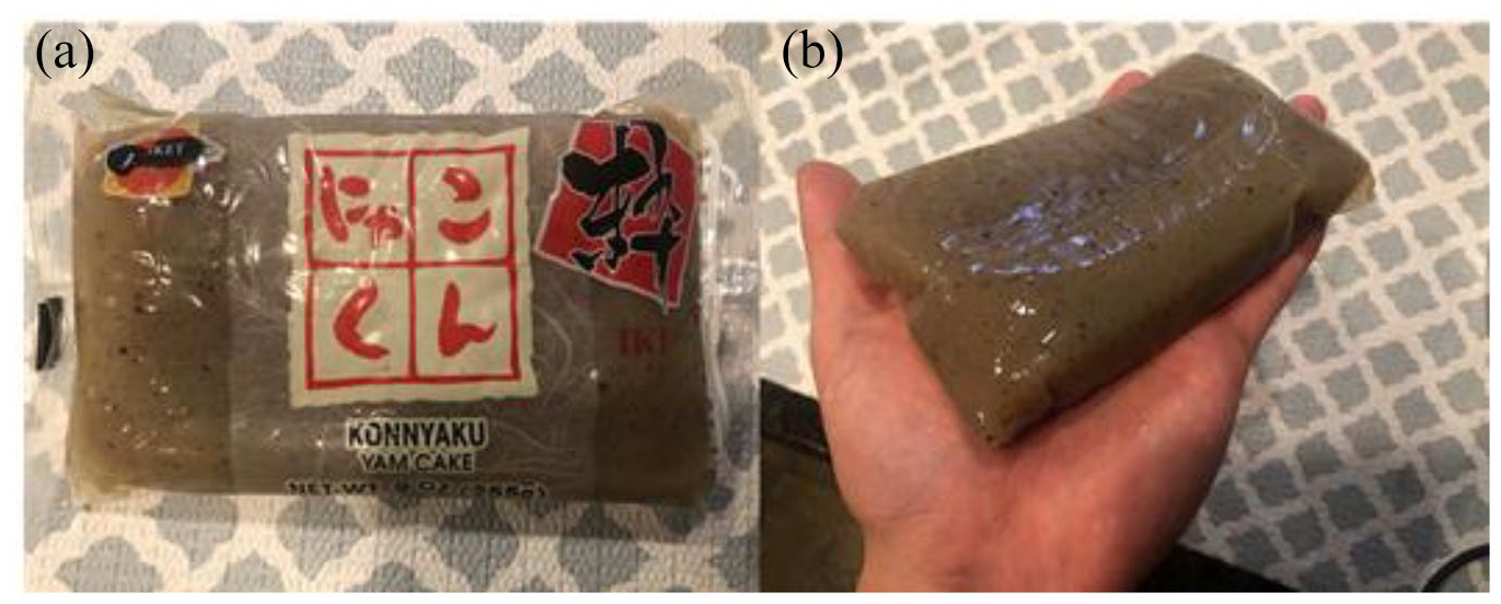
(a) Konnyaku jelly in its packaging and (b) Konnyaku jelly out of its packaging.
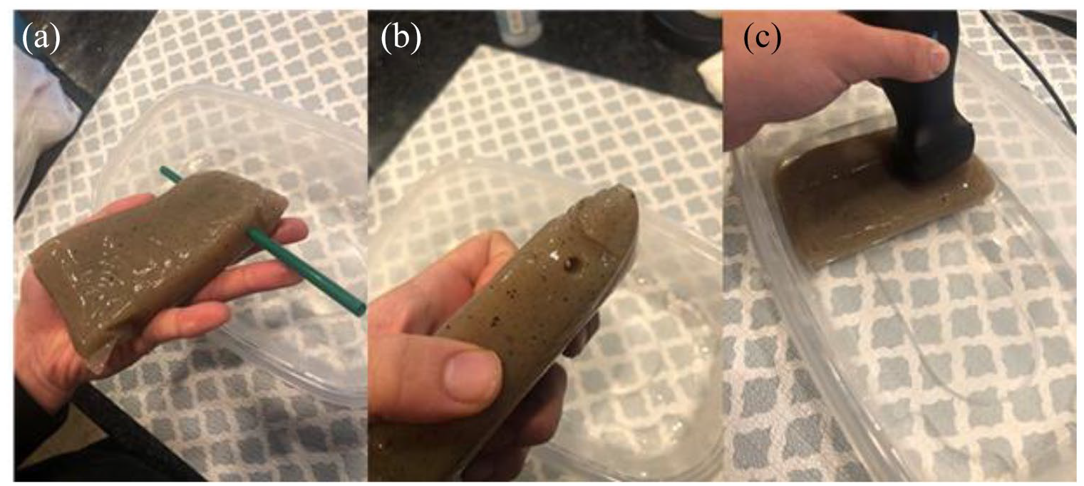
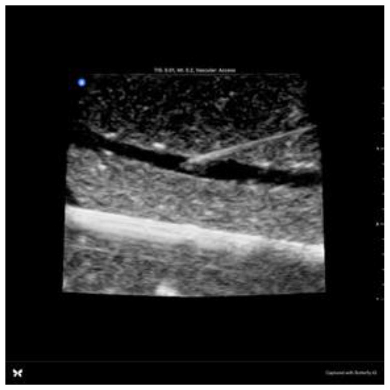
(a) Plastic straw boring through Konnyaku jelly to create a hollow lumen simulating a blood vessel, (b) Hollow vessel created through Konnyaku jelly and (c) Konnyaku jelly placed in a shallow container of water, with an ultrasound probe resting on the model.
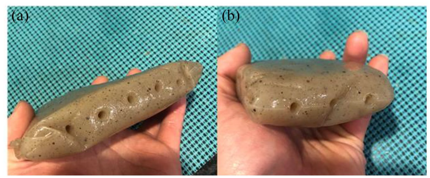
Multiple vessels can be created in parallel, allowing for multiple pierces. Here we show five vessels created approximately 2 cm apart along its length (a) and three vessels created approximately 3 cm apart along its width (b).
Testing
Images were obtained using a hand-held Butterfly (Butterfly Network, Inc. Guilford, CT) ultrasound probe on vascular settings (Images 4–6). To test its durability against repeated piercing, one Konnyaku jelly sample was repeatedly pierced in the same location. It was then evaluated for any deterioration to the sample or quality of images under ultrasound (Images 7 and 8).
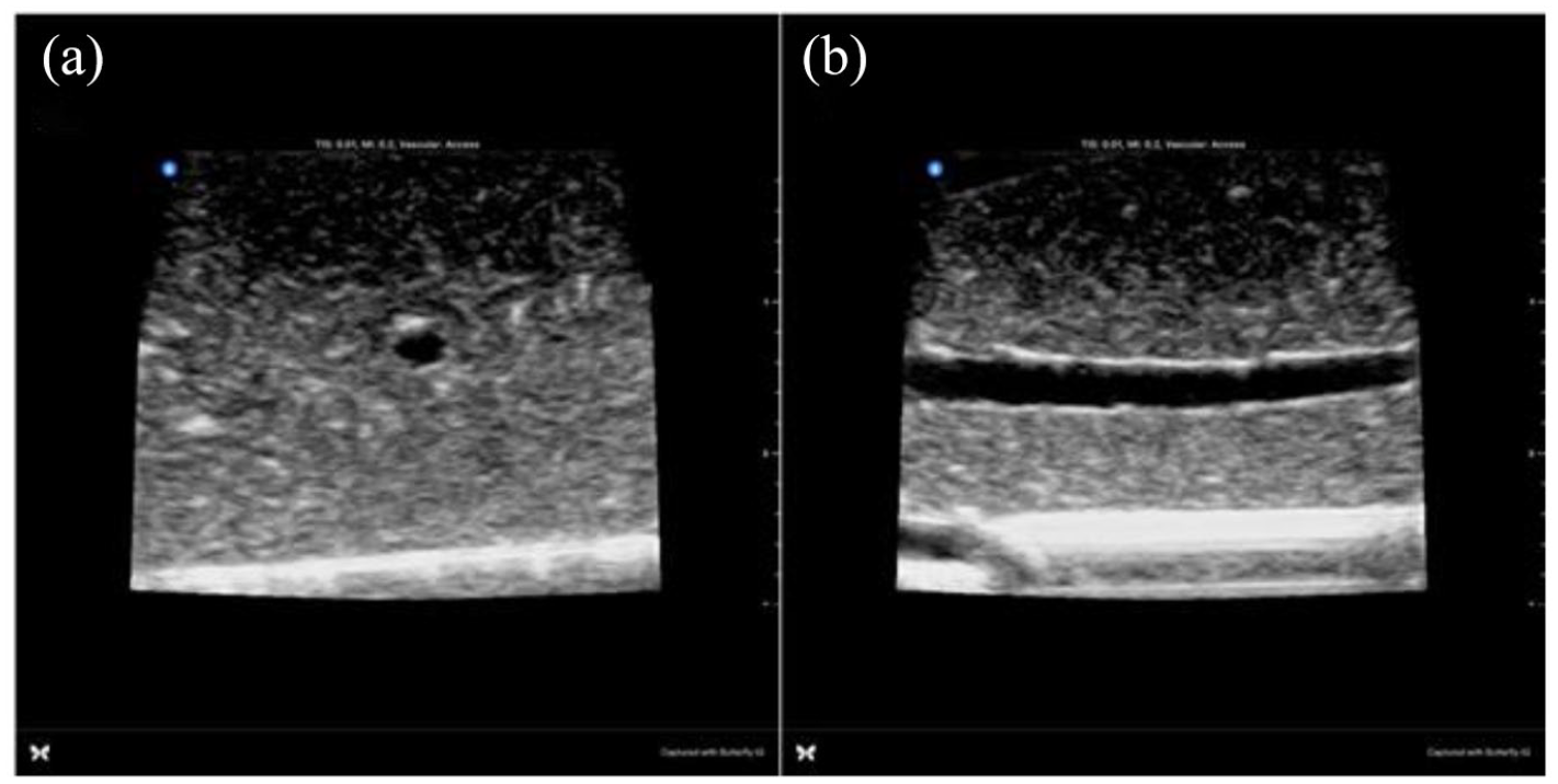
Out-of-plane (a) and in-plane (b) views of the vessel within the phantom using a Butterfly ultrasound transducer.
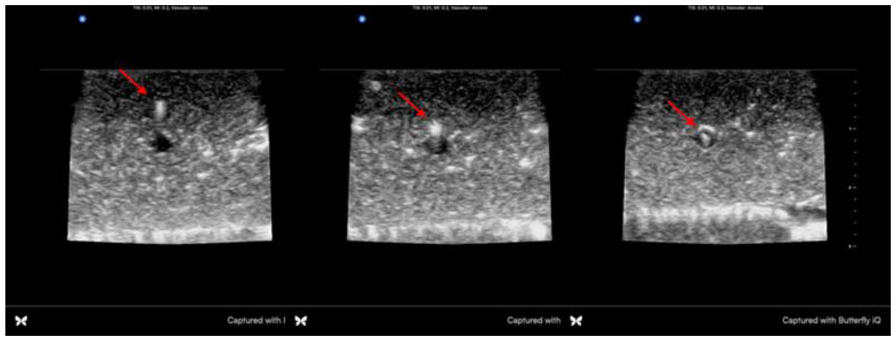
Demonstrating ultrasound-guided IV cannulation using the out-of-plane technique. Needle tip (red arrow) is visualized in the soft tissue, and then eventually in the vessel lumen.
In-plane view of the IV catheter with the needle tip within vessel.
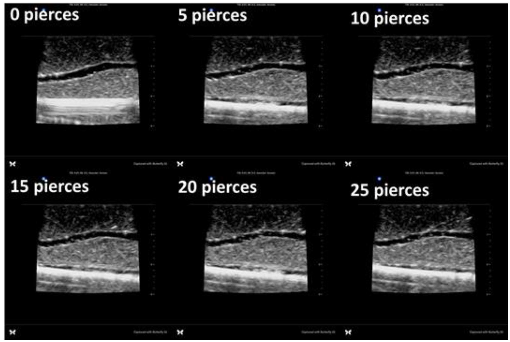
In-plane views of the vessel after 0, 5, 10, 15, 20, 25 pierces with an 18g IV catheter.
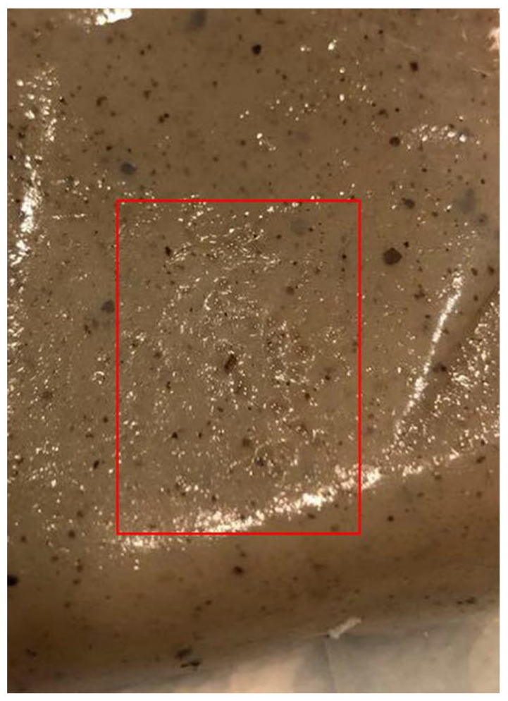
Surface of Konnyaku jelly after 25 pierces in one location with an 18g IV catheter.
To test for degradation in storage, two Konnyaku jelly samples were refrigerated after initial use, one in a dry plastic container, and the other submerged in water. After 7 days, images were obtained to evaluate for simulator breakdown or changes to the shape, texture or quality of images under ultrasound (Image 9).
In-plane and out-of-plane views of the Konnyaku jelly model vessels after storage in a refrigerator for 7 days, either in a dry plastic container (a), or submerged in water (b). (c) Konnyaku jelly after 7 days of storage.
Results
The appearance of the needle tip on the screen adequately simulates soft tissue and blood vessel and ultrasound-guided IVs can be practiced in both the in-plane and out-of-plane approaches (Images 4–6). Given the firm nature of Konnyaku jelly, the “vessel” did not collapse and remained patent even with repeated compression of the model.
The model can be cannulated multiple times without any visible deterioration or destruction of the phantom. In our testing, the model was cannulated in a single location 25 times without significant deterioration of the model, or its appearance under ultrasound. With each model, multiple vessels can be created. In our example we were able to create at least five vessels along its length. This suggests that one Konnyaku jelly model can withstand at least 25 piercings × five vessels, or 125 piercings and still maintain quality images on ultrasound.
Konnyaku jelly can be stored in water or in a dry container for 7 days without significant changes in texture or image quality under ultrasound. There was no significant difference between the models stored in a dry container and the models submerged in water.
Discussion
We describe the use of Konnyaku jelly as a cost effective and quick-to-prepare ultrasound guided IV static stimulator for peripheral IV placement education and practice. The appearance of the material is similar to that of soft tissue and the vessel created using a plastic straw adequately simulates a blood vessel. The model can be used to practice IV insertion using both the in-plane and out-of-plane techniques. From a durability standpoint, Konnyaku jelly was able to sustain multiple piercings without significant deterioration of the ultrasound image. The Konnyaku jelly was found to last at least one week while stored in the refrigerator either dry or submerged in water. Finally, the Konnyaku jelly is affordable at less than $3 per item. There is no concern of damaging an expensive commercially-available phantom.
Konnyaku jelly is not without its drawbacks, however. It is not readily found in all grocery stores. If available, we recommend going to an Asian grocery store for purchase. Online purchase by searching “J-basket Konnyaku Yam Cake” is also an option. The jelly has a limited shelf life. While the jelly may in practice last longer than the 1 week we tested, it is a perishable foodstuff and its shelf life is on the order of days to weeks, not months to years. The Konnyaku jelly has a mild odor, which may deter some users. This can be reduced by rinsing with water. Also, the odor significantly diminishes if the Konnyaku jelly is boiled for 10 min (as is typically done when one cooks a dish with Konnyaku); however, this adds time to preparation of the model. Another disadvantage is that the texture of the model does not quite match that of human skin and soft tissue. Advancing the catheter off of the needle will usually pierce the back wall of the “vessel” instead of staying within it. Consequently, we recommend using the model to practice the in-plane and out-of-plane approaches, needle tip visualization, and not for threading the catheter off of the needle. Finally, the vessels themselves will have no wall and will not adequately simulate the feeling of puncturing the needle through the wall of a blood vessel. We consider these limitations similar to and overlapping with the limitations of other DIY phantoms.
Further study of this model including more rigorous comparison to commercially available models is required. Ideally, this model should be tested prospectively against industry standards to assess its efficacy as a teaching tool.
We believe that Konnyaku jelly is an inexpensive and user-friendly model that is an excellent alternative to both commercially available and homemade phantoms. Its appearance on ultrasound is sufficient to teach needle visualization and manipulation, and it is durable enough to sustain multiple punctures without degradation. Experimenting with different straw sizes may even allow use of this model for central venous access. We hope this provides training programs an affordable alternative when teaching ultrasound-guided IV placement.
Footnotes
Declaration of conflicting interests
The author(s) declared no potential conflicts of interest with respect to the research, authorship, and/or publication of this article.
Funding
The author(s) received no financial support for the research, authorship, and/or publication of this article.