
Abstract
Background:
Central venous catheterization may be required in patients with amyotrophic lateral sclerosis (ALS) for parenteral nutrition, antibiotic treatment, or blood sampling. Different venous access devices can be taken into consideration—centrally inserted central catheters (CICC), peripherally inserted central catheters (PICC), and femorally inserted central catheters (FICCs)—depending on the clinical conditions of the patients. Regardless of the type of access, the presence of paraplegia or tetraplegia is commonly considered a risk factor for catheter-related thrombosis (CRT).
Method:
This retrospective study analyzes the rate of CRT and other non-infectious complications associated with central venous access in a cohort of 115 patients with paraplegia or tetraplegia, most of them affected by ALS (n = 109).
Results:
In a period of 34 months, from January 2021 to October 2023, we inserted 75 FICCs, 29 CICCs, and 11 PICCs. PICCs were inserted only in patients with preserved motility of the upper limbs. All devices were inserted by trained operators adopting appropriate insertion bundles. We had no immediate or early complication. Though antithrombotic prophylaxis was adopted only in 61.7% of patients, we had no symptomatic CRT. Other non-infectious complications were infrequent (4 out of 115 patients).
Conclusion:
These results suggest (a) that the presence of paraplegia or tetraplegia is not necessarily associated with an increased risk of CRT, (b) that the adoption of well-designed insertion bundles plays a key role in minimizing non-infectious complications, and (c) that the insertion of FICCs by direct cannulation of the superficial femoral vein at mid-thigh in paraplegic/tetraplegic patients may have the same advantages which have been described in the general population.
Keywords
Introduction
Amyotrophic lateral sclerosis (ALS) is a progressive neurologic disease involving the motor neuron, with degeneration of the nerve cells responsible of the voluntary muscle movement. The disease causes the loss of mass and function of the skeletal and respiratory muscles. Dysphagia, dysarthria, fatigue, respiratory insufficiency, and cognitive impairment are the main symptoms. Prognosis is poor within 3–5 years. Patients with advanced ALS develop tetraplegia, often requiring tracheostomy, mechanical ventilation, and artificial nutrition. Measurement of the forced vital capacity (FVC)—defined as the volume of air that can be exhaled after a maximal inhalation—is useful for monitoring the disease progression and the indication to the mechanical ventilation.
To prevent malnutrition, guidelines recommend percutaneous endoscopic gastrostomy (PEG) when FVC is still >50%. When FVC drops <50%, there is indication to non-invasive mechanical ventilation and, eventually, invasive mechanical ventilation and tracheostomy, with subsequent PEG. Though, ALS patients without cognitive impairment sometimes voluntarily refuse invasive mechanical ventilation and tracheostomy: in these patients, PEG is not indicated and placement of a central venous line for home parenteral nutrition is usually considered, as alternative option for artificial nutrition. 1
Also, in hospitalized patients with ALS, central venous catheters are often indicated not only for parenteral nutrition but for blood sampling and antibiotic therapy. Some of these hospitalized patients may benefit of peripheral venous access devices, but direct cannulation of the superficial veins of the upper limbs may be difficult in many of them. Furthermore, insertion of midline catheters or peripherally inserted central catheters (PICC) may be contraindicated, because of the paresis of the upper limbs or because of the small caliber of the deep veins at mid-arm. Last, centrally inserted central catheters (CICC) may also be relatively contraindicated by the presence of the tracheostomy or of the mask for non-invasive ventilation; abundant oral/tracheostoma secretions may increase the risk of contamination of the exit site when a non-tunneled CICC is inserted in this area. Femorally inserted central catheters (FICC) are often an alternative option in ALS patients, when insertion of a PICC or a CICC is potentially challenging or overtly contraindicated. 2 Recent studies have shown that FICCs inserted by cannulation of the superficial femoral vein at mid-thigh in non-paretic lower limbs carry a low risk of thrombosis or infection.2,3 Though, there are no clinical studies on the incidence of thrombosis when FICCs are inserted by cannulation of the superficial femoral vein in patients with paraplegia or tetraplegia, such as ALS patients.
In our ALS Unit, we insert different types of central venous access devices, choosing between PICC, CICC, and FICC depending on several factors (the clinical state of the patient, the risk associated with venipuncture, the availability of deep veins at the pre-procedural ultrasound scan, the risk of late complications related to the location of the exit site, etc.). In this retrospective study, we analyzed our experience on central venous catheters inserted in ALS patients during their hospital stay, for both intra and extra-hospital use, focusing specifically on the incidence of catheter-related thrombosis (CRT).
Method
A retrospective study was conducted in the NeMO (Neuro Muscular Omnicenter) ward of a large university hospital from January 2021 to October 2023. The study investigated all the central venous access devices (CVAD) implanted in the NeMO ward and expected to stay in situ for at least 1 week. Approval from the local ethical committee was obtained, and a specific consent form was read and then approved by the patient or by her/his tutor.
The primary endpoint of the study was to evaluate the incidence of symptomatic CRT in all CVADs, either PICC, CICC, or FICC. The secondary endpoint was to evaluate the incidence of other non-infective catheter-related complications. We included all patients admitted to our ALS unit who required a central venous access. We excluded patients who already had CVADs inserted in the emergency room or in other hospitals; we also excluded dying patients or patients with an expected hospital stay of less than 1 week; dialysis catheters or lack of patient’s/tutor’s consent to retrieve the data needed for the study were also exclusion criteria. All CVADs were inserted by two trained operators of the hospital Vascular Access Team; the insertion technique followed the hospital policies and more specifically the insertion bundles developed by the GAVeCeLT (the Italian Group for Long Term Venous Access Devices), published as “SIC protocol” (SIC = Safe Insertion of CICCs), 4 “SIP protocol” (SIP = Safe Insertion of PICCs), 5 and “SIF protocol” (SIF = Safe Insertion of FICCs). 6
Each of the aforementioned insertion bundles included seven sequential steps: (a) pre-procedural ultrasound evaluation of the veins, with appropriate choice of the vein (adopting a 3:1 ratio between internal diameter of the vein and external diameter of the catheter) and appropriate choice of the exit site according to the RAVESTO protocol (RAVESTO = Rapid Assessment of Venous Exit Site and Tunneling Options) 7 ; (b) hand hygiene, skin antisepsis with 2% chlorhexidine in alcohol and maximal barrier precautions (full body drape sterile covering the whole patient, cap, mask, sterile gown sterile gloves, long sterile cover for the ultrasound probe); (c) ultrasound-guided venipuncture with micro-puncture kits; (d) ultrasound-based tip navigation; (e) intra-procedural tip location using intracavitary ECG (IC-ECG) and/or ultrasound-based tip location according to previously published protocols8,9; (f) securement of the catheter with sutureless device (preferably, subcutaneous anchorage); (g) protection of the exit site with cyanoacrylate glue and semipermeable transparent membrane.
All CVADs were inserted bedside, using a wireless ultrasound device with two transducers (3–12 MHz linear and 2–5 MHz convex: V-Scan, GE) or three transducers (7.5–10 MHz linear, 3.2–5 MHz convex, and 2.5–5 MHz sectorial: Cerbero 4.0, ATL). The linear transducer was used for pre-procedural scan, venipuncture, tip navigation, and control of the pleura (during CICC insertion), while the convex and the sectorial transducers were used for tip location. In all patients, pressure injectable, 55–60 cm long, non-valved polyurethane catheters of two different brands (Synergy CT, Healthline; Pro-PICC, MedComp) were used. Single lumen (4 Fr) or double lumen (5 Fr) catheters were chosen, depending on the clinical requirements. All procedures were performed using dedicated insertion packs.
During the maneuver, patients were supine, or in a semi-sitting position, or sometimes even on a lateral flank (in the case of forced postures due to spastic tetraparesis). In patients with preserved mobility of the arms, insertion of a PICC was the first choice. In patients with paretic or spastic arms, CICCs inserted by cannulation of the axillary vein in the infraclavicular area or FICCs inserted by cannulation of the superficial femoral vein at mid-thigh were considered. Ultrasound-guided venipuncture was performed either by the short-axis/out-of-plane approach, or by the oblique axis/in-plane approach. 2 After venipuncture, the catheter was inserted by the modified Seldinger technique, trimmed for CICCs or PICCs but not trimmed for FICCs. Intra-procedural ECG tip location was used for CICCs and PICCs while tip location of FICCs was verified by ultrasound trans-hepatic visualization of the subdiaphragmatic tract of the inferior vena cava (IVC). 9 This method allows direct identification of the catheter tip inside the IVC. If the IVC was too small or collapsed because of hypovolemia, the catheter tip was indirectly localized by the “bubble test” method. 9
The diagnosis of CRT was based on ultrasound examination of the veins, by the same two operators who performed the procedures. Ultrasound scan was considered only in case of local signs or symptoms suggestive for venous thrombosis.
Results
From January 2021 to October 2023, 809 patients were admitted to the NeMO ward of our hospital; 115 patients out of 809 had CVADs inserted during hospitalization, thus meeting the criteria for inclusion in this retrospective analysis. Mean age of the patients included in the study was 60 years (ranging from 40 to 64 years). About half of the patients were male (64 patients out of 115). All patients were either tetraplegic (88%) or paraplegic (12%): most of them had diagnosis of ALS (109), two had spinal muscular atrophy (SMA), three had Duchenne disease, and one had advanced Parkinson disease. The mean length of hospital stay was 3 weeks.
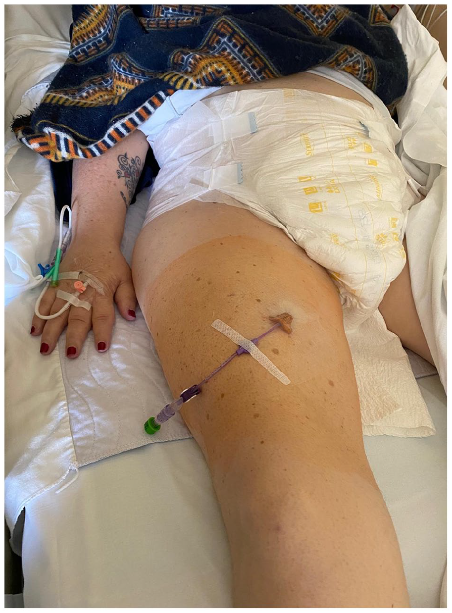
One hundred and fifteen CVADs were analyzed: 29 CICCs (25.2%), 11 PICCs (9.5%), and 75 FICCs (65.2%; see Table 1). Most of the catheters were single lumen: 20/29 in the CICC group, 10/11 in the PICC group, and 74/75 in the FICC group. PICCs were inserted only in 10 ALS patients with movement of the arms still preserved, and in one patient with Duchenne disease.
Central venous access devices inserted in the study period.
As regards CICC insertion, the thoracic tract of the axillary vein was accessed by the short-axis/out-of-plane approach in 21 patients, and with an oblique-axis/in-plane approach in eight patients. All PICCs were inserted in brachial or basilic veins by the short-axis/out-of-plane approach. As regards FICC insertion, the venipuncture of the superficial femoral vein (SFV) was performed as short-axis/out-of-plane in 40 patients and as oblique-axis/in-plane in 35 patients. The right SFV was cannulated in 43 patients while in 32 patients the left SFV was cannulated. The depth of the SFV ranged between 2 and 4 cm, with a caliber ranging between 6 and 8 mm.
All catheters were secured by subcutaneous anchorage (Secur-a-Cath, Interrad), and the exit site sealed with glue (octyl-butyl-cyanoacrylate) and covered with semi-permeable transparent dressing.
The mean duration of CVADs was 21 days (range 10–40). Forty-eight patients (44%) went home with the CVAD, since they required home parenteral nutrition.
Treatment with anti-thrombotic drugs (direct oral anticoagulants or low molecular weight heparin) was overall reported in 71 patients out of 115 (61.7%), somehow less frequently in the FICC subgroup (37 patients out of 75: 49.3%).
There were no intra-procedural complications. As regards late complications, there was no symptomatic CRT in any group of patients. We had two secondary malpositions, one in the PICC group (tip migration into the contralateral brachiocephalic vein) and one in the FICC group, and both were corrected by guidewire replacement of the catheter. We recorded two persistent withdrawal occlusions (PWO), both in the FICC group. One FICC was accidentally removed by the patient.
As shown in Table 1, while the number of patients admitted to the ward remained approximately the same, from 2021 to 2023 there was a shift from a prevalence of CICCs to a prevalence of FICCs, and an increase in the number of CVADs inserted yearly.
Discussion
Patients with ALS are often admitted to the hospital for respiratory insufficiency, necessity of artificial nutrition, or infection.10,11 In these patients, CVADs are often necessary for intra and extra-hospital intravenous treatments (such as antibiotic therapy and parenteral nutrition) and for blood sampling. Home parenteral nutrition (HPN) is usually indicated when the PEG is not feasible for respiratory problems, anatomical difficulties, patient’s refusal, or advanced dementia.
Clinical data on the choice of CVAD and on catheter-related complications in ALS patients are very scarce.12,13 In particular, there are few data about the incidence of catheter-related thrombosis in this population of patients. In a clinical study on 25 PICCs inserted in ALS, 12 only one CRT was reported; though, the authors provided no details regarding the choice of the vein, the exit site, the method of securement; fluoroscopy was used for tip location. In an older study, 13 73 totally implanted venous access devices were used for HPN in ALS patients, and there was a 36% incidence of complications (3.11 complications/1000 catheters days), most of them being infections (1.93 episodes/1000 catheters days), with only one CRT; data regarding the technique of the venipuncture or the tip location were not reported.
The most interesting result of the present study is the absence of any event of symptomatic CRT in 115 CVADs inserted in a span of almost 3 years. This might be explained by several factors:
- all CVADs were inserted according to insertion bundles which already include the main strategies for prevention of CRT (choice of a vein with an internal diameter at least three times the external diameter of the catheter; ultrasound-guided venipuncture with micro-puncture kits; accurate intra-procedural tip location by IC-ECG and/or ultrasound; proper securement of the catheter);
- PICCs were inserted only in ALS patients with preserved movements of the upper limbs;
- anti-thrombotic prophylaxis might also have played a role, though it was used in only in 61.7% of patients.
Interestingly, in this retrospective study including 75 consecutive FICCs in patients with different degrees of paresis, the femoral approach—which is often regarded at high risk for CRT—was not associated with any symptomatic thrombotic event; this may be explained by the systematic adoption of SFV cannulation at mid-thigh rather than cannulation of common femoral vein at the groin, considering that recent clinical studies suggest that the SFV approach is not associated with high risk of CRT. 2
This study strongly supports the importance of an appropriate insertion bundle, designed to minimize all catheter-related complications, including CRT. In a recent systematic review and meta-analysis on 5914 PICCs inserted with ultrasound guidance for venipuncture, 14 catheter tip location and a catheter size selection strategy, the rate of symptomatic PICC-related venous thrombosis was of 2.4% in the general population, 2.2% in oncologic patients, and 5.9% in hematologic patients. The importance of adhering to appropriate insertion bundle to minimize CRT was also confirmed by another recent meta-analysis 15 which reported an incidence of PICC-related thrombosis of 2.3%. Use of ultrasound for the choice of the vein, adoption of a vein to catheter ratio of 3:1 or more, appropriate choice of exit site through the tunneling technique, intraprocedural tip location and adequate stabilization are of paramount importance in reducing the risk of thrombosis, and they are all included in the GAVeCeLT insertion bundles. Adherence to the GAVeCeLT bundle in a cohort of 486 CVADs (both CICC and PICC) in a neurological intensive care unit resulted in zero incidence of symptomatic thrombosis. 16
As regards the secondary endpoints, immediate and early catheter-related complications were also absent, and this may be explained by the optimal performance of the two operators (one physician and one nurse), both specifically and appropriately trained in insertion of CVADs, as well by the consistent adoption of the above-mentioned insertion bundles, which are designed to minimize all insertion-related complications.
Late mechanical complications occurred rarely (4/115), and they were not associated to any damage to the patient.
Another interesting information derived from this retrospective analysis is the shift from a prevalence of CICCs in 2021 (60%) to a prevalence of FICC in 2023 (65%; Table 1). In our NeMO ward, FICCs have become the first choice CVAD in ALS patients, also because of the increasing experience of our vascular access team in the technique of SFV puncture at mid-thigh and in the ultrasound-based method of tip location. The SFV approach has some specific advantages in ALS patients:
- similarly to PICCs (which are often contraindicated in ALS patients), FICCs inserted by SFV puncture can be regarded as a non-invasive procedure: they can be safely inserted even in patients receiving anti-thrombotic drugs, 17 and they are not associated with cardiopulmonary complications that may occur during CICC insertion (pneumothorax, hemothorax, cardiac arrhythmias, air embolism);
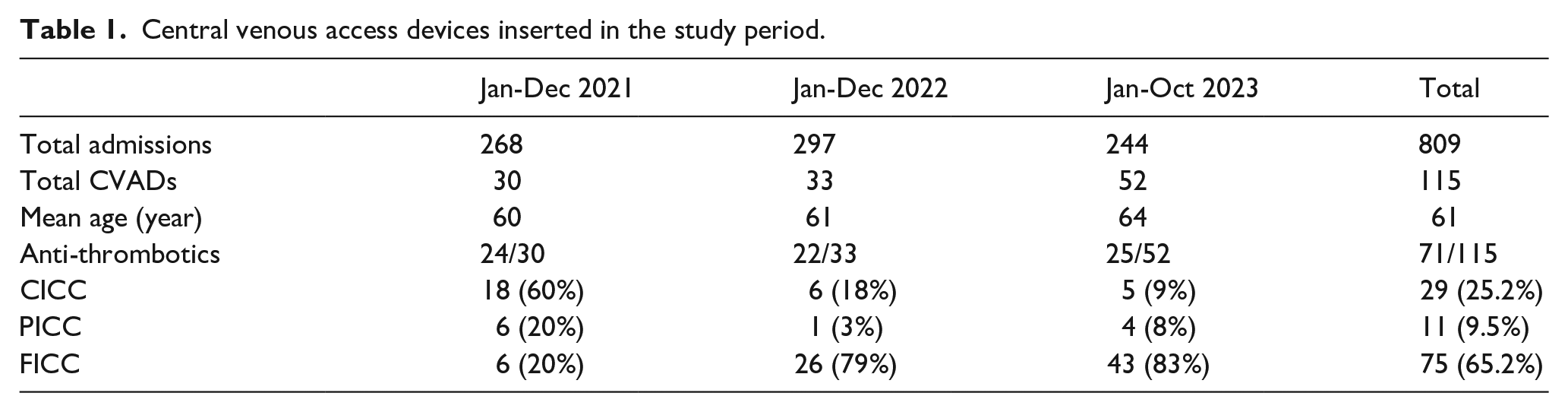
- due to the reduced muscle mass of the thigh, the SFV is less deep in ALS patients (2–4 cm) if compared with the general population (3–6 cm), 2 and thus easier to puncture and cannulate (Figure 1); its caliber is quite large (6–8 mm), and thus appropriate for the adoption of 4 Fr or 5 Fr venous catheters;
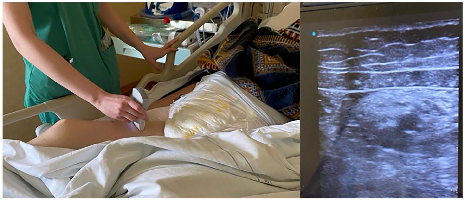
- ultrasound-based intra-procedural tip location using the trans-hepatic window with visualization of the catheter inside the inferior vena cava (Figure 2) is easy and consistently feasible, even in patients with spastic lateral decubitus, while a subxiphoid window may be very difficult in ALS patients due to the presence of gastrostomy, or air in the stomach secondary to non-invasive ventilation 18 ;
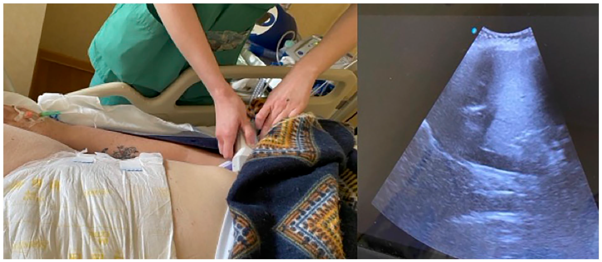
- the exit site at mid-thigh is far from the tracheal secretions, and also far from the groin area, which is contaminated by bacteria; also, dressing is quite stable and easy to manage (Figure 3).
Ultrasound visualization of the superficial femoral vein at mid-thigh.
Ultrasound visualization of the sub-diaphragmatic tract of the inferior vena cava, for verifying the location of the tip of the femoral catheters.
Exit site at mid-thigh, after placement of a central catheter by cannulation of the superficial femoral vein.
Limitations
This study has several limitations:
- it is a retrospective study;
- the incidence of infective complications could not be assessed, due to the unavailability of some key information regarding the extra-hospital history of the CVAD;
- all CVADs were inserted by a small team of two operators particularly expert and competent, adopting systematically the GAVeCeLT bundles, so that the optimal results in terms of complications may not be easily repeated elsewhere.
Conclusion
The retrospective analysis of this experience strongly suggests that the presence of paraplegia or tetraplegia is not necessarily associated with an increased risk of CRT: in this study on 115 CVADs, no symptomatic thrombosis was reported. This may be explained by the wise choice between different options (PICC or CICC or FICC, depending on the clinical conditions and on the status of the vasculature, as evaluated by ultrasound) and—most importantly—by the adoption of well-designed insertion bundles including appropriate strategies for CRT prevention.
The consistent adoption of such insertion bundles designed by GAVeCeLT may also explain the absence of puncture-related complications and of catheter dislodgments, as well as the very low incidence of other non-infective complications (two cases of tip migration and two cases of PWO).
An additional clinical implication of the study lies in the safety of the femoral route in patients with paretic lower limbs, provided that the exit site of the catheter is located at mid-thigh. The insertion of FICCs by direct cannulation of the SFV may have in paraplegic/tetraplegic patients the same advantages which have been described in the general population.
Footnotes
Declaration of conflicting interests
The author(s) declared no potential conflicts of interest with respect to the research, authorship, and/or publication of this article.
Funding
The author(s) received no financial support for the research, authorship, and/or publication of this article.