
Abstract
Introduction:
The distal radial artery presents a particular challenge for puncture and catheterization due to its diminutive size, tortuous path, and tendency to spasm, increasing the risk of procedural failure and injury. Ultrasound guidance improves success rates and reduces risk in radial artery catheterization. This study evaluates the efficacy and safety of a refined dynamic needle tip positioning technique for distal radial artery access.
Methods:
One hundred twelve patients were randomized to either the modified dynamic needle tip positioning technique (MDNTP) or palpation guidance groups (palpation group), each with 56 participants. The primary outcomes were the success rate of the initial puncture and overall puncture success rate, while secondary outcomes included procedural time and complications such as puncture site hematoma and radial artery occlusion within 24 h.
Results:
The MDNTP group exhibited superior initial puncture success (71.43% vs 46.43%, p < 0.05) and fewer puncture attempts (median 1 (1, 2) vs 2 (1, 4), p < 0.05), resulting in a higher overall puncture success rate (98.21% vs 87.50%, p = 0.028). Notably, sheath insertion times were significantly shorter (17 (12, 21) s vs 57 (32, 100) s, p = 0.001) and the Sheath insertion success rate was higher (96.43% vs 82.14%, p = 0.015) in the MDNTP group. Furthermore, the incidence of puncture site hematomas was reduced (5.36% vs 19.64%, p = 0.022), although puncture time was longer (60 (28, 116) s vs 40 (15, 79) s, p = 0.033). Despite these differences, total procedural time and the incidence of radial artery occlusion at 24 h postoperatively were comparable between the two groups.
Conclusion:
The MDNTP technique boosts the success of distal radial artery puncture and catheterization, reducing the risk of complications associated with the procedure.
Keywords
Introduction
Distal radial access offers benefits like rapid hemostasis and patient comfort but faces challenges due to small vessels, spasm susceptibility, and low catheterization success rates.1–3 Expert consensus recommends ultrasound guidance to improve these outcomes, although clinical evidence is limited. 4 Two techniques, the Dynamic Needle Tip Positioning Technique (DNTP) and Acoustic Shadowing, have emerged from the short-axis out-of-plane method, with their operational methods detailed by Clemmesen et al. 5 and Quan et al. 6 These techniques have demonstrated improved puncture success and reduced complications in challenging cases, with DNTP achieving the highest success rate in pediatric scenarios, as evidenced by a Meta-analysis.7–10
However, our preliminary trials revealed challenges with DNTP, such as difficulty distinguishing the needle tip on ultrasound due to its similarity in echo with subcutaneous tissue. The Acoustic Shadowing Technique facilitated rapid puncture but limited depth of entry. Additionally, spasmodic or tortuous vessels could complicate wire delivery. To address these issues, MDNTP utilizes Acoustic Shadowing for initial vessel puncture and DNTP for more profound needle advancement, placing the needle tip in a region rich in blood flow for optimal ultrasound guidance. This study aims to evaluate the application, efficacy, and safety of MDNTP in distal radial artery puncture and catheterization.
Study protocol
Methods
This prospective randomized controlled clinical trial was approved by the Ethics Committee of Guangdong Provincial Hospital of Traditional Chinese Medicine (Ethical Approval Number: BF2022-140-01, Approval Date: July 27, 2022) and registered at ClinicalTrials.gov, Registration Number: NCT06196749. The study was conducted at an 850-bed tertiary-grade A hospital in Zhuhai, Guangdong Province, China. All procedures followed the principles of the Declaration of Helsinki and obtained written informed consent from the patients.
Sample
This research enrolled patients planning to undergo coronary angiography or percutaneous coronary intervention procedures from August 2022 through December 2023. The inclusion criteria included participants aged between 18 and 85 years who provided informed consent to participate in the study. Exclusion criteria were defined as follows: (i) Absence of a palpable distal radial artery pulse; (ii) An anomalous result on Allen’s test; (iii) A distal radial artery diameter, as measured by ultrasound, that was smaller than 1.8 mm; and (iv) Hemodynamic instability.
Intervention
Participants received study details from hospital physicians and provided informed consent. Randomization was overseen by a statistician, with allocations concealed in sealed envelopes. Interventional cardiologists with at least 50 successful catheterizations under either palpation or ultrasound guidance assigned patients to MDNTP or palpation groups based on odd/even numbers and performed the procedures. Specialized nurses in the catheterization suite managed data collection. In the catheterization lab, patients were positioned supine with arms extended to orient the anatomical snuffbox superiorly, a triangular concave area formed by the tendons of the abductor pollicis longus, extensor pollicis longus, extensor pollicis brevis, and the radial styloid process. The site was sterilized and draped, and a 22G × 45 mm puncture needle (Braidin™ II Vascular Sheath Kit, APT Medical, China) was used for distal radial artery puncture within the snuffbox. The experimental group underwent MDNTP-guided puncture, while the control group employed palpation guidance. Following vascular sheath insertion via the Seldinger technique or its modified version, all patients received 200 µg of nitroglycerin and a heparin bolus at 3000 units. PCI patients received a dosage based on body weight. Hemostasis was maintained using a radial artery compression device (TR-Band, Terumo, Japan) for 3 h post-angiography, extended to 4 h post-PCI for PCI patients. If bleeding occurred, compression was prolonged an additional 30 min before release. A Doppler ultrasound assessment of the radial artery was performed by the ultrasound department 24 h post-procedure on all patients.
Modified dynamic needle tip positioning technique
MDNTP improves DNTP by employing the Acoustic Shadowing Technique for initial vessel visualization and needle tip tracking, utilizing a linear array probe (12–2 MHz, Hitachi L441) on an ultrasound machine (ATIETTA60, Hitachi, Japan). The probe is coated with coupling gel and enclosed in a sterile sheath. We position the probe to visualize the radial artery lumen under a short-axis view between the two acoustic shadows. We direct the puncture needle at a 30° angle toward the sutures, with the needle tip visualized around or within the vessel lumen. The probe is then repositioned toward the proximal end to guide the needle tip further into the vessel, and the tip reappears within the lumen as it advances. This process is repeated to advance the needle as deeply as possible into the vessel. Figure 1 illustrates the detailed operational steps.
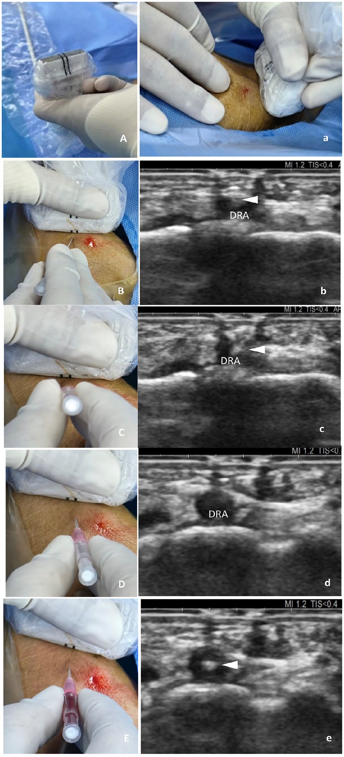
Modified dynamic needle tip positioning ultrasound guidance technique. (A) Silk sutures are positioned parallel to the probe’s center. (a) The ultrasound aligns the distal radial artery (DRA) between the sutures’ shadows. (B) The puncture needle targets the midpoint between the sutures. (b) The needle tip (indicated by a triangle) becomes visible above the vessel. (C) Move the probe proximally, and adjust and advance the needle. (c) The tip enters the vessel, marked by a hyperechoic spot. (D) Continue to move the probe proximally. (d) The needle tip vanishes from view. (E) Adjust and advance the needle. (e) The needle tip reappears within the center of the vessel. Repeat as necessary to ensure the tip remains within the lumen.
Palpation technique
Interventionists guide the needle insertion for radial artery puncture by palpating the location with the most vital arterial pulse. They angle the needle at 30°–45° and proceed until they observe blood return in the needle hub. Then, they adjust the angle slightly and advance the needle a few more millimeters before carefully removing the stylet. If necessary, they slowly withdraw the cannula until pulsatile blood flow indicates correct placement. Throughout the procedure, they actively avoid hitting the scaphoid or trapezium bones to prevent pain-related vasospasms.
Observation indicators and definitions
The study’s observed metrics include first-pass puncture success rate, overall puncture success rate, puncture time, number of puncture attempts, sheath insertion success rate, arterial sheath insertion time, total procedure time, incidence of hematoma at the puncture site, 24-h post-procedure radial artery occlusion rate. First-pass puncture success rate: The proportion of successful radial artery punctures in the first attempt. Puncture success rate: The proportion of successful radial artery punctures among all attempts. Puncture time: The duration from needle insertion until a pulsatile blood spray is observed from the puncture needle sheath, recorded as 1200 s in case of failure. Number of puncture attempts: The number of times the needle is retracted to beneath the skin and reinserted since the first insertion. Sheath insertion time: Measured from successful puncture to arterial sheath insertion and blood withdrawal, or set to the average of successful cases if puncture fails, and 1200 s if catheterization fails after a successful puncture. Sheath insertion success rate: The proportion of successful arterial sheath insertions. Total procedure time: The sum of the puncture time and sheath insertion time. Incidence of hematoma at the puncture site: The incidence of swelling and bruising larger than 1 cm around the puncture site. 24-h post-procedure radial artery occlusion rate: The rate of radial artery occlusion, as detected by Doppler ultrasound within 24 h, is divided into proximal (above the anatomic snuffbox) and distal (within the snuffbox) radial artery occlusion. Puncture and sheath insertion failure is classified as taking over 20 min or when the operator decides to use an alternative access route. Additionally, the body surface area (BSA) for each participant was calculated as part of the study using the Du Bois formula: BSA (m2) = weight (kg)0.425 × height (cm)0.725 × 0.007184.
Sample size estimation and statistical analysis
Based on our preliminary trial results, the first-pass puncture success rate with MDNTP was 70%, compared to 40% with the palpation guidance method. To achieve a test power of 80% and a confidence level of 95%, we estimated a sample size of 56 cases per group. Statistical analysis was conducted using SPSS software (version 27.0, IBM, USA). All attempted cases and successful cases were included in the analysis. The Shapiro-Wilk test was employed to test normal distribution. Normally distributed continuous data are presented as mean ± standard deviation (±s), and non-normally distributed data are expressed as median (interquartile range; M (Q)). The t-test or Mann-Whitney U test was used for intergroup comparisons of continuous data. Categorical data are presented as counts and percentages, with the χ2 test used for comparison. A p value less than 0.05 was considered statistically significant.
Results
Patient characteristics
A total of 122 patients were recruited from August 2022 to December 2023. After excluding five who refused to participate, one with unstable hemodynamics, one with an abnormal Allen test, and three with a distal radial artery diameter less than 1.8 mm, a total of 112 patients were included in the study, with 56 in each group (Figure 2). Baseline characteristics, including age, gender, body mass index, body surface area, medical history, medication history, heparin dosage, sheath size, and distal radial artery diameter, were similar between the two groups, with no statistically significant differences (p > 0.05). Detailed data are presented in Table 1.
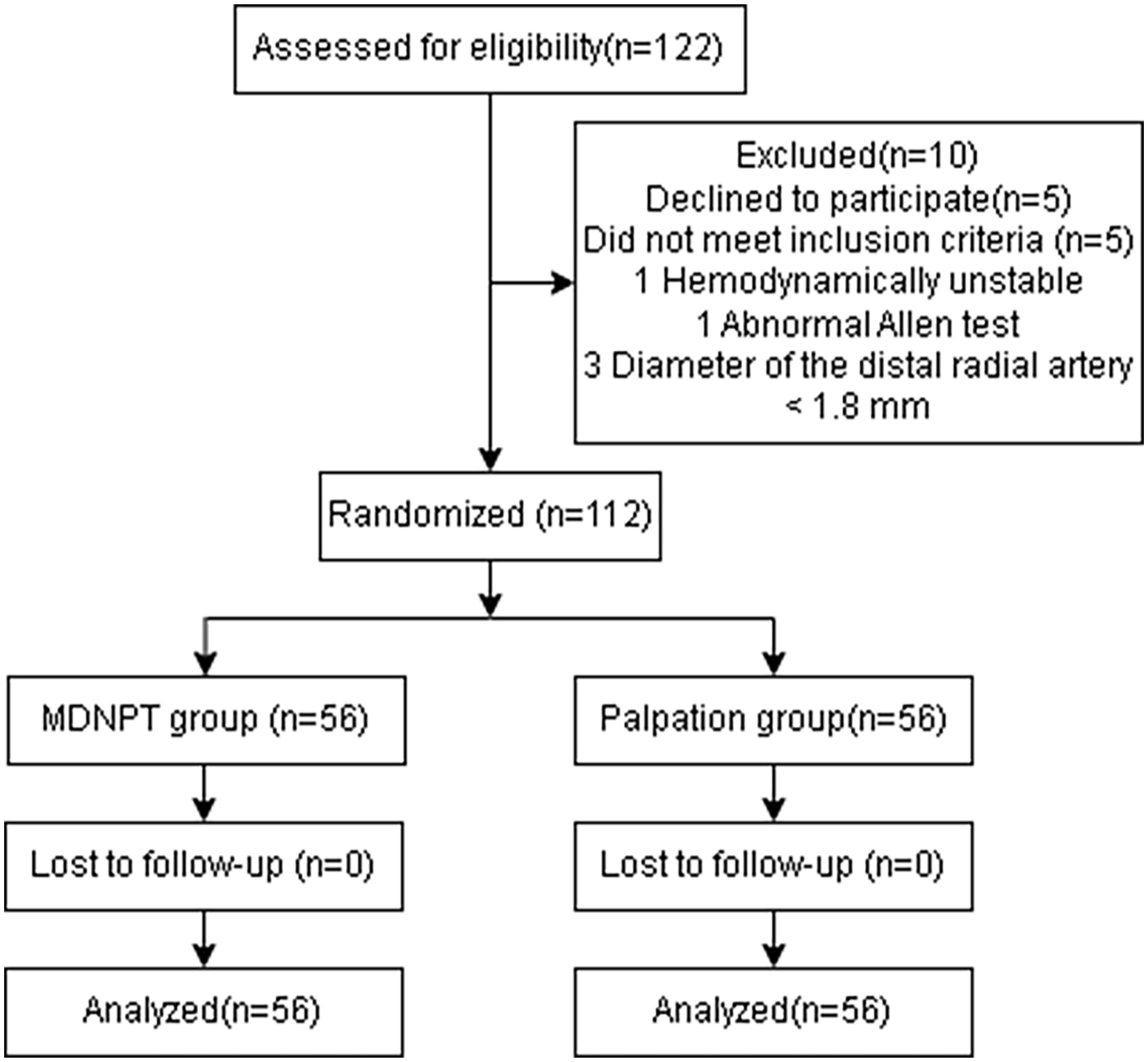
Flow diagram of patient recruitment and exclusion criteria for the study.
Patient characteristics.
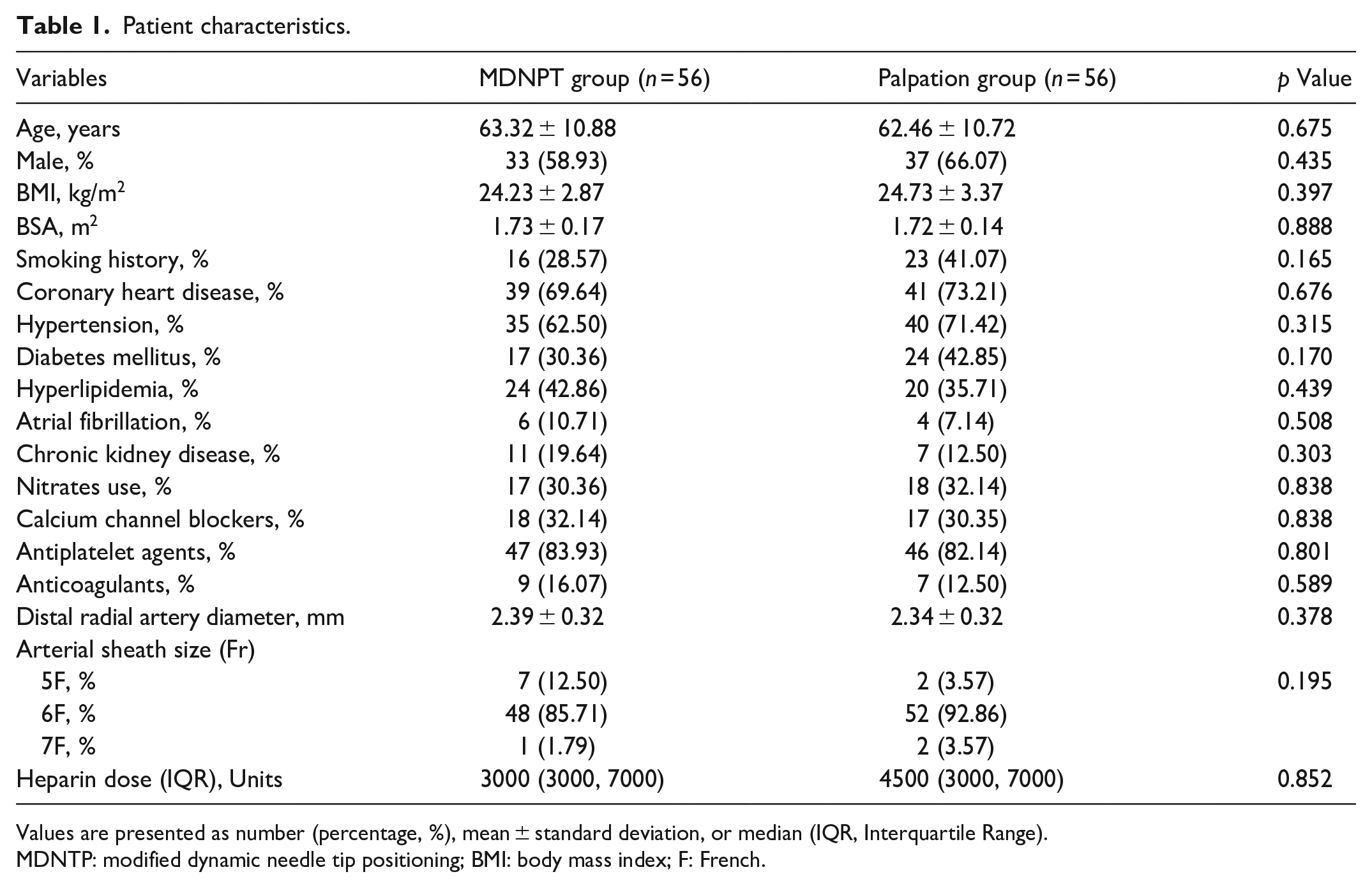
Values are presented as number (percentage, %), mean ± standard deviation, or median (IQR, Interquartile Range).
MDNTP: modified dynamic needle tip positioning; BMI: body mass index; F: French.
Study outcomes
Compared to the palpation group, the MDNTP group demonstrated a higher first-pass puncture success rate (71.43% vs 46.43%, p = 0.007), fewer puncture attempts (1 (1, 2) needles vs 2 (1, 4) needles, p = 0.004), a higher overall puncture success rate (98.21% vs 87.50%, p = 0.028), a shorter sheath insertion time (16 (12, 22) s vs 70 (33, 105) s, p = 0.001), a higher sheath insertion success rate (96.43% vs 82.14%, p = 0.015), and fewer puncture site hematomas (5.36% vs 19.64%, p = 0.022). Among patients who successfully underwent catheterization, the MDNTP group had a longer puncture success time compared to the palpation group (60 (28, 116) s vs 40 (15, 79) s, p = 0.033). The number of puncture attempts, total procedure time, hemostasis time, and the incidence of radial artery occlusion at 24 h were similar between the two groups, with no statistically significant differences. Detailed study outcomes are presented in Table 2.
Clinical outcomes.
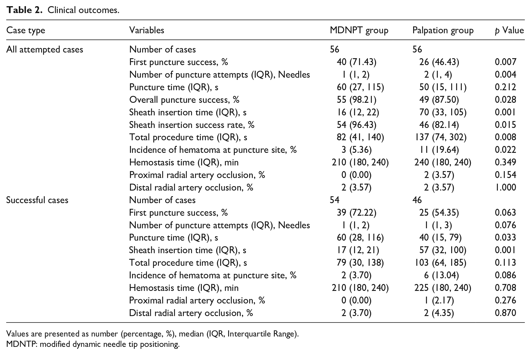
Values are presented as number (percentage, %), median (IQR, Interquartile Range).
MDNTP: modified dynamic needle tip positioning.
Among the 10 cases that experienced failure in the palpation group, a secondary intervention utilizing MDNTP was employed in six instances, with four of these resulting in successful navigation. The remaining two cases were unsuccessful due to complications arising from arterial spasm and excessive vessel tortuosity. Across the entire study population, a total of eight patients were crossed over to the proximal radial artery approach, all of whom achieved successful cannulation. Figure 3 exemplifies such a scenario where, following an unsuccessful attempt under palpation guidance, ultrasonographic imaging disclosed the development of a vessel dissection hematoma, which was effectively navigated and managed using MDNTP technology.
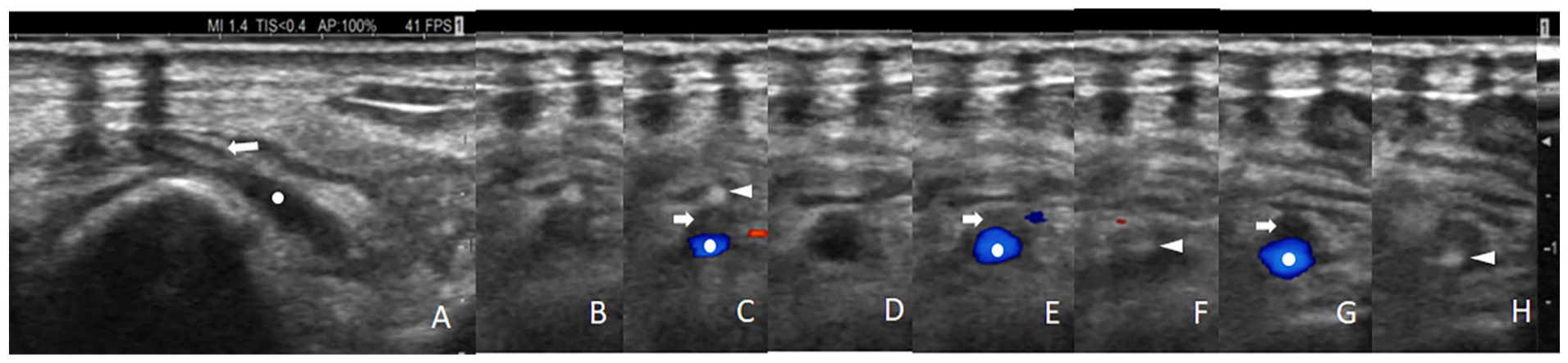
Application of the modified dynamic needle tip positioning technique in a distal radial artery catheterization case failed by palpation guidance. (A) The long-axis view shows a dissection in the anterior wall of the distal radial artery, (B) the needle tip imaging above the lumen, (C) color Doppler reveals the true lumen located slightly below the lumen, (D) moving the probe proximally, the needle tip disappears, (E) the dissection hematoma is above the lumen, while the true lumen is below, (F) adjusting the needle tip toward the true lumen, (G) moving the probe proximally again, the needle tip disappears, with the true lumen located slightly lower left in the lumen, (H) adjusting the needle tip to reach the lower left true lumen. Arrows represent the dissection hematoma, dots represent the true lumen, and triangles represent the needle tip.
Discussion
This study evaluated the effectiveness and safety of MDNTP for guiding distal radial artery puncture and catheterization, demonstrating that MDNTP significantly improved first puncture, total puncture, and catheterization success rates compared to palpation guidance and was associated with a lower incidence of puncture site hematomas, boosting procedure success and lowering injury risk.
Our study found the MDNTP group had superior success rates in first puncture attempts, total puncture success, and catheterization results. Previous studies report 94%–70% success rates for DRA catheterization using palpation guidance.11–13 Our lower success rate of 82.14% may be due to the cohort’s demographics, especially the smaller body surface area common in Eastern populations, which can reduce the chances of successful punctures. 14 Recent studies highlight improved puncture success and reduced complications with ultrasound guidance, especially in infants, elderly, and obese patients.7,10,15 Ultrasound-guided techniques offer benefits like visualizing vessel lumens and blood flow. Mori et al.’s team achieved a 97% success rate for DRA procedures with ultrasound, surpassing the 87% rate for palpation guidance, and resolving all palpation failures. 16 The distal radial artery’s narrow diameter, tortuous path, and spasm susceptibility make puncture challenging. The DNTP technique tracks the needle tip at the subcutaneous tissue layer, but accurate identification can be challenging for ultrasound due to the similar echogenicity between the tissue and the needle tip. 17 The MDNTP technique, aided by a bifilar thread marker, improves needle tip visibility for precise positioning either by causing a lumen depression (see Figure 1(b)) or by presenting as a white dot within the hypoechoic lumen (see Figure 1(e)), guaranteeing accurate needle insertion into the vessel.
Our study also found the MDNTP group had a shorter sheath insertion time and higher success rate, likely due to reduced wire delivery time and increased success. Previous studies have indicated that wire delivery failure accounts for 69%–100% of unsuccessful DRA catheterizations, primarily due to vessel spasms at the exit of the puncture needle sheath or the presence of a vessel dissection.18–20 MDNTP mitigates this, as shown in Figure 3, by adjusting the needle tip to regions with robust blood flow signals, facilitating successful wire insertion.
In terms of procedural complications, the MDNTP group required fewer needle passes and exhibited a significantly lower incidence of hematoma compared to the palpation group. This aligns with a study by Liu et al., showing a lower hematoma rate in the DNPT group (3.3%) than in the palpation group (26.7%). 17 The reduced puncture attempts and posterior wall perforations in MDNTP may account for this. 21 The 24-h radial artery occlusion rate was similar in both groups, with an overall rate of 5%, matching the TENDERA trial’s 4%. 12 Overall, the MDNTP technique demonstrates good safety profiles.
Limitations
This study has several limitations, including its non-blind, single-center design and the non-continuous inclusion of samples during the COVID-19 pandemic. The research pause during the pandemic peak likely excluded sicker patients, skewing the sample toward less severe cases, which may introduce subjective and selection biases, affecting the generalizability and external validity of the results. Additionally, all operators had more than 50 cases of experience with both ultrasound and palpation guidance before the study, indicating that MDNTP may require a specific learning curve, and its efficacy may not be immediately reproducible in other centers.
Conclusion
The results of this study indicate that compared to palpation guidance, MDNTP enhances the success rate of wire delivery into the vessel by guiding the needle tip to regions rich in luminal blood flow, significantly improving the success rate of distal radial artery puncture and catheterization, and reducing the risk of injury. Therefore, MDNTP, as a safe and efficient guiding technique for distal radial artery puncture, has the potential for widespread application in clinical practice and serves as a reliable alternative when other guiding methods fail.
Footnotes
Acknowledgements
The submitter would like to express sincere gratitude to Professor Ding Chunhua for his valuable suggestions on the writing of this paper.
Declaration of conflicting interests
The author(s) declared no potential conflicts of interest with respect to the research, authorship, and/or publication of this article.
Funding
The author(s) disclosed receipt of the following financial support for the research, authorship, and/or publication of this article: This work was supported by the Guangdong Province Medical Science and Technology Research Foundation Project [Number B2021044]; the Zhuhai City Science and Technology Plan Project [Number ZH22036201210077PWC]; the Guangdong Provincial Hospital of Traditional Chinese Medicine Traditional Chinese Medicine Science and Technology Research Special Project [Number YN2020QN09].
Ethical approval
This study was approved by the Research Ethics Committee of Guangdong Provincial Hospital of Traditional Chinese Medicine (Approval Number: BF2022-140-01) on July 27, 2022.
Trial registration
Due to the impact of the COVID-19 pandemic, the trial registration process for this study was retrospectively registered at Clinicaltrials.gov, Registration Number: NCT06196749.