
Abstract
Tryptophan is an essential amino acid required for protein synthesis and a precursor of key bioactive metabolites produced through the kynurenine, indole and serotonin pathways. Beyond its nutritional role, tryptophan metabolism critically regulates the nervous, immune and endocrine systems, coordinates circadian rhythms via melatonin, and exerts antioxidant effects. These processes are profoundly remodeled during pregnancy. Pregnancy represents a unique physiological state characterized by systemic adaptations, including changes in immunity and erythropoiesis, while the placenta emerges as a central organ coordinating fetal development, maternal–fetal exchanges, immune tolerance, and protection against external infections. Increasing evidence indicates that tryptophan availability and its downstream metabolites play key roles in maintaining maternal–fetal and placental homeostasis. In particular, serotonin, traditionally viewed as a neurotransmitter, has emerged as a locally produced signaling molecule in the placenta, impacting trophoblast development, placental vascularization, and immune modulation. Dysregulation of tryptophan metabolic pathways has been associated with pregnancy complications and adverse developmental outcomes. This review aims to provide an overview of tryptophan metabolism during gestation, with a specific focus on serotonin as a key factor in placental homeostasis. Targeting tryptophan-derived pathways may offer novel opportunities to improve maternal and neonatal health.
Overview of Tryptophan Metabolism
Tryptophan
In 1901, Hopkins and Cole identified a previously unknown substance:
Beyond its fundamental role in

Tryptophan and the kynurenine, indole and serotonin pathways.
Kynurenine Pathway
Tryptophan is primarily degraded via 3 major metabolic pathways: the
Among them, the kynurenine pathway stands out as the dominant route of tryptophan catabolism. 9 For a long time, it was considered the unique metabolic pathway of tryptophan. This pathway plays a pivotal role in the immune system, notably by promoting immune tolerance and regulating inflammation.
The first step involves the degradation of tryptophan into N-formylkynurenine, by the enzymes indoleamine 2,3-dioxygenase (
N-formylkynurenine is then converted into
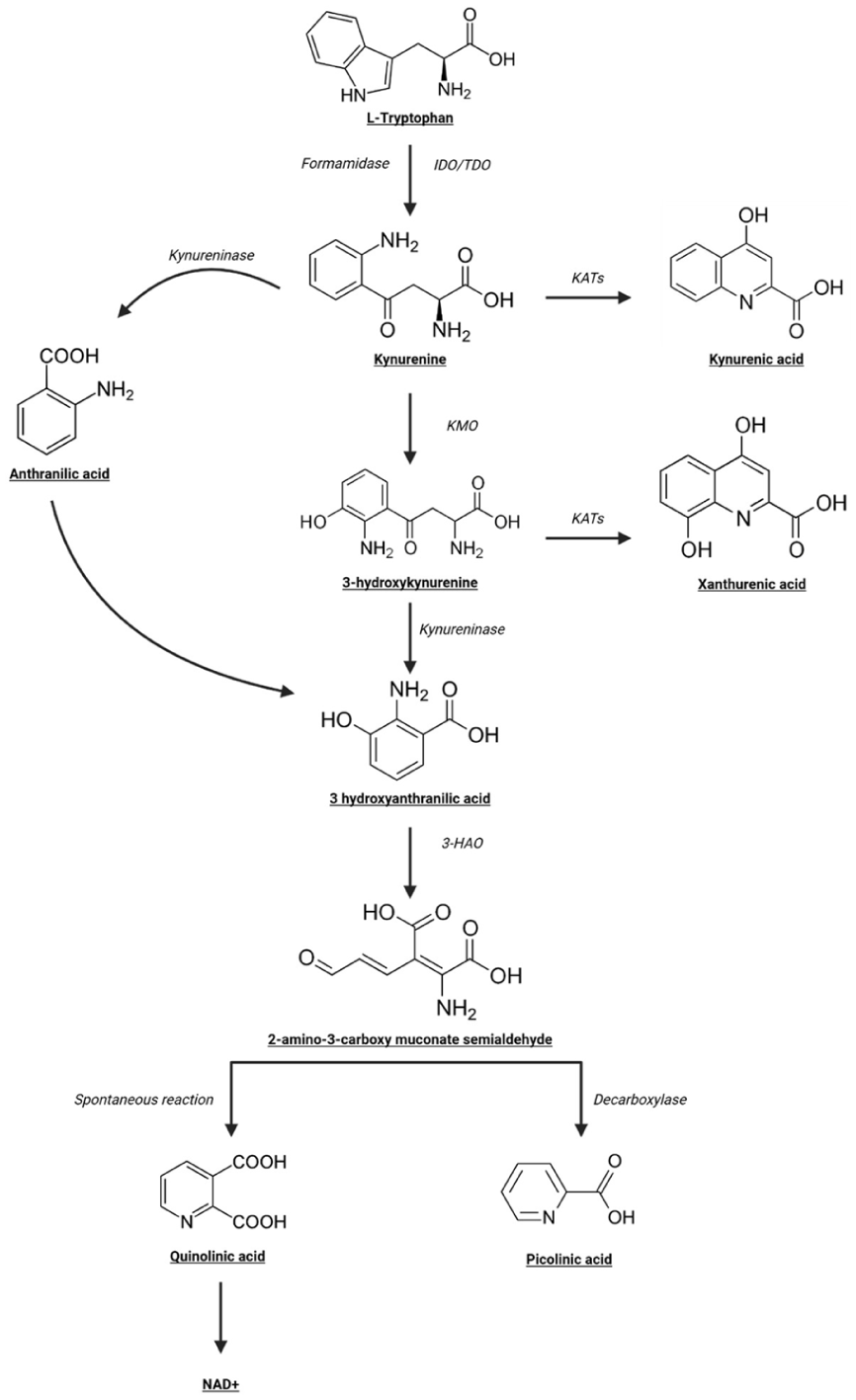
Overview of the kynurenine pathway. (3-HAO: kynurenine 3-monooxygénase; IDO: indoleamine 2,3 dioxygenase; KAT: kynurenine-amino transferase; KMO: Kynurenine 3-monooxygenase; NAD+: nicotinamide adenine dinucleotide; TDO: tryptophan 2,3-dioxygenase).
Under physiological conditions, kynurenine metabolites primarily play a role in the central nervous system (CNS). However, in peripheral organs, these metabolites may reflect a dysregulation of pathways such as inflammation or immune response. 15 Several metabolites derived from the kynurenine exert important physiological functions:
-
-
-
-
-
Indole Pathway
Around 5% of tryptophan is metabolized by the

Overview of the indole pathway.
Certain bacterial species of the microbiota can convert tryptophan into tryptamine via the enzyme tryptophan decarboxylase.
32
Recent discoveries on the importance of the microbiota for overall health have highlighted new insights into the indole pathway, linking gut microbiota and tryptophan metabolism. Metabolites from this microbial pathway play diverse
Serotonin Pathway
Approximately 5% of tryptophan is metabolized via the serotonin pathway, as shown in Figure 4. This pathway produces several key metabolites, including

Overview of the serotonin pathway.
Serotonin (5-HT)
Once synthesized, serotonin is stored into synaptic vesicles by vesicular monoamine transporter (

Serotonin synthesis and metabolism.
Fifteen 5-HTRs have been identified, grouped into 7 families (5-HT1 to 5-HT7). All are GPCRs, also known as 7-transmembrane domain receptors, except for 5-HT3, which is a ligand-gated ion channel. The activation of these receptors regulates a wide range of physiological and behavioral functions, depending on the receptor, tissue localization and cellular context.
In the
A second isoform of Tph,
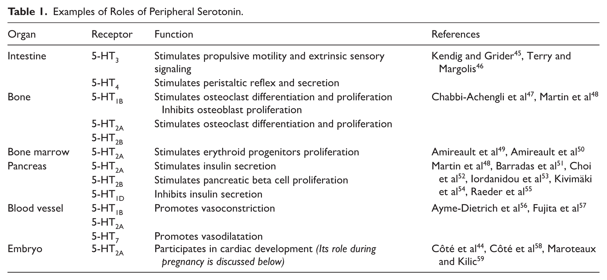
Examples of Roles of Peripheral Serotonin.
Melatonin
Roles of Melatonin in the Central Nervous System.

Several
Roles of Melatonin in Peripheral Organs.

Specific physiological conditions can modulate the action of these different tryptophan-derived metabolites especially during pregnancy: we will now explore how tryptophan metabolism is regulated throughout the 9 months of gestation.
Tryptophan Pathways Throughout Pregnancy
The
Tryptophan
Tryptophan and Pregnancy Immunity
Pregnancy presents an
It has been reported that circulating tryptophan levels decrease during pregnancy, and further decrease with gestational age.88,89 Abnormal tryptophan level has been associated with an increased risk of
Impact of Altered Tryptophan Levels during Pregnancy, Role of LAT
The fetal uptake of essential amino acids during pregnancy is critical for protein synthesis, as well as for neurotransmitters and nucleotides synthesis. Tryptophan crosses the placenta via the active L-type amino acid transporter (LAT), 98 a sodium-independent system composed notably of 2 exchangers, LAT1 and LAT2. 99 LAT1 and LAT2 are both expressed in human placenta on the syncytiotrophoblast,6,100 while only LAT2 is present on the fetal endothelial cells. 101
Some studies suggest that LAT expression levels may correlate with pregnancy parameters such as
Nonetheless, the causal relationship between LAT expression and pregnancy complications remains unclear, and it is not yet established whether changes in LAT expression are a cause or a consequence of these outcomes.
The impact of dietary tryptophan has also been studied in different models. A high-tryptophan diet before conception was associated with increased body weight, brain weight, and blood pressure. Alterations in embryonic hormonal balance, increased protein synthesis, and a decreased number of serotonergic neurons were also reported.110,111 A tryptophan-free diet before conception was associated with hypoandrogenism, hypoprolactinemia, and cardiorespiratory alterations.112-114 Finally, elevated systemic tryptophan levels were associated with a lower risk of preeclampsia and pregestational diabetes mellitus. 115 These effects may be explained by the role of tryptophan in protein synthesis, but are also likely largely driven by its downstream effects on serotonin, indole, and kynurenine synthesis.
Kynurenine
Kynurenine Levels during Pregnancy
As previously mentioned, plasma tryptophan levels decrease during pregnancy. According to Munn et al , this decline is attributed to the overexpression in placenta of IDO, which suggests a concurrent increase in circulating kynurenine.
90
However, studies have shown that kynurenine levels in maternal blood
The placenta produces and metabolizes kynurenine, expressing both key enzymes
Kynurenine Metabolites during Pregnancy
The kynurenine can cross the placenta to reach the fetal circulation. Oral administration of kynurenine has been shown to markedly increase its concentration in maternal plasma, placenta, fetal plasma and fetal brain tissues.
127
However, not all kynurenine metabolites are equally transferred. For instance,
Several enzymes of the kynurenine pathway – such as kynurenine monooxygenase (KMO), kynurenine aminotransferases (KYAT), kynureninase (KYNU), and quinolinate phosphoribosyltransferase (QPRT) – have also been identified in the placenta. Their expression decreases during the third trimester.
122
The authors suggest that placental kynurenine is mainly used for the production of metabolites such as
Impact of Altered Kynurenine Metabolite Levels during Pregnancy
Proper regulation of kynurenine-derived compounds is crucial for fetal development. Indeed, abnormal levels of kynurenine metabolites in placenta or fetal tissues have been associated with adverse pregnancy outcomes, such as abnormal birth weight or impaired central nervous system development.131,132
Several studies reported that elevated levels of kynurenic acid, hydroxykynurenine, and kynurenine are associated with preeclampsia and gestational diabetes mellitus.115,133 Overactivation of the kynurenine pathway may be related to the inflammatory environment observed for instance in preeclampsia. Lipopolysaccharide injection, an inflammatory stimulus, has been shown to overactivate the kynurenine pathway and increase the production of its metabolites. Excess levels of certain metabolites can lead to fetal alterations; for example, quinolinic acid has neurotoxic effects when overproduced.134-136 Upregulation of this pathway may also alter NAD⁺ production, contributing to cellular dysfunction.
The kynurenine pathway has also been linked to hormonal regulation. Estrogens can decrease kynurenic and picolinic acids, whereas progesterone increases kynurenic and quinolinic acid levels.137,138 It is well established that the large decrease in progesterone and estrogen levels after delivery contributes to the normalization of the kynurenine pathway postpartum. Endocrine alterations may contribute to kynurenine dysregulation and increased inflammatory responses. 139
Finally, in a uninephrectomized mouse model with an impaired adaptive increase of kynurenine, this failure to increase kynurenine led to preeclampsia-like symptoms, including impaired placentation. 140 This finding is consistent with reports linking abnormal kynurenine levels to fetal growth restriction, a common consequence of preeclampsia. 141 These effects may be explained by the role of the kynurenine pathway in trophoblast migration and invasion, which are critical steps in placental development; their impairment can contribute to fetal rejection. Most mechanistic studies have been conducted in placental cell lines and in vivo data remain limited. Nevertheless, current evidence suggests that disruption of the kynurenine pathway may contribute to abnormal placental development. 142
Indole Pathway
Little is known about the potential roles of indoles on placenta and pregnancy. However, it has been reported that during pregnancy, levels of indole-3-lactic acid (
Serotonin (5-HT)
Serotonin Levels During Pregnancy
The regulation of plasma serotonin levels during pregnancy remains poorly understood. Very few studies have investigated plasma serotonin under various physiological and pathological conditions. In 2011, Brand and Anderson conducted a comprehensive review of studies measuring plasma serotonin levels and highlighted the
Source of Serotonin in the Placenta
While it is generally accepted that the placenta synthesizes serotonin, whether placental synthesis alone can provide sufficient levels of serotonin during pregnancy remains a subject of debate. In 2011, Bonnin et al proposed that the placenta produces serotonin during gestation, influencing embryonic development.
153
However, subsequent studies, including Kliman et al,
154
suggested that most of the serotonin needed during gestation comes from maternal blood, and is transported by platelets. Evidence supports the idea that maternal serotonin is crucial for embryo development,
58
and that the placenta is equipped to uptake maternal serotonin.122,155 Thus,
Expression of Components of the Serotonergic System in the Placenta
It is well established that the placenta expresses components of the serotonergic system.
156
Studies have demonstrated the
The placenta, via the serotonin transporter (
Expression of serotonergic components in the placenta varies with gestational age. Tph gene expression declines, while MAO expression increases during pregnancy. However, Tph enzymatic activity remains unchanged, in contrast to MAO activity, which rises over time. This suggests that the influence of serotonin on the placenta decreases toward the end of gestation. 122 MAO regulation is particularly important to control placental serotonin levels and avoid vasoconstriction caused by elevated serotonin.157,161,162 However, serotonin transporter expression increases toward term,122,155,163 likely reflecting the growing serotonergic needs of the developing fetus.
Impact of Altered Serotonin Levels during Pregnancy
Given the well-established function of serotonin in the central nervous system, several studies have investigated its role in
For instance, altered serotonin levels have been observed in children with neurodevelopmental disorders such as autism, suggesting a potential role for serotonin in neurodevelopment.164-167
Dysregulation of the serotonergic system during gestation has been associated with long-term consequences, including behavioral disorders such as hyperactivity, depression, and anxiety.165,168
A study using a Tph2 KO mouse model has highlighted abnormalities in neuroplasticity mechanisms. 169
Impaired fetal brain development has been reported in pregnancy with dysregulations in placental serotonergic components, notably increased serotonin synthesis and transport, and reduced degradation. 170 These last findings suggest that placental serotonin signaling, rather than total serotonin levels, may play a key role in early fetal brain development.
Regarding peripheral serotonin, analysis of the Tph1 KO mouse model (deficient in peripheral serotonin) has revealed
Several hypotheses have been proposed to explain this developmental delay. Key reproductive tissues contain serotonin and components of the serotoninergic system, including the uterus and the oocyte microenvironment (cumulus cells, oocytes and granulosa cells).171,172 Serotonin receptors expressed in reproductive tissue can activate the cyclic adenosine monophosphate (cAMP) signaling pathway, which is critical for hormonal synthesis, particularly steroidogenesis. 173 Dysregulation of serotonin levels may lead to abnormal steroidogenesis which has been associated with IUGR or impaired implantation.174,175
Moreover, serotonin-mediated activation of the cAMP pathway is involved in cell proliferation, a process essential for normal embryonic development. 176
In addition, serotonin is known to be present in stem cells, where it contributes to stem cell survival. Reduced serotonin levels can increase stem cell apoptosis via the protein kinase B (AKT)-Forkhead box O1 (Foxo1) signaling pathway.177,178
Another hypothesis that could explain developmental delay involves alterations at the maternal-fetal interface. Serotonin also plays a crucial role in
Finally, peripheral serotonin is essential for embryonic
SSRI Exposure during Pregnancy
In clinical settings, serotonin reuptake inhibitors (SSRIs), which block SERT, are commonly prescribed for anxiety or depression. Some studies report a small risk of
Furthermore, it has been shown that SSRI treatment on BeWo cells and placental explants affects endocrine function, cell proliferation and differentiation.181,185
The evidence argues that the use and dosage of SSRIs during pregnancy should be carefully evaluated and adjusted according to the severity of maternal depression. 186
Melatonin
Melatonin Levels during Pregnancy
Placental serotonin is metabolized into melatonin. The placenta expresses both enzymes involved in melatonin synthesis (AANAT, HIOMT) and the receptors involved in its signaling (MT1A and MT1B (MT2).187-189 Melatonin present in the maternal circulation – coming from either pineal or extra-pineal sources – can cross the placenta and enter the fetal circulation.
190
During pregnancy,
Impact of Altered Melatonin Levels during Pregnancy
While global melatonin homeostasis during pregnancy remains insufficiently characterized, several studies have highlighted the crucial role of placental melatonin. First, melatonin seems to contribute to
A negative correlation has been observed between MT1A receptor expression and adverse pregnancy outcomes such as
Beyond its role in placentation, melatonin exerts protective effects in the primary human trophoblast cells by modulating autophagy under hypoxic conditions, improving cellular respiration and mitochondrial activity as well as potent anti-inflammatory and antioxidant.188,190,196 It has notably
Melatonin appears particularly critical during the early stages of
Finally, melatonin also plays a role in
In the female organism, melatonin modulates granulosa cell function by reducing oxidative stress and protecting cells from apoptosis through decreased p53 expression.206,207 Similar signaling pathways have been described in oocytes. 208
Altogether, the effects of melatonin on reproductive tissues lead to improved embryo quality and fertilization rates.
Conclusion
Tryptophan metabolism plays a crucial role in pregnancy, acting through multiple downstream pathways that contribute to fetal development, placental development and function, and maternal adaptation. Among its metabolites, serotonin and melatonin stand out as key modulators of embryonic and placental development. Several studies have highlighted the importance of peripheral serotonin, particularly during early gestation, in various processes such as placental structure, hormone secretion, immune tolerance, and oxidative stress responses. The placenta is both a regulator and a potential source of serotonin and melatonin, reflecting the complexity of tryptophan-derived signaling in pregnancy.
However, the current body of evidence is subject to several limitations. Many findings are derived from animal models, which may not fully recapitulate human pregnancy, and human studies are often observational, limiting causal inference. In addition, methodological heterogeneity in the assessment of tryptophan metabolites, differences in gestational timing, and limited longitudinal data complicate direct comparisons across studies. Furthermore, the interactions between the serotonin, melatonin, and kynurenine pathways remain incompletely understood.
Altogether, these findings still underscore the significance of maintaining balanced tryptophan metabolism during gestation. Disruptions in its pathways – whether due to genetic, environmental, or pharmacological factors – may contribute to pregnancy complications or long-term developmental outcomes.
Future research should focus on longitudinal and integrative approaches to better characterize the dynamic regulation of tryptophan metabolism throughout pregnancy and to evaluate its potential as a therapeutic target to support maternal and fetal health.
Footnotes
Abbreviations
3-HAO: kynurenine 3-monooxygénase
5-HIAA: acide 5-hydroxyindoleacétique
5-HT: 5-hydroxytryptophan = serotonin
5-HTR: 5-HT receptor
AAD: aromatic amino acid decarboxylase
AhR: aryl hydrocarbon receptor
AMPA receptor: α-amino-3-hydroxy-5-methyl-4-isoxazolepropionic acid receptor
AANAT: Serotonin N-acetyltransferase
ASMT: Acetylserotonin O-methyltransferase
IDO: indoleamine 2,3 dioxygenase
KAT: kynurenine-amino transferase
KMO: Kynurenine 3-monooxygenase
KO: knock-out
LAT: L-type amino acid transporters
NAD+: nicotinamide adenine dinucleotide
NAT: serotonin-N-acetyltransferase
NMDA receptor: N-methyl-D-aspartate receptor
MAO: monoamine oxidase
MT1 and MT2: melatonin receptors
PD-1: programed cell death protein 1
SERT: serotonin transporter
SSRI: selective serotonin reuptake inhibitors
TDO: tryptophan 2,3-dioxygenase
Tph: tryptophan hydroxylase
Tregs: regulatory T cells
VMAT: vesicular monoamine transporter
WT: wild type
Author Contributions
Funding
The authors received no financial support for the research, authorship, and/or publication of this article.
Declaration of Conflicting Interests
The authors declared no potential conflicts of interest with respect to the research, authorship, and/or publication of this article.