
Abstract
Importance:
Radiation-induced angiosarcoma after conservative treatment of breast cancer affects a small percentage of patients but has a significant impact on survival. Early detection requires a high index of suspicion and is important for optimal management of this aggressive disease.
Observations:
The patient reported here presented with radiation-induced angiosarcoma of the left breast 14 years after radiation therapy. Histopathology was positive for anti-CD31, anti-CD34, D2-40, and anti–factor VIII (von Willebrand). She underwent a total mastectomy and is still in remission 20 months later. The authors present a review of the clinical presentation, diagnostic methods, and treatment options.
Conclusions:
This case report demonstrates the importance of long-term follow-up and investigation of even the subtlest cutaneous changes in the breast after radiation treatment, because radiation-induced angiosarcoma is a very aggressive disease that could benefit from early diagnosis and management.
Keywords
Since the 1990s, many women have benefited from breast conservation therapy, which includes radiotherapy for early-stage single-quadrant breast cancer at a site not previously irradiated and surgically resectable with R0 margins. 1 In the past, radiation fields used for cancer treatments were not as precise as they are today, causing irradiation of adjacent structures. With new technology allowing the combination of imaging and advanced radiation delivery modalities, the likelihood of projecting radiation onto adjacent structures is much lower. This technological evolution has allowed a reduction of late toxicity to adjacent organs, but there is still a risk for radiation side effects. 2
In this report we present a case of radiation-induced breast angiosarcoma and describe the clinical presentation, diagnostic modalities, and treatment options.
Case Report
A 72-year-old woman was referred to dermatology for evaluation of cutaneous alterations on her left breast. Fourteen years previously, she had been diagnosed with infiltrative ductal carcinoma of the breast (grade I/III) and had undergone partial mastectomy, as well as radiotherapy (50 Gy) over a 5-week period to the left breast. Six months prior to her evaluation, she developed slowly progressing cutaneous lesions on her breast. Different sites of the breast were affected, such as periareolar, superior internal quadrant, and inferior external quadrant. Mammography performed prior to the referral to dermatology revealed no change in the breast tissue compared with mammography performed in 2012. Breast ultrasound showed multiple ill-defined subcutaneous nodules varying in size from 5 to 15 mm and showing internal vascularity.
Upon examination, macules and patches of different colors, such as blue, green, and yellow, were seen in the right superior and inferior quadrants (Figure 1A). A few infiltrated erythematous papules (2-4 mm) were felt in the periareolar region, as well as 2 firm, reddish nodules in the inferior portion of the breast (Figure 1B).
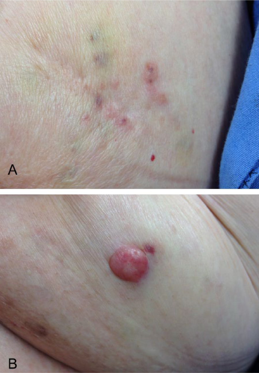
(A) Ecchymotic yellowish-blue macular cutaneous lesions and erythematous infiltrated papules. (B) Erythematous firm nodule.
The differential diagnosis included recurrence of breast carcinoma, cutaneous metastases, adnexal carcinoma, radiation-induced angiosarcoma, Kaposi sarcoma, or leiomyosarcoma. A 4-mm punch biopsy was performed from 3 different types of skin lesions mentioned above. Histopathology analysis revealed the same characteristics at these 3 sites: malignant proliferation in the dermis of ramified anastomotic vascular canals bordered by very atypical and highly mitotic endothelial cells. Anti–Pan Keratin antibodies were negative. Vascular markers such as anti-CD31, anti-CD34, D2-40, and anti–factor VIII were positive.
A total mastectomy was performed shortly afterward, and the patient is still in complete remission 20 months later.
Discussion
Angiosarcoma is a rare (<1% of all sarcomas) and highly malignant tumor of the vascular endothelium. Breast angiosarcomas, the second most common site for angiosarcoma, are commonly divided into 2 groups, primary and secondary. Primary breast angiosarcoma tends to arise without a recognized associated factor and usually affects younger women in their 20s to 40s. Secondary breast angiosarcoma is seen in older patients who have undergone radiotherapy for breast cancer or non-Hodgkin lymphoma. 3
In the literature, the interval between radiation and diagnosis ranges from 3 to 25 years, with a median latency period of 7 years. 4 The prevalence ranges between 0.002% and 0.005% per year, 5 but the incidence is thought to be about 0.3%. 6 Approximate incidence in breast cancer patients is about 0.9 in 1000. 4
A clear dose-response relationship for radiation-associated malignancies is not established, although it is generally accepted that sarcomas are induced in heavily radiated tissues in or close to the radiation fields. At the edge of the radiation fields, the dose of radiation is heterogeneous and may enable genetic mutations to progress into developing tumors. 7
Clinical Presentation
The clinical manifestation of this disease usually affects the dermis of the breast. Radiation-induced breast angiosarcoma may present as a single but more typically as multiple ecchymotic macular and/or purplish papular cutaneous lesions. 8 It can also present as a hematoma-like lesion resembling a bruise, palpable tumors, or erythematous nodules. 9 Less often, patients may present with metastatic disease, with possible spread to the contralateral breast, bones, brain, and skin. 10 When comparing primary and secondary breast angiosarcoma, as was done in a study of 70 patients with breast angiosarcoma, it was observed that primary angiosarcomas were more frequently well differentiated and localized at presentation compared with radiation-induced breast angiosarcomas. 3 Cahan et al 11 and later Arlen et al 12 proposed the following diagnostic criteria: (1) an initial malignant tumor of a different histology, (2) development of the sarcoma in an irradiated field, (3) a latency period of more than 3 to 4 years, and (4) the second malignancy must histopathologically be a sarcoma. Differential diagnosis includes benign hemangiomas, atypical vascular lesions after radiation, Kaposi sarcoma, epithelioid hemangioendothelioma, and hemangiopericytoma.
Investigation
Mammography may reveal nonspecific cutaneous thickening. Ultrasound may demonstrate hypervascularity and heterogeneous and hyperechoic masses of the breast. 13 Both mammography and ultrasound have high false-negative rates. 3 The best imaging option seems to be magnetic resonance imaging, which would reveal blood lakes and a rapidly enhancing heterogeneous mass.3-13 Magnetic resonance imaging also helps define the disease better than computed tomographic imaging.
Diagnostic confirmation is preferably made by an incisional punch biopsy at more than 1 site, to provide adequate material for histologic architectural and immunohistochemical evaluation.
Histologically well differentiated areas show irregular, anastomosing vascular channels lined by endothelial cells with different degrees of atypia and mitotic activity, with a characteristic dissection of collagen pattern that may alternate with less well differentiated areas of closely packed large pleomorphic cells with a high mitotic index and focally epithelioid and/or spindle cell morphology. 14 Suggested poor prognostic histologic features are mitotic rate, depth of invasion, and positive tumor margins. 15 Radiation-induced breast angiosarcoma typically expresses endothelial markers such as factor VIII–related antigen (von Willebrand), CD-34, CD-31, Ulex europaeus agglutinin 1, vascular endothelial growth factor, and, more recently, anti-Fli-1 protein antibody (a nuclear marker, as opposed to the others).13-15 On electronic microscopy, ovoid laminated organelle-like Weibel-Palade bodies may be seen. 15 Until 2010, the cytogenetics of angiosarcoma were poorly characterized. Manner et al 16 demonstrated that despite their identical morphology, secondary angiosarcomas are genetically different from primary angiosarcomas and are characterized by a higher frequency of amplification of MYC on chromosome 8q24.21. MYC regulates genes involved in cell growth, division, apoptosis, and angiogenesis. In 2015, Cornejo et al 17 confirmed that MYC gene amplification is a valuable ancillary tool, with sensitivity of 77% and specificity of 100% for radiation-induced breast angiosarcoma. A high level of FLT4 (VEGFR3) gene amplification was also related to radiation-induced breast angiosarcoma. 18
Treatment
Because of the rarity of this disease, there are no randomized controlled trials and few small prospective studies. Evidence to guide clinical practice is based mostly on single-institution cohort studies, retrospective studies, and case reports of heterogeneous groups of angiosarcomas not necessarily radiation induced.
The size of the primary lesion as well as the presence of distant metastasis are the main determinants for treatment options. Predictive factors for local recurrence consist of tumor size greater than 5 cm, previous radiation, histology with necrosis, mitotic figures per 10 high-power field, and combined morphologic score. These factors could help select patients most likely to benefit from more aggressive local or systemic adjuvant therapy. 19
Surgery
Radiation-induced breast angiosarcoma tends to have low-grade changes at the margins of the tumor that can be difficult to differentiate with histology from benign radiation changes. It is also known that it has a high probability of local recurrence. 20 For these 2 reasons, total mastectomy and local aggressive resection with widely negative margins are the first choices in therapeutic modalities. 21 Retrospective studies have shown that surgical margins of 1 cm provide successful local control and that 2- to 4-cm margins are necessary for proper disease clearance.22-24 Many studies noted no lymph node involvement, although it can happen, so sentinel-node biopsy is not routinely recommended. However, the local recurrence rate with surgery alone is high (50%-60%). 22
Chemotherapy
Cytotoxic chemotherapy is reserved for inoperable locally advanced or metastatic angiosarcoma. Globally different regimens demonstrate only short-lived response. A few trials displayed that doxorubicin and paclitaxel have some activity against different types of angiosarcoma.19-25 Doxorubicin, ifosfamide, and paclitaxel were studied for general soft tissue sarcoma, including a few nonspecified angiosarcomas, with some response but no analysis of survival. The addition of cisplatin, vinorelbine, and gemcitabine is of no proven benefit in angiosarcoma. 25 In a retrospective study with various regimens of doxorubicin for patients with advanced angiosarcoma, partial response but no improved survival was reported. 25 Over the past decade, there has been growing interest in taxane-based chemotherapy in angiosarcoma, but partial response and survival for only a few months were noted. 19
Recently, molecularly targeted therapies have been investigated as alternatives to conventional chemotherapy for angiosarcomas. More specifically, antiangiogenic therapies such as sorafenib (a tyrosine kinase inhibitor) and bevacizumab (a humanized monoclonal antibody against vascular endothelial growth factor A) have shown some activity in angiosarcoma.18,19 A few case reports have shown some response with other antiangiogenic agents, including thalidomide 26 and metronomic trofosfamide. 27
Radiotherapy
Because few studies have evaluated radiotherapy in these patients, the role of adjuvant radiotherapy remains controversial. In a recent retrospective cohort study, Palta et al 28 demonstrated overall 5-year survival of 86% in 14 patients with radiation-induced breast angiosarcoma who had undergone hyperfractionated and accelerated radiotherapy. Hyperthermia could be a strategy to augment the benefits of radiotherapy. 19
A second course of radiotherapy raises concerns about toxicity, such as rib fracture, pneumonitis, and soft tissue necrosis.
Prognosis
Overall, radiation-induced breast angiosarcoma has poor outcomes, with very few patients surviving for 5 years. Exact numbers vary in the current literature, and data come from studies of heterogeneous groups of angiosarcoma not necessarily radiation induced.
Conclusions
We report a case of radiation-induced breast angiosarcoma, a complication of breast radiotherapy that is important to recognize. Histopathologic markers reported in the literature, such as factor VIII–related antigen (von Willebrand), CD-34, CD-31, Ulex europaeus agglutinin 1, and vascular endothelial growth factor, help confirm the diagnosis. Cytogenetic analysis seems to demonstrate high levesl of MYC and FLT4 gene amplification. Because radiation-induced breast angiosarcoma can develop many years after radiation therapy, it is important to remain vigilant to any cutaneous changes and to educate patients about presenting signs and symptoms. Surgery is the main curative treatment, and there is no compelling evidence for adjuvant radiotherapy or chemotherapy. Chemotherapy is given mainly in a palliative context. Antiangiogenic therapies offer hope, but further prospective studies are needed.
Footnotes
Declaration of Conflicting Interests
The author(s) declared no potential conflicts of interest with respect to the research, authorship, and/or publication of this article.
Funding
The author(s) received no financial support for the research, authorship, and/or publication of this article.