
Abstract
Background:
Ki-67 is an immunohistochemical stain used as a nuclear proliferation marker. It is nonspecific, and is expressed in all active phases of the cell cycle. Vulvar vestibular papules in women and pearly penile papules in men are benign fibrous papules on the genitals, are noninfectious, and do not require treatment. However, these lesions can be clinically confused with condylomata acuminata induced by human papillomavirus (HPV), which have medical and social implications.
Objective:
Because HPV infection is known to induce expression of proliferation markers, we propose that Ki-67 be used to differentiate condylomata acuminata from vulvar vestibular papules or pearly penile papules on pathologic examination.
Methods:
We reviewed a total of 26 lesions from 18 patients of previously pathologically diagnosed lesions, including condylomata acuminata (11 lesions), vulvar vestibular papules (10 lesions), and pearly penile papules (5 lesions). All slides were stained with Ki-67, reviewed, and categorized as positive or negative for Ki-67 staining by 1 investigator who was unaware of the original diagnosis.
Results:
Eleven out of 11 cases of condylomata acuminata were identified as positive for Ki-67 staining. Ten out of 10 cases of vulvar vestibular papules were negative for Ki-67. Five out of 5 cases of pearly penile papules were negative for Ki-67.
Conclusion:
Ki-67 is a reliable marker to pathologically distinguish benign vulvar vestibular papules in women, or pearly penile papules in men, from HPV-induced condylomata acuminata.
Introduction
Ki-67 is an immunohistochemical stain used as a nuclear proliferation marker. It is nonspecific and is expressed in all active phases of the cell cycle. Vulvar vestibular papules (VVP) in women and pearly penile papules (PPP) in men are benign, noninfectious, fibrous papules on the genitals and do not require treatment. However, these lesions can be clinically confused with condylomata acuminata induced by human papillomavirus (HPV), which have medical and social implications.1,2
Condylomata acuminata may be overdiagnosed when morphologic and pathologic examination is equivocal, and Ki-67 can be helpful for more accurate diagnosis. 3 The current standard for diagnosis is haematoxylin and eosin (H&E) staining only, traditionally based predominantly on the difficult judgement whether or not koilocytes are present. Koilocytes are often absent in condylomata, and various artefacts can mimic koilocytes in non–HPV-induced conditions. Because HPV infection is known to induce expression of proliferation markers, we propose that Ki-67 may be used to reliably differentiate condylomata acuminata from VVP or PPP on pathologic examination.
Methods
We reviewed 26 lesions from 18 patients of previously pathologically diagnosed lesions, including condylomata acuminata (11 lesions), VVP (10 lesions), and PPP (5 lesions). Cases were identified by searching the Vancouver Coastal Health Authority laboratory database for patients with the phrase “vestibular papillomatosis,” “pearly penile,” or “condyloma” in the final diagnosis on pathology reports. Cases with a compatible clinical history and preferred pathologic diagnosis of the corresponding search were included. Only cases from Vancouver General Hospital were included. Four cases were excluded (2 VVP, 1 PPP, and 1 condylomata acuminata) because paraffin-embedded material was no longer available. One case of PPP was excluded because features suspicious for condylomata acuminata were mentioned on review of the pathology report.
All slides were stained with Ki-67, and reviewed by 1 investigator who was unaware of the original diagnosis. The slides were then categorized as positive or negative for Ki-67 staining. A positive Ki-67 stain was defined as having positive Ki-67 staining of nuclei in the upper half of the epithelium, larger than the average nucleus for the same level (Figure 1); H&E staining of condyloma acuminata immediately adjacent to the section in Figure 1 is shown for comparison (Figure 2). The stain was considered negative if it stained only in the lower half of the epithelium, as would be expected for normal proliferative cell staining of the basal and suprabasal level (Figure 3). Ki-67 staining of normal skin is shown for comparison (Figure 4). Institutional review board approval was waived.
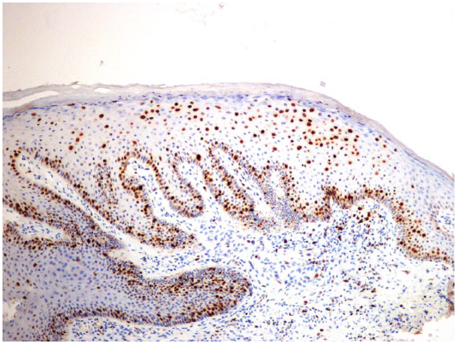
Condyloma acuminata. Ki-67 ×100.
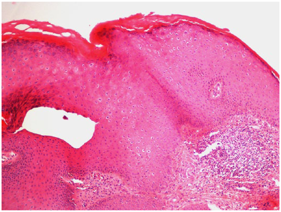
Condyloma acuminata. Haematoxylin and eosin ×100.
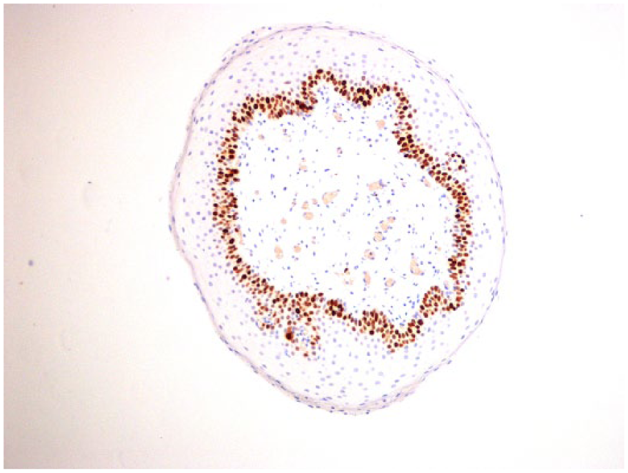
Vulvar vestibular papilloma. Ki-67 ×100.
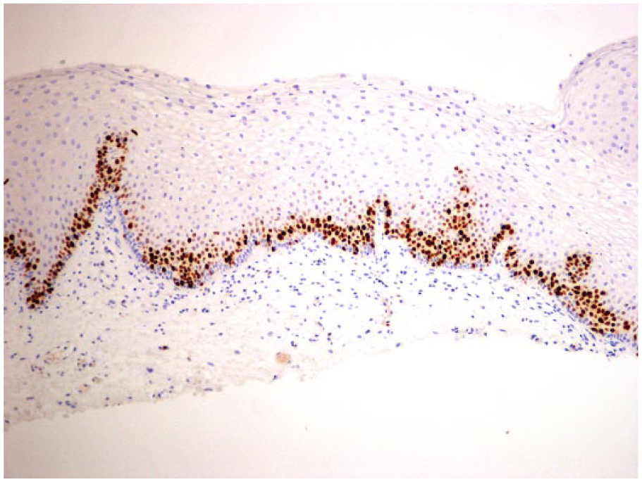
Normal skin. Ki-67 ×100.
Results
Eleven out of 11 cases of condylomata acuminata were identified as positive for Ki-67 staining. Ten out of 10 cases of VVP were negative for Ki-67. Five out of 5 cases of PPP were negative for Ki-67.
Discussion
Ki-67 staining was 100% accurate in differentiating condylomata acuminata from VVP and PPP. Special attention must be paid to not erroneously identify a positive result when a dermal papilla rises high in the epithelium because of a tangential cut. This is a risk for a false-positive result; however, care can be taken when examining slides to look for increased density and configuration, as well as smaller size of cells, which suggest basal epithelium rather than a true positive Ki-67 stain. One limitation was the relatively small sample size. Another limitation is the lack of corroboration by a second gold standard (such as in situ hybridization or polymerase chain reaction for specific HPV subtypes). In situ hybridization and polymerase chain reaction can have limited clinical use because of their low sensitivity and high cost, respectively.
Our results indicate that Ki-67 is a reliable marker to pathologically distinguish benign VVP in women, or PPP in men, from HPV-induced condylomata acuminata. Ki-67 is a simple, quick, inexpensive test that is widely used and available on site in almost any laboratory.
Footnotes
Declaration of Conflicting Interests
The author(s) declared no potential conflicts of interest with respect to the research, authorship, and/or publication of this article.
Funding
The author(s) received no financial support for the research, authorship, and/or publication of this article.